This article contains AI generated content.
Non-Ossifying Fibroma Genes And Biomarkers: 3 Genes And 6 Biomarkers To Track
If your child (or you, as an adult reminiscing about an old x-ray) has been told about a "non-ossifying fibroma," you've probably already noticed the strange gap in available information. Radiology reports use confident language — "benign," "incidental," "no treatment needed" — while parents are left wondering what actually caused the lesion, whether it says anything about their child's biology, and whether there's anything meaningful to monitor beyond "wait and see."
Generic reassurance isn't wrong, but it's often incomplete. "It's benign, it'll go away" is true for most cases, yet it skips over the more interesting and more useful question: what is actually happening at the cellular level, and are there specific markers — genetic or biochemical — worth understanding while the lesion runs its natural course. Most patient handouts don't go there, not because the science doesn't exist, but because it's newer, more technical, and harder to summarize in a two-minute clinic visit.
This article goes deeper. Over the last several years, researchers have identified the specific molecular pathway driving non-ossifying fibroma, reclassifying it from a passive "developmental quirk" into what is now understood as a true, low-grade neoplasm with identifiable genetic drivers. That single shift in understanding changes how the condition should be thought about, monitored, and discussed with an orthopedic team.
None of this replaces medical care, and none of it turns a benign bone lesion into something it isn't. But better information — about the genes involved, and about the practical biomarkers and imaging metrics that matter for fracture-risk monitoring — tends to produce calmer, more informed decisions. This article covers both angles: the genetics and pathway biology behind non-ossifying fibroma first, in depth, followed by a shorter, practical rundown of the blood and imaging markers worth knowing about, a look at how a broader longevity framework applies to growing, resilient bones, and a review of which supportive therapies actually have evidence behind them for children going through this.
Summary
Non-ossifying fibroma was long taught as a "leave it alone" developmental defect — not a real tumor, just a quirk of growing bone. Genetic sequencing published in the last several years overturned that idea. Three genes — KRAS, FGFR1, and NF1 — turn out to drive the lesion through a single shared signaling pathway, and knowing which one is involved (and whether it's a one-off tissue mutation or part of a broader inherited condition) actually changes the monitoring conversation. Beyond the genetics, there's a practical layer most families never hear about: the blood tests and imaging measurements that give a clearer picture of bone strength and fracture risk than "it looks fine" ever could. This article walks through both — what the genes suggest, what can realistically be done in response, which six biomarkers are worth tracking, how a proactive longevity framework applies to a growing skeleton, and which supportive therapies have real evidence behind them for kids navigating imaging, anxiety, or recovery. The goal isn't to alarm anyone about a lesion that resolves on its own in the overwhelming majority of cases — it's to replace vague reassurance with an actual understanding of what's going on, and what's worth watching.
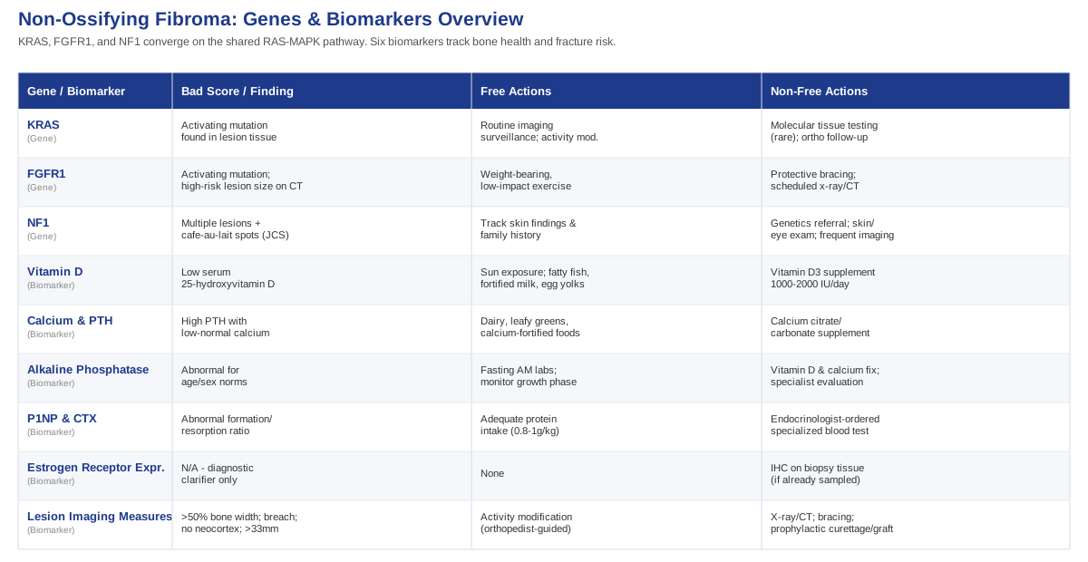
What the RAS-MAPK Pathway Reveals About Non-Ossifying Fibroma
The field of personalized genomics — the kind of work researchers like Ali Torkamani have built careers on, and the kind that figures like Gary Brecka have popularized for a general audience — has taught a useful lesson that applies well beyond common inherited variants: a "concerning" gene finding is rarely a fixed sentence. Context, cell type, and biological environment usually matter more than the mutation label itself. Non-ossifying fibroma is a good example of a condition where that lesson applies in an unusual way, because the mutations involved aren't the common inherited variants that a 23andMe-style report would flag. They're somatic — meaning they occur in the cells of the lesion itself, not in every cell of the body — which changes what "fixing" the gene can even mean.
A Quick Primer: Why Non-Ossifying Fibroma Is Now Considered a True Neoplasm
For decades, non-ossifying fibroma (and its smaller cousin, the fibrous cortical defect) was taught as a benign, self-limited quirk of bone growth — not a real tumor, just disorganized fibrous tissue that would ossify and disappear as a child's skeleton matured. That changed with a 2019 whole-exome sequencing study led by Baumhoer and colleagues, which found that a large majority of these lesions carry activating mutations in a single shared signaling cascade known as the RAS-MAPK pathway — the same broad family of pathways implicated in neurofibromatosis and several other "RASopathies" (Non-ossifying fibroma: a RAS-MAPK driven benign bone neoplasm, J Pathol 2019). That finding reclassified non-ossifying fibroma from a reactive process into a genuine, if low-grade and self-limited, neoplasm — one driven by identifiable mutations in three genes: KRAS, FGFR1, and NF1.
KRAS: What It Does and Why It Shows Up in the Lesion
KRAS is one of the most well-studied genes in all of cancer biology because it acts as a molecular "on switch" for cell growth signaling. In non-ossifying fibroma, activating hotspot mutations in KRAS were found in a substantial share of lesions, and they behave in a mutually exclusive pattern with FGFR1 mutations — meaning a given lesion is typically driven by one or the other, not both. The mutation locks the growth signal in the "on" position within the affected tissue, encouraging the fibrous, non-mineralized tissue that defines the lesion. Evidence here is strong for the association (multiple independent sequencing cohorts, replicated in the same 2019 paper), though it remains a relatively young field — there is no long-term outcomes data yet tying specific KRAS variants to fracture risk or recurrence.
If a KRAS mutation is identified on tissue analysis (something that would only happen if a biopsy or curettage sample is sent for molecular testing, which is uncommon for a routine, low-risk lesion), the plan without supplements is straightforward: continued clinical and radiographic surveillance, typically every six to twelve months until the lesion shows sclerotic healing or the growth plate closes, per natural-history data on how these lesions resolve over time (Non-ossifying fibroma: natural history, stage-related growth, and fracture risk, Skeletal Radiol). Activity modification — avoiding high-impact collision sports if the lesion is large relative to the bone — is the other lever, guided by an orthopedic surgeon rather than self-directed.
The plan with supplements or equipment is really a bone-health support plan rather than a gene-targeting one, since nothing ingested changes a somatic mutation confined to lesion tissue. Ensuring adequate dietary calcium (roughly 700mg/day for ages 1-3, 1000mg/day for ages 4-8, and 1300mg/day for ages 9-18, per standard pediatric nutrition guidelines) and correcting any vitamin D insufficiency supports the surrounding bone that will eventually remodel the defect. Vitamin D3 is typically dosed at 600-1000 IU/day for maintenance in children, or 1000-2000 IU/day for 8-12 weeks if a blood test shows deficiency, then rechecked — chronic doses above 4000 IU/day in children risk hypercalcemia and should only be used under pediatric supervision. Equipment-wise, the only genuinely useful "device" is the follow-up imaging itself (plain x-ray, occasionally low-dose CT for pre-surgical planning), not a supplement stack or a bone-growth gadget — there is no equipment shown to accelerate resolution of the lesion itself.
FGFR1: The Other Half of the Mutually Exclusive Pair
FGFR1 (fibroblast growth factor receptor 1) sits upstream of the same RAS-MAPK cascade. Activating mutations here produce a similar downstream effect to KRAS mutations — persistent growth signaling within the lesion — and, as noted above, the two mutations essentially never occur together in the same lesion, suggesting either one is sufficient to drive the fibrous overgrowth. This is again strong molecular evidence from the same sequencing cohort, but clinical correlation (does an FGFR1-driven lesion behave differently in terms of size, fracture risk, or recurrence than a KRAS-driven one) is still an open, actively studied question rather than settled fact.
The plan without supplements mirrors the KRAS approach almost exactly: scheduled imaging rather than reactive imaging, and orthopedic-guided activity modification if the lesion meets higher-risk size criteria — specifically, lesions occupying more than half the bone's width on both the coronal and sagittal CT views, showing any cortical breach, or lacking a "neocortex" (a thin rim of new bone reinforcing the defect) are considered higher risk for pathologic fracture and warrant closer follow-up or prophylactic treatment (Nonossifying fibromas: a CT-based criteria to predict fracture risk).
The plan with supplements or equipment again centers on general skeletal support rather than pathway-specific correction. Weight-bearing, low-impact activity (walking, swimming transitioning to light jogging, resistance-band work) three to five times a week supports bone mineral accrual in a growing child, but should be cleared by the treating orthopedist first if the lesion falls into a higher fracture-risk category. Protective bracing or activity restriction — genuine "equipment" in this context — is sometimes recommended for a period of weeks to months for large lesions before a return to contact sports, based on the surgeon's assessment rather than a fixed protocol. There is no supplement, herbal compound, or peptide with evidence for altering FGFR1 signaling in this context, and claims to the contrary should be treated skeptically.
NF1: When a Single Lesion Might Be Part of a Bigger Picture
NF1 is a different category of finding entirely, and this is the one place where the distinction between a somatic (tissue-only) mutation and a germline (inherited, whole-body) mutation actually matters for a family. Isolated non-ossifying fibromas can show inactivating NF1 mutations confined to the lesion, which — like KRAS and FGFR1 — simply removes a brake on the same RAS-MAPK pathway. But when a child has multiple non-ossifying fibromas together with café-au-lait skin macules, and sometimes with jaw giant-cell lesions, that combination has a name: Jaffe-Campanacci syndrome, and it overlaps substantially with neurofibromatosis type 1 (NF1), a germline condition. A 2020 case report specifically documented an NF1 gene mutation detected within non-ossifying fibroma tissue in a child with this exact phenotypic overlap (Jaffe-Campanacci syndrome or neurofibromatosis type 1: a case report with NF1 gene mutation in non-ossifying fibroma), and a broader clinical review has examined the biologic overlap between the two conditions in detail (Non-ossifying fibroma, fibrous cortical defect, and Jaffe-Campanacci syndrome: a biologic and clinical review). Multiple lesions in this pattern also carry a meaningfully higher combined fracture risk, since more than one weak point can exist in the same or different bones (Multiple non-ossifying fibromas as a cause of pathological femoral fracture in Jaffe-Campanacci syndrome).
Here, the plan without supplements is not a wellness plan at all — it's a referral. A single non-ossifying fibroma needs nothing beyond routine orthopedic follow-up. Multiple lesions, especially alongside café-au-lait spots, freckling in the armpits or groin, or a family history of neurofibromatosis, warrant referral to a clinical geneticist and a comprehensive skin and eye exam, because NF1 carries implications well beyond bone (including monitoring for other tumor types) that no lifestyle change addresses.
The plan with supplements or equipment, in this specific case, is really about supportive bone health during a longer surveillance window rather than any attempt to influence the NF1 finding itself. Because multiple NOFs raise cumulative fracture risk, more frequent imaging (often every six months rather than annually) is the main "equipment" involved, alongside firmer activity guidance from the orthopedic team. Vitamin D and calcium sufficiency remain reasonable general supports, dosed and monitored exactly as described above, with blood levels rechecked roughly every three to six months until stable. Nothing here should be self-directed — this is the one branch of the three genes where specialist coordination, not a supplement plan, is the actual answer.
Putting the Three Genes in Context
Taken together, KRAS, FGFR1, and NF1 tell a consistent story: non-ossifying fibroma is driven by one pathway, activated through different genetic doors, almost always confined to the lesion itself, almost always self-limited, and only occasionally a signal of something broader. The practical takeaway for a parent or adult patient isn't to chase molecular testing for a routine, low-risk incidental finding — that's rarely done and rarely necessary. It's to recognize the pattern that would justify a closer look: multiple lesions, skin findings, or a family history, any of which shifts the conversation from "watch and wait" to "let's involve genetics."
Beyond Genetics: Blood and Imaging Markers Worth Watching
Because the genetic drivers of non-ossifying fibroma live inside the lesion tissue rather than circulating in blood, there's no blood test that diagnoses or tracks the mutation itself. But that doesn't mean there's nothing to measure. The markers below aren't NOF-specific in the way a tumor marker is specific to a cancer — they're general bone-health and imaging metrics that, taken together, give a far more concrete picture of fracture risk and skeletal resilience than a verbal "it looks stable" ever could, echoing the broader philosophy that figures like Peter Attia, Thomas Dayspring, and Allan Sniderman have pushed in adult preventive medicine: measure the mechanism, not just the outcome.
Serum 25-Hydroxyvitamin D
This is the standard measure of vitamin D status and it matters because vitamin D governs how efficiently a growing skeleton absorbs and uses calcium — directly relevant to how well the bone around a fibroma remodels and reinforces itself. How to measure it: a simple blood draw, widely available through primary care or direct-to-consumer labs, typically costing $40 to $80 out of pocket if not covered by insurance. Interestingly, research in prepubertal children found that low vitamin D levels didn't clearly worsen bone turnover markers in that age group (Low serum 25-hydroxyvitamin D level does not adversely affect bone turnover in prepubertal children), a useful reminder not to over-interpret a single low value in a young child.
If the score is bad, the plan without supplements starts with sun exposure guidance (fifteen to twenty minutes of midday skin exposure several times a week, balanced against skin-cancer-prevention advice) and dietary sources — fatty fish, fortified milk, and egg yolks. If the score is bad, the plan with supplements is a measured one: vitamin D3, 1000-2000 IU/day for eight to twelve weeks, rechecked with a follow-up blood draw, then tapered to a maintenance dose of 600-1000 IU/day. Side effects are rare at these doses but include gastrointestinal upset; toxicity (hypercalcemia, nausea, kidney strain) is a real risk only at sustained high doses and should prompt stopping supplementation and consulting a pediatrician.
Calcium and Parathyroid Hormone (PTH)
Calcium and PTH work as a pair — PTH rises when calcium is low, pulling calcium out of bone to protect blood levels, which is the opposite of what you want during a phase of active bone remodeling. How to measure it: a basic metabolic panel plus an intact PTH test, typically $30 to $70 combined, available through most labs. A high PTH with normal or low calcium is the pattern worth flagging to a physician, since it suggests the body is compensating for inadequate intake or absorption.
If the score is bad, the plan without supplements is largely dietary: dairy, leafy greens, and calcium-fortified foods, spread across the day rather than in one large dose, since absorption is more efficient that way. If the score is bad, the plan with supplements uses calcium citrate or carbonate, split into two doses of 500mg or less taken with food (the body absorbs calcium poorly in larger single doses), continued until follow-up labs normalize, generally rechecked in three months. Side effects include constipation and, at excessive intake, a small increase in kidney stone risk — reasons to treat calcium supplementation as a correction, not a default, and to favor food sources first.
Alkaline Phosphatase (Bone-Specific ALP)
Alkaline phosphatase is a marker of osteoblast activity — the cells actively building new bone — and it's naturally elevated in growing children, which is exactly why it's useful here: a level that's unusually high or low relative to age-adjusted norms can flag unusually active or unusually sluggish bone remodeling around a lesion. How to measure it: included in most standard metabolic panels, often $20 to $40 as a standalone test, though it requires age- and sex-specific reference ranges to interpret correctly given how much it shifts through childhood and puberty.
If the score is bad, the plan without supplements is mostly about ruling things out rather than fixing something directly — persistent abnormal ALP warrants a conversation with the treating physician about whether it reflects normal growth-spurt physiology or something to investigate further, since ALP is a nonspecific marker. If the score is bad, the plan with supplements loops back to the same vitamin D and calcium sufficiency approach described above, dosed identically, since both nutrients are cofactors in the mineralization process ALP reflects. There's no equipment upgrade here beyond ensuring labs are drawn fasting in the morning, since bone markers show meaningful diurnal variation.
P1NP and CTX (Bone Turnover Markers)
P1NP reflects bone formation and CTX reflects bone resorption; together they give a formation-versus-breakdown ratio that's more sensitive to short-term change than a bone density scan, which only shows accumulated results months later. These markers are strongly age-dependent in children, with reference ranges still being refined in pediatric research (Bone turnover markers, growth, and bone parameters in infants participating in a vitamin D intervention study). How to measure it: a specialized blood draw, typically $80 to $150 combined, usually ordered through an endocrinologist rather than a general practitioner, and best drawn fasting in the early morning due to diurnal swings.
If the score is bad, the plan without supplements involves working with a pediatric endocrinologist to interpret the ratio in context rather than reacting to a single number, since these markers are influenced by growth velocity, puberty stage, and recent fractures, not just the fibroma. If the score is bad, the plan with supplements is again nutritional — adequate protein intake (roughly 0.8-1g per kg of body weight daily, more during growth spurts) supports the collagen matrix these markers are measuring, alongside the vitamin D and calcium approach already described. This is a monitoring tool for specialists managing complex or multiple lesions, not a routine test for an isolated, low-risk finding.
Estrogen Receptor Expression on Tissue Biopsy
This one is unusual: it's not a blood test but a tissue-based biomarker used by pathologists. Recent research found that non-ossifying fibroma reliably expresses estrogen receptor (ER) on immunohistochemistry, a feature that helps distinguish it from other giant-cell-containing bone tumors that can look similar under the microscope (Oestrogen receptor expression distinguishes non-ossifying fibroma from other giant cell containing bone tumours). How to measure it: only relevant if tissue is already being taken for another reason (biopsy or curettage before bone grafting), where it adds no extra cost or risk since it's run on the same sample. There's no "plan" to improve this marker — it's a diagnostic clarifier, not a target — but it's worth knowing it exists, since it's part of why a pathologist may sound confident distinguishing a benign fibroma from a lesion that needs more aggressive management.
Imaging-Based Lesion Measurements
The single most clinically useful "biomarker" for non-ossifying fibroma isn't in blood at all — it's the size and shape of the lesion relative to the bone on plain x-ray or CT. Lesions occupying more than half the bone's width in both the coronal and sagittal planes, longer than roughly 33mm, showing a cortical breach, or lacking a protective neocortex are associated with meaningfully higher fracture risk, and staging systems built around these features help decide whether watchful waiting or prophylactic curettage and grafting is the safer path (Non-ossifying fibroma: natural history with an emphasis on stage-related growth and fracture risk). How to measure it: a plain x-ray costs roughly $50 to $150; a CT scan for more precise pre-surgical measurement runs $300 to $1,500 depending on region and insurance.
If the score is bad, the plan without supplements is activity modification set by the orthopedic surgeon — avoiding contact sports and high-impact loading on the affected limb until repeat imaging shows sclerotic healing or growth of a protective cortex. If the score is bad, the plan with equipment includes protective bracing during a defined window and, for the higher-risk category, prophylactic curettage and bone grafting, which is a surgical decision made jointly with an orthopedic surgeon rather than something to pursue independently. Follow-up imaging every six to twelve months remains the backbone of monitoring regardless of which path is chosen.
What Peter Attia's Outlive Teaches About Protecting a Growing Skeleton
Peter Attia's book Outlive: The Science and Art of Longevity isn't written about non-ossifying fibroma, and it doesn't need to be to be useful here. Its core argument — that most of medicine reacts to disease that's already advanced, while a smarter approach tracks and trains the body's structural resilience years before a crisis — maps unusually well onto how a family should think about a growing skeleton with a known area of relative weakness. The following ten ideas are drawn from the book's framework and applied specifically to fracture-risk thinking and long-term bone health.
Medicine 3.0 Means Acting Before the Fracture, Not After
Attia's central distinction between "Medicine 2.0" (treat disease once it appears) and "Medicine 3.0" (identify and reduce risk long before a crisis) is exactly the mindset a fracture-risk scoring system for non-ossifying fibroma already embodies — using imaging criteria to intervene before a bone breaks, rather than waiting for a fracture to force the decision.
The Centenarian Decathlon Reframes What "Healthy Bones" Are For
Attia asks readers to define the physical tasks they want to still perform decades from now, then train backward from that goal. For a growing child, this translates into thinking about lifelong bone strength and injury resilience, not just clearing the current lesion — the goal is a skeleton that serves an active life for the next seventy years, not just a clean-looking x-ray next year.
Bone Density Is an Overlooked Pillar of Longevity
One of the book's more surprising arguments is that bone mineral density deserves the same attention as cardiovascular risk, because fractures — especially in older age — are a major driver of loss of independence. Building strong bone-health habits early, while a family is already paying close attention because of an incidental lesion, is a genuine long-term asset.
VO2 Max Is Framed as the Single Most Powerful Longevity Marker
While not a bone marker directly, Attia argues cardiorespiratory fitness predicts long-term mortality risk more strongly than almost any other single measure — a reminder that once fracture-risk activity restrictions lift, returning to varied cardiovascular activity (running, cycling, swimming) matters for whole-body health, not just the healed bone.
Grip Strength and Muscle Mass Predict Resilience
Attia treats muscle strength, and grip strength specifically, as an accessible proxy for overall physical robustness. For a child recovering activity levels after a period of restriction, rebuilding strength gradually (resistance bands, bodyweight work) supports the muscles that protect bone during everyday falls and missteps.
Zone 2 Training Builds the Metabolic Base Bone Repair Depends On
Attia's emphasis on low-intensity, sustained aerobic training (Zone 2) as a metabolic foundation applies loosely here: consistent, moderate activity supports the circulation and metabolic health that underlie efficient tissue repair, complementing rather than replacing higher-intensity training once cleared.
Stability and Balance Training Prevent the Falls That Cause Fractures
A recurring theme in the book is that most serious fractures in older adults stem from falls, not disease — meaning balance and coordination training is a fracture-prevention tool in its own right. For an active child, agility and balance work (which many youth sports naturally include) plays a similar protective role around a healing lesion.
Track Objective Data Over Time Instead of Waiting for Symptoms
Attia is emphatic that waiting for symptoms means waiting too long. Scheduled follow-up imaging for a non-ossifying fibroma is a direct application of this principle — checking the lesion on a calendar, not just when something starts to hurt.
Protein Intake Deserves More Attention Than Most People Give It
The book pushes back on outdated protein guidelines, arguing most people — especially active, growing individuals — benefit from more dietary protein than commonly assumed, since it's the raw material for the collagen matrix bone is built on.
Sleep and Emotional Health Are Force Multipliers for Physical Recovery
Attia closes much of his practical advice by returning to sleep and emotional well-being as underrated multipliers of every other intervention. For a family managing the stress of a diagnosis, monitoring, or a recovery period after a procedure, this is a genuine and easily overlooked lever.
Complementary Approaches That Support Kids (and Parents) Through the Process
None of the following therapies treat non-ossifying fibroma itself, and no clinical trial has tested any complementary approach against the lesion directly — that evidence simply doesn't exist for this specific condition. What does exist is meaningful pediatric evidence for reducing the anxiety, pain, and stress that surround imaging, monitoring, and, for higher-risk lesions, surgical procedures like curettage and bone grafting. These three have the clearest support.
Guided Imagery
Guided imagery uses a scripted, sensory-rich mental visualization — imagining a calm, specific scene in detail — to redirect attention away from anxiety and pain signals. For a child facing repeated follow-up x-rays, an MRI, or a surgical procedure for a larger lesion, this is directly relevant: procedural anxiety is one of the most common real-world burdens of a "just monitor it" plan that still involves needles, scanners, and unfamiliar rooms.
A randomized study of sixty children aged six to twelve undergoing minor surgery found that a relaxation-guided imagery session before anesthesia induction produced significantly less anxiety and less pain than standard care alone (Relaxation-guided imagery reduces perioperative anxiety and pain in children: a randomized study), and a broader systematic review of non-pharmacological therapies used by nurses in children undergoing surgery found consistent support across guided imagery, distraction, and related techniques (A systematic review of the effectiveness of non-pharmacological therapies used by nurses in children undergoing surgery).
In practice, a fifteen-minute recorded script, played through headphones in the waiting room or pre-op area, costs nothing beyond a free app or a hospital's own child-life resources — many pediatric hospitals already have child-life specialists trained in exactly this. It's low-risk and worth requesting specifically before any procedure related to a fibroma, even a routine follow-up MRI that requires a child to lie still.
Massage Therapy
Massage therapy, applied to unaffected areas around a surgical site, is sometimes used in pediatric post-operative recovery to reduce anxiety and perceived pain, working through relaxation and touch rather than any direct effect on healing bone.
Evidence is mixed but promising: a pilot study at a children's hospital found that massage therapy reduced anxiety scores and lowered total benzodiazepine use in pediatric surgical patients compared to standard care, while a smaller pilot in children with cerebral palsy undergoing orthopedic surgery found more modest effects, underscoring that results vary by context and that this remains an adjunct, not a primary therapy.
Realistically, this means asking a hospital's child-life or integrative care team whether pediatric massage is available post-operatively, rather than seeking it out independently during the routine, non-surgical monitoring phase of a small, low-risk fibroma — it earns its place around a procedure, not around a lesion that's simply being watched.
Mindfulness-Based Approaches
Mindfulness and related relaxation-based techniques teach a child (or an anxious parent) to notice anxious thoughts without being swept up in them — useful for the specific, recurring stress of "the next scan is in six months" that comes with monitoring any bone lesion over a period of years.
A recent systematic review and meta-analysis of mindfulness interventions in children and adolescents with mental health and psychiatric concerns found generally positive, though still maturing, evidence across randomized trials (Mindfulness in mental health and psychiatric disorders of children and adolescents: a systematic review and meta-analysis of randomized controlled trials), with the caveat that pediatric-specific chronic pain and procedural-anxiety trials remain fewer and smaller than the adult literature.
A practical starting point is a short, age-appropriate guided mindfulness app (many pediatric hospitals now recommend specific ones) used for five to ten minutes before an appointment, not as a daily therapeutic regimen but as a targeted tool for the specific moments — scan day, pre-op morning — when anxiety tends to spike.
The Bottom Line
Non-ossifying fibroma remains, in the overwhelming majority of cases, exactly what it's always been described as: a benign lesion that resolves on its own as a child's skeleton matures. What's changed is the depth of understanding underneath that reassurance. Three genes — KRAS, FGFR1, and NF1 — converge on one shared growth pathway, explaining why the lesion forms and clarifying the rare situations (multiple lesions, skin findings, family history) where a broader genetic evaluation is actually warranted. Alongside that, a handful of practical blood and imaging markers give a far clearer read on bone strength and fracture risk than a verbal reassurance ever could, and a long-term, proactive framework for skeletal health pays off well beyond the lesion itself.
The next useful step isn't more worry — it's more precision. Ask the treating orthopedist where the current lesion falls on standard fracture-risk imaging criteria, request a vitamin D and calcium check if none has been done recently, and, if more than one lesion or any skin findings are present, ask directly about a genetics referral. Small, specific questions like these tend to produce far more useful answers than a general "is everything okay."
Musculoskeletal Endocrine & Metabolic
Musculoskeletal: Bone Conditions