This article contains AI generated content.
· UpdatedOccupational Overuse Arthritis — 6 Genes And 7 Biomarkers To Track
Introduction
If you spend most of your working day performing the same motions — gripping, lifting, typing, using vibrating tools, or kneeling on hard surfaces — and you have started noticing joint stiffness, tenderness, or swelling that simply does not go away with a weekend of rest, you already know something is wrong. What most people are not told is that the line between temporary overuse strain and the onset of chronic occupational arthritis is not the same for everyone. Two people doing the same job for the same number of years can have completely different outcomes, and that gap is not just explained by posture or workload.
Generic advice about ergonomics and anti-inflammatories is useful as a starting point, but it rarely explains why some people recover quickly while others continue to accumulate joint damage. The truth is that your cartilage repair capacity, your baseline inflammatory response, your collagen quality, and your metabolic tendencies all interact in ways that vary significantly from person to person. Understanding your individual biology is not a luxury — it is the missing layer that makes the rest of the guidance actually work.
This article takes a more targeted approach. Rather than reviewing what everyone already knows about rest and ice packs, it looks at two layers of biology that most clinical advice leaves out: specific inflammatory and structural biomarkers you can measure with a blood or urine test, and the genetic variants that shape how your joints respond to mechanical stress and inflammation over time. Both levels of information are actionable. Knowing where you stand on each gives you something to track, something to correct, and a clearer framework for making decisions with a qualified professional.
The goal here is not to promise a cure or replace medical evaluation. It is to help you ask better questions and act on better information. The biomarker section shows you seven measurable signals that tell you what is currently happening in your joints. The genetics section shows you the deeper tendencies that explain why it is happening. Taken together, they offer a more complete map — and a clearer path forward.
Summary
This article covers seven biomarkers you can actually measure — including hsCRP, IL-6, urinary CTX-II, uric acid, vitamin D, homocysteine, and ferritin — with clear guidance on how to test them, what a bad score means, and what to do about it, both with and without supplements. It then covers six gene variants that shape your joint vulnerability at a structural and inflammatory level: COL1A1, IL1B, VDR, MMP3, GDF5, and TNFA. For each gene, there is a practical correction plan. Beyond that, you will find a breakdown of Andrew Huberman's collagen synthesis protocol and what it means for joint recovery, along with evidence-based complementary therapies — including low-level laser therapy, tai chi, massage, and MBSR — that have specific clinical support for this condition. If you have been managing occupational joint pain without knowing your numbers, this article gives you the framework to start doing so.
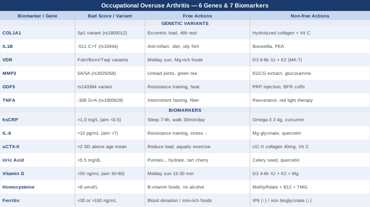
7 Biomarkers to Track for Occupational Overuse Arthritis
Biomarkers do not diagnose occupational arthritis on their own, but they tell you what is happening inside the joint environment right now. They reveal whether inflammation is active or dormant, whether cartilage is being broken down faster than it is being repaired, whether metabolic triggers are driving flares, and whether nutrient deficiencies are leaving your joint structures vulnerable. The seven below are selected for their clinical relevance to overuse-related joint pathology, their affordability, and their actionability.
Biomarker 1: hsCRP (High-Sensitivity C-Reactive Protein)
Why it matters. C-reactive protein is produced by the liver in response to inflammation signals, and the high-sensitivity version detects low-grade chronic inflammation that the standard CRP test misses. In occupational arthritis, the joint damage is often driven by persistent low-grade inflammation rather than acute flares, making hsCRP a more reliable early warning signal than symptoms alone. Peter Attia consistently highlights hsCRP as one of the most cost-effective inflammatory markers for tracking metabolic and musculoskeletal health. Elevated hsCRP in workers with repetitive strain patterns correlates with accelerated synovial inflammation and slower cartilage recovery.
How to measure it. A simple blood draw, available through most primary care physicians or direct-to-consumer labs. Cost ranges from $15 to $45. Optimal target: below 0.5 mg/L. Levels above 1.0 mg/L suggest active low-grade inflammation; above 3.0 mg/L indicates significant systemic inflammation requiring investigation. Test fasting in the morning and retest every 3 to 6 months while actively managing inflammation.
If the score is bad, the plan without supplements. Focus on eliminating the most potent lifestyle drivers first. Reduce or eliminate refined seed oils (canola, sunflower, corn oil) from cooking, as their high omega-6 content chronically elevates CRP. Prioritize 7 to 9 hours of sleep per night — sleep restriction alone can raise CRP by 25 to 40%. Add structured daily walking of 20 to 30 minutes: low-intensity aerobic exercise reliably lowers systemic CRP over 8 to 12 weeks. Reduce exposure to occupational vibration during joint recovery windows where possible.
If the score is bad, the plan with supplements or equipment. Omega-3 fatty acids (EPA+DHA) at 2 to 4 grams per day have strong evidence for CRP reduction; take with the largest meal to improve absorption and minimize GI side effects; no cycling required for long-term use. Curcumin with piperine (500 to 1000 mg curcumin, 5 to 10 mg piperine) taken twice daily with fat can reduce hsCRP meaningfully over 8 to 12 weeks; cycle 8 weeks on, 2 weeks off to avoid tolerance. Consider a cold/contrast therapy device (cold plunge or contrast shower protocol: 2 minutes cold, 2 minutes warm, repeated 3 to 4 times) three times per week; evidence suggests this lowers CRP and supports recovery from repetitive mechanical stress.
Biomarker 2: IL-6 (Interleukin-6)
Why it matters. IL-6 is the upstream cytokine that drives CRP production, but it also acts directly on the synovial membrane, stimulating the cells that degrade cartilage matrix. In occupational overuse patterns, mechanical trauma to joint structures triggers IL-6 release locally and systemically. What makes IL-6 particularly important is that it can be elevated well before CRP rises to detectable levels, making it a more sensitive early-warning signal. Elevated IL-6 in people with occupational hand, knee, and shoulder arthritis has been documented in multiple cohort studies, and it correlates with pain intensity and functional decline more closely than CRP alone.
How to measure it. Available as a serum test through specialized labs (LabCorp, Quest Diagnostics, or European equivalents). Cost: $40 to $120 depending on the lab. Normal range: typically below 7 pg/mL; levels above 10 pg/mL suggest active inflammatory activity. Requires a fasting morning blood draw; retest every 3 to 6 months during active intervention.
If the score is bad, the plan without supplements. Excess visceral adipose tissue is one of the most potent drivers of chronically elevated IL-6. A caloric-appropriate, whole-food diet with emphasis on lean protein and non-starchy vegetables reduces IL-6 independent of weight change by reducing inflammatory lipopolysaccharide exposure from gut-derived endotoxins. Structured resistance training (2 to 3 sessions per week, moderate load) reduces resting IL-6 over 12 to 16 weeks — though acute exercise transiently raises it, the chronic adaptation is anti-inflammatory. Stress reduction matters: chronic psychological stress maintains elevated IL-6 through glucocorticoid resistance pathways.
If the score is bad, the plan with supplements or equipment. Magnesium glycinate (300 to 400 mg before bed) has emerging evidence for IL-6 reduction via its role in inflammatory transcription factor regulation; long-term use is safe. Quercetin (500 mg twice daily with fat) inhibits NF-κB, the transcription factor upstream of IL-6 production; cycle 8 weeks on, 2 weeks off; generally well tolerated. Infrared sauna sessions (20 to 30 minutes, 3 to 4 times per week) have shown meaningful reductions in circulating IL-6 in rheumatoid and OA populations over 8 to 12 weeks.
Biomarker 3: uCTX-II (Urinary C-Telopeptide of Type II Collagen)
Why it matters. This is the most specific biomarker available for cartilage breakdown. When type II collagen in articular cartilage is degraded by enzymes like MMP-13, fragments are released into the urine as CTX-II. Unlike CRP or IL-6, which reflect general inflammation, uCTX-II tells you directly that your cartilage matrix is currently being dismantled. In occupational overuse populations, elevated uCTX-II predicts radiographic joint damage years before it becomes visible on imaging. The EULAR-endorsed research on CTX-II as an osteoarthritis biomarker has established it as one of the strongest prognostic signals for cartilage loss progression.
How to measure it. Urine sample, collected from the second morning void. Available through specialty labs; cost ranges from $60 to $150. Reference ranges vary by lab and age-sex group; values more than 2 standard deviations above the age-matched mean are considered elevated. Retest every 4 to 6 months to assess trajectory.
If the score is bad, the plan without supplements. Mechanical load reduction during the active degradation phase is critical — this means modifying the specific occupational movement patterns driving overuse, even temporarily. Joint-sparing technique adjustments (ergonomic tools, split keyboards, anti-vibration gloves, cushioned kneeling pads) reduce peak compressive forces that activate MMP enzymes. Low-load swimming or aquatic exercise preserves joint mobility and synovial fluid quality without continuing the degradation cycle. Adequate dietary protein (1.6 to 2.0 g/kg body weight daily) supports the repair side of the cartilage turnover equation.
If the score is bad, the plan with supplements or equipment. Undenatured type II collagen (UC-II) at 40 mg daily has shown in randomized trials a statistically significant reduction in cartilage degradation markers including CTX-II; take on an empty stomach in the morning; no cycling required; effects emerge at 3 to 4 months. Vitamin C (500 to 1000 mg daily) is necessary for collagen crosslinking — without it, newly synthesized collagen is structurally weak; take with a meal; long-term use at these doses is safe. Strontium ranelate (a prescription option in some countries) has documented CTX-II-lowering effects in OA; discuss with a rheumatologist. Continuous passive motion devices used for 30 to 60 minutes daily can improve synovial fluid distribution and reduce enzymatic cartilage degradation.
Biomarker 4: Uric Acid
Why it matters. Most people associate uric acid only with gout, but elevated serum urate acts as a metabolic accelerant for joint inflammation across multiple arthritis types. In occupational overuse contexts, repetitive mechanical joint trauma combined with elevated uric acid creates a particularly damaging combination: the crystals deposit preferentially in previously damaged cartilage, and the resulting NLRP3 inflammasome activation amplifies the IL-1β and IL-18 response far beyond what mechanical damage alone would produce. Workers in physically demanding jobs who also have elevated uric acid show significantly worse joint function outcomes over time. The target is below 5.5 mg/dL for joint protection purposes — well below the clinical gout threshold of 6.8 mg/dL.
How to measure it. Standard blood panel, available in most metabolic panels; cost: $10 to $30. Fasting morning draw recommended. Retest every 3 to 6 months if above 5.5 mg/dL.
If the score is bad, the plan without supplements. Dietary purines are the primary driver: red meat, organ meats, shellfish, and beer should be significantly reduced. High-fructose corn syrup is one of the strongest dietary uric acid drivers independent of purines and should be eliminated. Adequate hydration (minimum 2 to 2.5 liters of water daily) facilitates renal uric acid excretion. Tart cherry juice (240 mL twice daily) has Level B evidence for uric acid reduction and is one of the most cost-effective food-based interventions for this marker. Reducing alcohol intake is often the single fastest intervention.
If the score is bad, the plan with supplements or equipment. Celery seed extract (1000 to 1500 mg daily) has traditional use and some clinical evidence for uric acid reduction via xanthine oxidase inhibition; cycle 8 weeks on, 2 weeks off; generally safe but avoid in pregnancy. Quercetin (500 mg twice daily) inhibits xanthine oxidase and has complementary anti-inflammatory effects; stack with vitamin C for enhanced effect. Low-dose lithium orotate (2.5 to 5 mg daily) is explored in emerging research for uric acid metabolism but evidence remains limited — discuss with a physician before using.
Biomarker 5: 25-OH Vitamin D
Why it matters. Vitamin D receptors are present on chondrocytes, synoviocytes, and immune cells relevant to joint inflammation. Deficiency directly impairs the chondrocyte's ability to produce the extracellular matrix proteins that make up cartilage, and it dysregulates the immune response in the synovium toward a more pro-inflammatory phenotype. In occupational arthritis research, vitamin D deficiency has been consistently associated with faster cartilage loss, more severe synovitis, and worse pain scores. The functional threshold for joint protection appears to be above 50 ng/mL (125 nmol/L), higher than the conventional deficiency cutoff of 20 ng/mL, a distinction Peter Attia has argued for based on the mechanistic and epidemiological data.
How to measure it. Standard blood test: 25-hydroxyvitamin D. Cost: $30 to $60. Optimal for joint protection: 50 to 80 ng/mL. Retest at 3 months after any supplementation change, then annually once stable.
If the score is bad, the plan without supplements. Daily outdoor sun exposure of 15 to 30 minutes around midday (depending on latitude and skin tone) on arms and legs provides meaningful vitamin D synthesis — though this rarely achieves optimal levels in most climates north or south of 35 degrees latitude. UVB tanning beds (use judiciously) can provide a supplemental source for those with limited sun access.
If the score is bad, the plan with supplements or equipment. Vitamin D3 at 4,000 to 6,000 IU daily is the standard evidence-based approach for raising levels into the protective range; always take with K2 (menaquinone-7, 100 to 200 mcg) to guide calcium appropriately and avoid arterial calcification; no cycling needed for maintenance; retest after 3 months to calibrate. Magnesium is required for vitamin D activation — without adequate magnesium (300 to 400 mg daily), supplemental D3 has blunted effects. For severely deficient individuals (below 20 ng/mL), a physician may prescribe a loading dose protocol.
Biomarker 6: Homocysteine
Why it matters. Homocysteine is a metabolic byproduct of methionine that, when elevated, damages collagen crosslinks and impairs the structural integrity of connective tissues. High homocysteine also promotes oxidative stress in the synovium and has been linked to accelerated cartilage degradation in multiple epidemiological studies. In occupational overuse contexts, the joints are already under mechanical stress — compromised collagen architecture makes them far more vulnerable to cumulative damage. Optimal target is below 7 to 8 μmol/L, not the conventional clinical cutoff of 15 μmol/L which only catches severe cases.
How to measure it. Fasting plasma homocysteine. Cost: $25 to $60 depending on lab. Measure alongside folate, B12, and B6 to identify the likely driver of elevation. Retest every 3 to 6 months during active correction.
If the score is bad, the plan without supplements. Increase dietary intake of B-vitamin-rich foods: leafy greens (folate), eggs and meat (B12), fish and poultry (B6). Reduce alcohol, which depletes B12 and folate. Adequate protein intake ensures methionine cycle intermediates are not bottlenecked. In some cases, MTHFR gene variants (see genetics section) explain persistent elevation that dietary changes alone cannot correct.
If the score is bad, the plan with supplements or equipment. Methylfolate (400 to 800 mcg daily), methylcobalamin (B12, 500 to 1000 mcg daily), and pyridoxal-5-phosphate (B6, 25 to 50 mg daily) is the standard corrective protocol — use the methylated forms to bypass MTHFR variants; no cycling required; monitor B6 for sensory neuropathy risk at doses above 50 mg. Trimethylglycine (betaine) at 1.5 to 3 grams daily provides an alternative methylation pathway and can dramatically lower homocysteine even in MTHFR-affected individuals; safe for long-term use; take with meals.
Biomarker 7: Ferritin
Why it matters. Serum ferritin is both a storage form of iron and an acute-phase reactant — meaning it rises with inflammation. In occupational overuse arthritis, ferritin serves a dual purpose as a biomarker: it tracks iron-mediated oxidative stress in the joint (excess free iron catalyzes hydroxyl radical production that destroys cartilage) and also reflects systemic inflammation independent of CRP. Elevated ferritin in the range of 150 to 300 ng/mL in women and 200 to 400 ng/mL in men, even within conventional normal limits, is associated with higher inflammatory burden and worse joint outcomes. Very low ferritin (below 30 ng/mL) indicates iron deficiency that impairs tissue repair and energy metabolism needed for joint healing.
How to measure it. Standard blood test included in most iron panels. Cost: $15 to $40. Optimal functional range: 40 to 120 ng/mL for most individuals (lower end for women of reproductive age is acceptable). Measure alongside transferrin saturation and serum iron for the full picture.
If the score is bad, the plan without supplements. For elevated ferritin driven by inflammation: address the upstream inflammatory drivers (hsCRP, IL-6 as above). For genuinely elevated iron stores (confirmed by high transferrin saturation), regular blood donation (every 2 to 3 months) is the safest and most effective iron reduction strategy. Avoid cooking acidic foods in iron cookware if ferritin is above optimal. For low ferritin: iron-rich foods (liver, red meat, lentils with vitamin C to improve absorption) combined with reducing tea and coffee around meals.
If the score is bad, the plan with supplements or equipment. For elevated ferritin: IP6 (inositol hexaphosphate) at 1 to 2 grams daily on an empty stomach chelates excess iron and has shown clinical efficacy; cycle 3 months on, 1 month off; do not use if ferritin is already low. For low ferritin: iron bisglycinate at 25 to 50 mg daily (better tolerated than ferrous sulfate); take with vitamin C and away from calcium; retest after 8 to 12 weeks; stop once within optimal range to avoid overshooting.
With a clear picture of your inflammatory and structural status through these seven biomarkers, the next layer worth understanding is the genetic architecture beneath them — the variants that make some people inherently more vulnerable to joint degradation under occupational stress.
6 Gene Variants That Shape Your Joint Vulnerability
Genetic testing through consumer platforms (23andMe, AncestryDNA) or clinical services (GenomePro, SelfDecode) can reveal variants in the genes below. Understanding these variants does not change the diagnosis, but it explains why your biomarkers trend the way they do, and it allows you to target interventions more precisely. Gary Brecka's work on nutrigenomics, particularly his emphasis on MTHFR and methylation, and Ali Torkamani's population genomics research at Scripps both underscore how individual gene variants explain divergent outcomes in people exposed to the same stressors.
Gene 1: COL1A1 (Collagen Type I Alpha 1)
What it does. COL1A1 encodes the primary chain of type I collagen, the structural protein found in tendons, ligaments, and the fibrous cartilage of weight-bearing joints. Variants in COL1A1 — particularly the Sp1 binding site polymorphism (rs1800012) — alter collagen fiber geometry, reducing tensile strength in tendons and ligaments. Workers with this variant who perform repetitive gripping or lifting tasks accumulate micro-damage faster than they can repair it. Evidence for this variant's role in overuse injuries and early arthritis comes from multiple human studies in athletes and manual workers alike.
If the gene is bad, the plan without supplements. Prioritize tendon-loading exercises that stimulate collagen synthesis without exceeding the lower mechanical tolerance threshold: isometric and slow eccentric exercises (e.g., 3 to 5 second negative phase) 3 to 4 times per week. Avoid high-frequency impact loading without adequate recovery windows — at minimum 48 hours between sessions targeting the same joints. Ergonomic interventions (anti-vibration gloves, split-force tools) become more important, not optional.
If the score is bad, the plan with supplements or equipment. Hydrolyzed collagen peptides (10 to 15 grams daily) provide the amino acid substrate for collagen synthesis; take 30 to 60 minutes before tendon-loading exercise to exploit the synergy between mechanical stimulus and substrate availability (this specific timing was studied in Keith Baar's research); no cycling needed. Vitamin C (500 mg taken alongside collagen) is mandatory for prolyl hydroxylase activity — the enzyme that crosslinks collagen strands. Silica (from horsetail extract, 150 to 300 mg daily) supports the mineral crosslinking of collagen; cycle 8 weeks on, 2 weeks off.
Gene 2: IL1B (Interleukin-1 Beta)
What it does. IL-1β is one of the most potent cartilage-degrading cytokines known. Variants in the IL1B gene — particularly rs16944 (the -511 C>T polymorphism) — are associated with higher baseline IL-1β production in response to mechanical and immunological stimuli. Individuals with this variant produce a significantly larger inflammatory response to the same joint stress compared to those without it, leading to faster synovial inflammation, greater MMP activation, and accelerated cartilage matrix breakdown. In occupational populations, IL1B variants are among the strongest genetic predictors of early-onset joint disease.
If the gene is bad, the plan without supplements. An anti-inflammatory dietary pattern is particularly important for IL1B-variant carriers: eliminate or dramatically reduce ultra-processed foods, refined carbohydrates, and seed oils, which all amplify IL-1β signaling through pattern-recognition receptors. Prioritize cold-water fatty fish (sardines, mackerel, salmon) at least 3 times weekly. Ensure adequate sleep and manage chronic stress — both independently suppress IL-1β overactivation.
If the gene is bad, the plan with supplements or equipment. Boswellia serrata extract (standardized to 65% boswellic acids, 200 to 400 mg twice daily) specifically inhibits the 5-LOX pathway that feeds into IL-1β-driven inflammation in joint tissue; use for 12 weeks, then assess; no known serious side effects at these doses. Cryotherapy or ice application (15 to 20 minutes on affected joints post-work shift) attenuates local IL-1β release in a way that oral anti-inflammatories sometimes cannot; use daily as a recovery tool. PEA (palmitoylethanolamide) at 600 to 1200 mg daily has Italian clinical trial support for IL-1β modulation in joint pain; cycle 12 weeks on, 4 weeks off.
Gene 3: VDR (Vitamin D Receptor)
What it does. The VDR gene encodes the receptor through which vitamin D exerts its effects on cells. Even with adequate serum vitamin D, individuals with specific VDR polymorphisms — particularly the FokI (rs2228570), BsmI (rs1544410), and TaqI (rs731236) variants — have reduced receptor sensitivity, meaning that more vitamin D is needed to achieve the same chondroprotective and immunomodulatory effect. Research from multiple populations has linked unfavorable VDR genotypes to higher rates of knee and hand OA, worse cartilage thickness, and greater inflammatory marker elevation in joint tissue.
If the gene is bad, the plan without supplements. Maximize natural vitamin D synthesis through daily midday sun exposure on as much skin surface as practical. Optimize magnesium status through food (dark chocolate, pumpkin seeds, leafy greens) since magnesium is needed to convert vitamin D into its active form. For VDR variant carriers, achieving optimal serum levels (above 60 ng/mL) may require significantly more sun exposure than average.
If the gene is bad, the plan with supplements or equipment. VDR variant carriers typically need higher supplemental D3 doses to achieve the same tissue-level effect: 6,000 to 8,000 IU daily may be appropriate; always pair with K2 (200 mcg MK-7) and magnesium (300 to 400 mg glycinate); retest serum levels at 3 months and target the upper end of the optimal range (60 to 80 ng/mL). Consider a full-spectrum UV lamp (narrowband UVB, 311 nm) for 5 to 10 minute sessions if supplementation alone is insufficient; this also converts vitamin D to active forms through skin contact.
Gene 4: MMP3 (Matrix Metalloproteinase-3)
What it does. MMP-3 (stromelysin-1) is an enzyme that degrades multiple components of the cartilage extracellular matrix, including proteoglycans, fibronectin, and type IV collagen. It also activates downstream MMPs including MMP-13, the primary collagenase that destroys type II collagen. The MMP3 gene promoter has a well-characterized 5A/6A polymorphism (rs3025058): individuals with the 5A/5A genotype have significantly higher MMP-3 expression, particularly in response to mechanical and inflammatory stimuli. In occupational overuse contexts, elevated MMP-3 activity is one of the key mechanisms linking repetitive stress to irreversible cartilage loss.
If the gene is bad, the plan without supplements. Active joint loading management is the first priority: the 5A/5A genotype means your cartilage matrix is degraded more aggressively per unit of mechanical stress. Structured active rest periods (15 to 20 minutes of joint unloading per 2 hours of repetitive work) have been shown to reduce cumulative MMP-3 activation. Anti-inflammatory diet emphasis remains important, particularly polyphenol-rich foods like blueberries and green tea, which have documented MMP inhibitory activity in vitro and some in vivo evidence.
If the gene is bad, the plan with supplements or equipment. Green tea extract (EGCG, 400 to 800 mg daily standardized to 50% catechins) has direct MMP-3 inhibitory activity in synovial tissue; take with food to reduce gastric irritation; cycle 12 weeks on, 4 weeks off; avoid high doses on an empty stomach. Doxycycline in sub-antimicrobial doses (20 to 40 mg daily) is a prescription MMP inhibitor used in periodontal medicine that has shown cartilage-protective effects in OA animal models — limited human trial data exists; discuss with a rheumatologist only. Glucosamine sulfate (1500 mg daily) reduces proteoglycan degradation downstream of MMP-3 activity and has the largest human trial evidence base for slowing structural OA progression.
Gene 5: GDF5 (Growth Differentiation Factor 5)
What it does. GDF5 is a growth factor essential for joint development, cartilage maintenance, and chondrocyte survival signals. The rs143384 polymorphism in the GDF5 promoter reduces GDF5 expression in joint tissue, and it is one of the most robustly replicated genetic risk factors for osteoarthritis across multiple populations. The original Nature Genetics publication identifying this variant reported significant associations with hip and knee OA across Asian and European populations. For occupational workers carrying this variant, the reduced cartilage maintenance signaling means cumulative joint stress is repaired less efficiently.
If the gene is bad, the plan without supplements. Exercise remains the most powerful GDF5 stimulator available without supplementation: moderate-load resistance training (not extreme loading) upregulates GDF5 expression in chondrocytes. A recent body of research supports blood flow restriction (BFR) training as a way to achieve this chondroprotective stimulus at lower absolute joint loads — particularly valuable for workers who need to preserve joint function while managing occupational exposure. Thermal stimulation (heat applied to affected joints for 20 to 30 minutes after work) also upregulates chondroprotective growth factor expression.
If the gene is bad, the plan with supplements or equipment. Platelet-rich plasma (PRP) injections are one of the few interventions shown to increase local GDF5 availability in joint tissue; this is a clinical procedure (1 to 3 injections, 4 to 6 weeks apart) with a growing body of evidence for early OA management. Strontium citrate (680 mg daily) supports chondrocyte anabolic activity and has shown some evidence for cartilage protection at the cellular signaling level. BFR cuffs (for blood flow restriction training) are widely available ($50 to $200) and allow high-muscle-recruitment training at low joint loads, preserving the GDF5-stimulating effect of resistance exercise without the compressive damage.
Gene 6: TNFA (Tumor Necrosis Factor Alpha)
What it does. TNF-α is a master regulator of joint inflammation. The TNFA gene promoter polymorphism rs1800629 (the -308 G>A variant) significantly increases TNF-α transcription in response to inflammatory stimuli. Carriers of the A allele at this position produce substantially more TNF-α in response to the same joint stress, driving faster synovial inflammation, greater IL-6 and IL-1β production downstream, and accelerated osteoclast activation at bone margins. In clinical rheumatology, TNF-α is the primary target of biologic therapies for inflammatory arthritis — understanding whether you have a variant that upregulates it naturally explains a great deal about your inflammatory phenotype.
If the gene is bad, the plan without supplements. Intermittent fasting (16:8 protocol or 24-hour fasts once weekly) is one of the most evidence-supported dietary interventions for reducing chronic TNF-α elevation — the effect appears through NF-κB downregulation during the fasting period. A whole-food, high-fiber diet supports gut barrier integrity, reducing lipopolysaccharide-driven TNF-α activation from the gut-immune axis. Adequate sleep and circadian alignment reduce TNF-α dysregulation driven by the sleep-immune interaction.
If the gene is bad, the plan with supplements or equipment. Resveratrol (250 to 500 mg daily, standardized trans-resveratrol) inhibits NF-κB and TNF-α signaling; take with a fat-containing meal for absorption; cycle 12 weeks on, 4 weeks off; avoid doses above 1 gram daily due to potential hormetic reversal effects. Luteolin (100 to 200 mg daily) is a flavonoid with potent NF-κB/TNF-α inhibitory activity and emerging clinical interest; well tolerated. Photobiomodulation (red/near-infrared light at 630 to 850 nm, applied to affected joints for 10 to 15 minutes per session, 4 to 5 sessions weekly) has shown direct TNF-α-lowering effects in joint tissue in multiple clinical studies.
Understanding both your biomarker status and your genetic tendencies gives you a complete picture of why your joints respond to occupational stress the way they do. The following sections look at the behavioral protocols and complementary therapies that build on this foundation.
The Huberman Lab Protocol That Can Change How You Think About Joint Recovery
Andrew Huberman's podcast episode featuring Dr. Keith Baar and subsequent discussions on tendon and cartilage health present a framework that challenges the default rest-and-wait approach to overuse joint injuries. The core insight is that connective tissue has a unique metabolic window and stimulus-response relationship that, when understood and used correctly, dramatically changes recovery outcomes.
1. The Collagen-Exercise Timing Principle
Keith Baar's research (published in American Journal of Clinical Nutrition and covered by Huberman) showed that consuming 15 grams of gelatin or hydrolyzed collagen with 50 mg of vitamin C approximately 60 minutes before a brief bout of connective tissue loading exercise increases collagen synthesis in tendons and ligaments by 2 to 3 fold compared to the same exercise without the pre-load. The exercise bout needs to be short (6 to 10 minutes of relevant movement) and not so intense that it triggers breakdown. This protocol is now used by elite sports programs globally.
2. Tendons Are Not Passive Structures
One of the most important reframes Huberman presents is that tendons and joint capsule tissue respond to mechanical loading signals in a frequency-specific way. Isometric contractions at moderate load (70% of maximum voluntary contraction) held for 30 to 45 seconds have been shown to reduce tendon pain and stimulate remodeling through a mechanism distinct from dynamic loading. This explains why standard rest fails while targeted isometric loading succeeds.
3. Insulin and Collagen Metabolism
Huberman and Baar discuss how high insulin levels (from high-glycemic diets) competitively inhibit the collagen synthesis signaling pathway. The practical implication is that the collagen + vitamin C pre-loading protocol should be done in a relative fasting state, not after a high-carbohydrate meal. Workers should take their collagen protocol in the morning before any significant carbohydrate intake.
4. Sleep Is the Primary Repair Window
Connective tissue repair occurs predominantly during deep sleep, driven by growth hormone release. Huberman emphasizes that for anyone managing overuse joint conditions, sleep optimization is not optional. Consistently less than 7 hours of sleep acutely suppresses growth hormone release and collagen synthesis rates. Practical tools covered include consistent sleep/wake timing, dark cool room, and the avoidance of food within 2 to 3 hours of sleep.
5. The Role of Vitamin C Timing
Huberman notes that high-dose vitamin C (1000 mg) taken with collagen is not just a general antioxidant strategy — it is specifically required for the enzyme prolyl hydroxylase to hydroxylate proline residues in the newly synthesized collagen triple helix. Without this step, collagen strands cannot crosslink properly and are structurally weak despite being synthesized. This makes vitamin C timing a precision intervention, not a general supplementation strategy.
6. Cold vs Heat for Different Phases
Huberman clarifies an important phase distinction that most people get wrong: cold application is appropriate for the acute inflammatory phase (first 24 to 72 hours after an overuse flare) to attenuate excessive IL-1β and TNF-α. Heat is appropriate during the repair and remodeling phase to increase blood flow, growth factor delivery, and collagen maturation. Using cold chronically in the remodeling phase may blunt the beneficial adaptive inflammatory response needed for tissue repair.
7. The Eccentric Loading Protocol for Tendons
Huberman and Baar both discuss the Alfredson eccentric loading protocol and its modifications as among the best-supported interventions for tendinopathies that overlap with occupational overuse. The key variables: slow eccentric phase (3 to 5 seconds lowering), 3 sets of 15 repetitions, once or twice daily, with pain levels up to 5/10 being acceptable during the exercise. This protocol has the highest quality evidence base in the overuse injury literature.
8. Peptides and Emerging Joint Repair Tools
Huberman has discussed BPC-157 (body protection compound) as a peptide with significant animal evidence and limited but emerging human evidence for accelerating tendon, ligament, and joint capsule repair. He is careful to note the current regulatory status (not approved as a therapeutic in most jurisdictions) and the absence of large human trials, but covers the proposed mechanisms including growth factor upregulation and collagen synthesis stimulation. This remains an area to watch for future clinical developments.
9. Nitric Oxide and Joint Blood Flow
Adequate joint perfusion is necessary for delivering repair substrates and clearing inflammatory mediators. Huberman discusses how dietary nitrates (from beetroot, leafy greens) and breathing patterns (nasal breathing during low-intensity exercise) support nitric oxide production that improves tissue blood flow. For occupational joint conditions, incorporating 20 to 30 minutes of nasal-only walking daily has both direct and indirect joint recovery benefits.
10. The NSAID Timing Caution
One of the most practically important points Huberman and his guests make is that chronic NSAID use, while effective for pain relief, directly inhibits the prostaglandin-mediated inflammatory response that is necessary for collagen synthesis and tendon remodeling. Using NSAIDs habitually during occupational overuse conditions may suppress pain while allowing structural damage to continue undetected. The evidence-based recommendation is to use NSAIDs acutely during severe flares, not as a daily pain management strategy, and to prioritize the upstream interventions above.
Complementary Approaches With Clinical Evidence
Low-Level Laser Therapy (Photobiomodulation)
Low-level laser therapy (LLLT) or photobiomodulation uses red and near-infrared light (typically 630 to 1000 nm) to penetrate joint tissue, where it is absorbed by mitochondrial cytochrome c oxidase. This initiates a cascade that reduces oxidative stress, lowers local IL-1β and TNF-α concentrations, stimulates mitochondrial ATP production in chondrocytes, and modulates the MMP activity that drives cartilage breakdown. For occupational overuse conditions, LLLT is particularly relevant because it addresses both the inflammatory and the structural components of joint damage simultaneously, and it can be applied at the specific joints involved.
A systematic review and meta-analysis of LLLT for musculoskeletal pain published in BMC Musculoskeletal Disorders found significant reductions in pain intensity and functional improvement in tendinopathy and osteoarthritis patients across randomized trials using wavelengths in the 780 to 860 nm range at doses of 2 to 6 J/cm². The evidence quality is moderate but consistent for pain and function outcomes in repetitive strain-related joint conditions.
Practically, LLLT can be applied using professional clinical devices (physiotherapy clinic, 3 to 5 sessions weekly for 3 to 4 weeks, then maintenance 2× weekly) or consumer-grade near-infrared panels (660 to 850 nm, 10 to 15 minutes per targeted joint area, 4 to 5 times weekly). Consumer devices range from $100 to $500. Safety profile is very good at recommended doses; avoid direct eye exposure; the intervention is contraindicated over malignant tissue. Results for pain and function typically appear within 4 to 6 weeks of consistent use.
Tai Chi
Tai chi is a low-impact mind-body practice that involves slow, controlled movements through joint range of motion combined with weight shifting and balance challenges. For occupational overuse arthritis, its relevance is threefold: it improves periarticular muscle strength and control, which reduces peak joint loading; it reduces psychological stress, lowering the cortisol-driven inflammatory amplification seen in chronic pain conditions; and the slow controlled movement patterns directly stimulate synovial fluid circulation, improving cartilage nutrition without exceeding joint mechanical tolerance.
A high-quality randomized trial published in Annals of Internal Medicine (Wang et al., 2016) compared 12 weeks of tai chi twice weekly to physical therapy in adults with knee osteoarthritis, finding equivalent pain reduction and greater improvement in depression and quality of life measures in the tai chi group. A subsequent Cochrane-adjacent systematic review on mind-body exercises for OA supported tai chi as a first-line adjunctive approach for joint pain and function.
Begin with a beginner's Yang-style tai chi class or online course (2 to 3 sessions weekly, 45 to 60 minutes each). The key for occupational overuse cases is to emphasize the weight-bearing lower body forms for knee/hip involvement and the upper body flowing forms for shoulder, elbow, and wrist involvement. Results for pain and stiffness typically emerge within 6 to 8 weeks of consistent practice. Tai chi is safe for most adults and has a very low injury risk — the main obstacle is adherence, which is improved by group classes over solo practice.
Massage Therapy
Massage therapy for occupational overuse arthritis works through several mechanisms: it reduces the muscular tension and fascial restriction that amplifies joint compression; it improves local circulation and lymphatic clearance, reducing the accumulation of inflammatory mediators in periarticular tissue; and it appears to modulate the descending pain inhibition pathways through mechanoreceptor stimulation. For workers performing repetitive tasks, the muscles surrounding the affected joints — the rotator cuff for shoulder, forearm flexors for wrist, quadriceps and hamstrings for knee — are often chronically shortened and hypertonic, mechanically overloading the joint even at rest.
A randomized controlled trial by Perlman et al. published in Archives of Internal Medicine found that 60-minute weekly massage sessions over 8 weeks produced significant and lasting improvements in pain and function in knee osteoarthritis patients compared to controls. The effect was maintained at the 16-week follow-up. Myofascial release techniques targeting the periarticular musculature appear to show particularly strong results for occupational overuse patterns where muscle imbalance is a primary driver.
The practical recommendation for occupational workers is 60-minute sessions with a licensed massage therapist trained in orthopedic or sports massage, once weekly during active pain phases, reducing to twice monthly for maintenance. Target work should be on the muscles directly surrounding and crossing the affected joint, not just the joint itself. Self-massage tools (foam rollers, percussive massage guns) can supplement professional sessions and allow daily application at low cost ($40 to $150 for quality devices). Caution: avoid deep tissue work directly over acutely inflamed joint capsules.
Mindfulness-Based Stress Reduction (MBSR)
MBSR is an 8-week structured program developed by Jon Kabat-Zinn that combines mindfulness meditation, body scan practices, and gentle movement. Its relevance to occupational overuse arthritis goes beyond simple pain coping — chronic pain states sensitize the central nervous system, amplifying pain signals beyond what tissue damage alone would predict. This central sensitization is driven in part by chronic psychological stress, which maintains elevated cortisol and IL-6 even when the peripheral joint condition is stable. MBSR directly targets this central component by reducing amygdala reactivity and cortisol output.
A systematic review of MBSR and mindfulness-based cognitive therapy (MBCT) for chronic pain conditions published in JAMA Internal Medicine (Hilton et al., 2017) found significant reductions in pain severity, depression, anxiety, and functional limitation compared to control conditions across 38 randomized trials. Evidence specific to arthritis-related pain shows that MBSR reduces pain catastrophizing — the cognitive amplification of pain experience — which is one of the strongest predictors of long-term disability in occupational joint conditions.
The standard MBSR format (8 weekly 2.5-hour sessions plus one all-day session) is available through hospitals, wellness centers, and online platforms. For occupational arthritis, the most relevant practices within MBSR are the body scan (developing awareness of tension patterns before they become pain) and mindful movement (which can serve as an early warning system for joint overload). Even a compressed self-directed version of 10 to 20 minutes of daily breath-focused meditation shows meaningful cortisol and inflammatory marker reductions within 8 weeks. The evidence base here is among the strongest of any non-pharmacological adjunct for chronic musculoskeletal pain.
Conclusion
Occupational overuse arthritis is not simply the result of working too hard — it is the result of a specific individual biology responding to specific mechanical and metabolic stressors in a way that outpaces the body's capacity for repair. The seven biomarkers covered here give you a measurable, actionable window into your current inflammatory and structural status. The six genetic variants explain the deeper tendencies that shape how quickly and severely those processes unfold. Neither layer of information is beyond reach — most of the tests are affordable, the interventions are practical, and the evidence for many of them is strong.
The most useful next step is not to act on everything at once, but to choose one measurement to start with. If you have no current data, hsCRP and vitamin D are the highest-yield, lowest-cost starting points. If you have had genetic testing done, review whether any of the six variants above appear in your results and begin matching the plan to your specific profile. Share the relevant findings with a rheumatologist, sports medicine physician, or occupational health specialist — these biomarkers and variants are increasingly recognized in clinical practice, and the conversation you have will be far more productive when you arrive with your own data.
The goal is not to manage symptoms indefinitely — it is to understand the mechanism well enough to address it at the root.
Musculoskeletal: Joint Conditions Tendon & Ligament Conditions
Autoimmune: Inflammatory Conditions Connective Tissue Conditions