This article contains AI generated content.
· UpdatedOllier Disease Genes and Biomarkers – 3 Genes and 6 Biomarkers to Track
Introduction
Ollier disease occupies an uncomfortable middle ground in medicine: it is rare enough to be poorly understood by most general practitioners, yet serious enough that the people living with it deal with bone deformity, chronic pain, leg length discrepancy, and a lifelong uncertainty about whether one of their enchondromas might be quietly turning malignant. If you have this diagnosis or are researching it for someone you care about, you have likely already encountered the standard advice — regular imaging, surgical intervention when deformity becomes severe, and watchful waiting for the rest. That is not wrong. But it leaves an enormous amount of molecular information on the table.
The reason generic bone health guidance falls short here is that Ollier disease has a very specific biological fingerprint. In the vast majority of cases, it is driven by somatic mosaic mutations in the IDH1 and IDH2 genes — mutations that do not just affect one bone tumor but alter the metabolic and epigenetic environment across affected tissues. This is fundamentally different from osteoporosis or a stress fracture. The pathology begins at the level of cellular chemistry, and monitoring it well requires looking at different targets than the standard bone health panel.
This article focuses on two connected layers of information. The first is a set of six biomarkers that offer meaningful, trackable signals in Ollier disease — from the oncometabolite produced directly by IDH-mutated cells, to bone turnover markers that can flag accelerated remodeling, to inflammation and malignant transformation indicators. For each biomarker, you will find measurement guidance, cost context, and specific protocols for when results fall outside a healthy range. The second layer is the genetic and epigenetic picture: what IDH1, IDH2, and PTH1R mutations actually do inside cells, and what evidence-supported steps may partially compensate for the downstream effects.
Neither section makes promises about reversing the condition. What they offer is a more precise map. Better information leads to sharper conversations with your medical team, earlier detection of concerning changes, and more targeted decisions about lifestyle and supplementation. That is a meaningful improvement over waiting for the next MRI.
Summary
Ollier disease is driven by somatic IDH1 and IDH2 gene mutations that produce an oncometabolite called 2-hydroxyglutarate, which silently rewires cellular epigenetics and elevates lifetime chondrosarcoma risk to roughly 25–30%. Standard monitoring tracks tumors by imaging, but six lab biomarkers — including serum 2-HG, bone turnover markers, LDH, and high-sensitivity CRP — can detect metabolic and inflammatory shifts much earlier. For each biomarker, this article provides measurement costs, target ranges, and both supplement-free and supplement-supported protocols to optimize results. The genetics section then maps the three key genes, explaining what each mutation does and what practical interventions have the best evidence behind them. A book summary on the metabolic framework behind IDH-mutated conditions and a section on evidence-supported complementary approaches round out the picture.
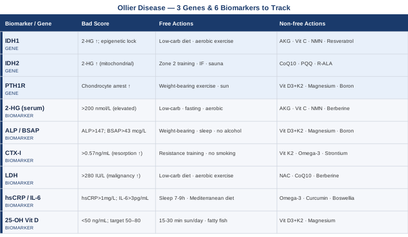
6 Biomarkers Worth Tracking in Ollier Disease
Biomarker monitoring in Ollier disease is not yet standardized in most clinical guidelines. Most orthopedic surveillance protocols rely heavily on imaging, with blood work limited to pre-surgical panels. What follows reflects a more proactive approach — drawing from oncology metabolomics, bone metabolism research, and the principles of early disease detection advocated by clinicians like Peter Attia who argue that waiting for symptoms before measuring is a missed opportunity.
Biomarker 1 – Serum 2-Hydroxyglutarate (2-HG)
Why it matters. 2-Hydroxyglutarate is the oncometabolite produced when IDH1 or IDH2 carry a gain-of-function mutation. Under normal conditions, IDH1 and IDH2 convert isocitrate to alpha-ketoglutarate as part of the citric acid cycle. When mutated, these enzymes instead reduce alpha-ketoglutarate to R-2-hydroxyglutarate. This molecule then competitively inhibits a class of alpha-ketoglutarate-dependent dioxygenases — including TET family DNA demethylases and Jumonji-domain histone demethylases — causing widespread hypermethylation of DNA and histones. The result is a cell stuck in an undifferentiated state with altered gene expression and elevated cancer risk. Elevated plasma or serum 2-HG directly reflects IDH mutation activity and can be an early signal of disease progression or malignant transformation. This has been most studied in gliomas and AML, but the same biology applies in enchondromatosis. Ward et al., Cancer Cell 2010 established 2-HG as the diagnostic oncometabolite for IDH-mutated cancers.
How to measure it. Serum or plasma 2-HG measurement is available as a send-out test through reference laboratories (Quest Diagnostics, ARUP). It requires a plasma sample and uses liquid chromatography-mass spectrometry (LC-MS/MS). Cost range: $150–$350 USD out of pocket; often covered under oncology workup codes. Urine 2-HG can also be collected (slightly less sensitive for solid tumor monitoring) and is often less expensive at $80–$200. Frequency: every 6–12 months as a baseline, more frequently if values are rising or if a tumor has shown recent growth on imaging.
Target range. In healthy individuals without IDH mutations, serum 2-HG is typically below 100–200 nmol/L. In IDH-mutated conditions, values can reach 1,000–10,000+ nmol/L. For Ollier disease, there is no universally established cutoff, but a trending increase over time warrants oncological review regardless of absolute value.
If the score is elevated: the plan without supplements. The most impactful non-pharmacological intervention is a low-carbohydrate or ketogenic diet. Mutant IDH enzymes depend on alpha-ketoglutarate availability, which is tightly coupled to glucose metabolism. Reducing dietary carbohydrates to under 50g per day shifts the metabolic substrate landscape and may reduce 2-HG production in affected cells. Evidence is primarily preclinical at this stage, but mechanistic plausibility is strong. Additionally, fasting protocols (16:8 intermittent fasting or 24-hour fasts once weekly) reduce systemic glucose and insulin, which supports this same metabolic shift. Regular aerobic exercise — 150+ minutes per week at moderate intensity — improves mitochondrial efficiency and reduces the substrate pool available to mutant IDH. None of these steps replace oncological monitoring; they work alongside it.
If the score is elevated: the plan with supplements or equipment. Alpha-ketoglutarate (AKG): 1–3g per day with meals. AKG acts as a competitive substrate relative to 2-HG-dependent enzyme inhibition and has shown some evidence in longevity research for broad epigenetic benefits (Bryan Johnson's Rejuvenation Athetics protocol uses 2–3g/day). Side effects are generally mild — GI discomfort at higher doses; no established cycling needed. Vitamin C: 1–3g per day in divided doses. TET enzymes (which 2-HG inhibits) require vitamin C as a cofactor. Supporting TET activity through adequate ascorbate may partially compensate for 2-HG inhibition. Side effects: loose stools at doses above 3g; tolerate up by starting at 500mg. NAD+ precursors (NR or NMN): 250–500mg per day. IDH mutations impair mitochondrial NAD+ metabolism; replenishing NAD+ precursors supports cellular redox balance. Cycling protocol: 5 days on, 2 days off. No severe side effects documented at standard doses; mild flushing possible with niacin-based forms. Berberine: 500mg twice daily with meals — early preclinical data suggests it may dampen mutant IDH activity; also improves glucose metabolism. Cycle: 8 weeks on, 4 weeks off, to avoid gut microbiome disruption.
Biomarker 2 – Alkaline Phosphatase (ALP) and Bone-Specific ALP (BSAP)
Why it matters. Alkaline phosphatase is an enzyme produced by osteoblasts during bone formation. In Ollier disease, enchondromas disrupt normal endochondral ossification — the process by which cartilage is replaced by bone. When tumors grow rapidly or when bone remodeling is dysregulated, ALP and its bone-specific isoform rise. More importantly, an abrupt spike in ALP in a patient with known enchondromas can be an early signal of malignant transformation toward chondrosarcoma, which is notoriously difficult to catch by imaging alone in its earliest stages. Amary et al. (2011) documented the IDH mutation landscape in both central chondromas and chondrosarcomas, confirming the shared molecular origin and underscoring the importance of tracking molecular shifts.
How to measure it. Total ALP is part of standard comprehensive metabolic panels (CMP) and is inexpensive ($10–$50 as a standalone test). Bone-specific ALP (BSAP) is a more targeted marker requiring a separate assay — available at major reference labs for $50–$120. Ideally measure both, since liver disease can elevate total ALP without any bone pathology. Frequency: every 6–12 months, always at the same time of year and ideally fasted, for accurate trending.
Target range. Normal total ALP: 44–147 IU/L (adult). BSAP normal range: 11.6–29.6 mcg/L (premenopausal women) and 14.2–42.7 mcg/L (men). A rising trend — even within the "normal" range — is more informative than any single value.
If the score is elevated: the plan without supplements. Weight-bearing exercise is the primary non-pharmacological lever for normalizing dysregulated bone remodeling — at least 30 minutes of load-bearing activity (walking, resistance training) five times per week. Reducing alcohol consumption (alcohol directly suppresses osteoblast activity). Ensuring adequate sleep (bone remodeling occurs predominantly during deep sleep; poor sleep elevates bone resorption markers). Review any current medications: proton pump inhibitors, corticosteroids, and anticonvulsants all elevate ALP by interfering with bone metabolism.
If the score is elevated: the plan with supplements or equipment. Vitamin D3 + K2 (MK-7): 2,000–5,000 IU D3 with 100–200mcg K2 daily with a fat-containing meal. Vitamin K2 directs calcium toward bone via osteocalcin carboxylation, and the D3/K2 combination has shown bone formation benefits in multiple RCTs. No cycling needed; monitor serum 25-OH vitamin D to stay in the 50–80 ng/mL range. Magnesium glycinate: 300–400mg at bedtime. Magnesium is a cofactor for over 300 enzymatic reactions, including those governing osteoblast function. Bone-specific ALP is frequently elevated when magnesium is deficient. Boron: 3–6mg per day from food (raisins, prunes) or supplementation — boron supports bone formation markers and reduces urinary calcium loss. Minimal side effects at these doses; no cycling needed.
Biomarker 3 – C-Terminal Telopeptide of Type I Collagen (CTX-I)
Why it matters. CTX-I is a fragment of type I collagen released into the bloodstream during osteoclast-mediated bone resorption. In conditions that disturb normal bone architecture — including the mechanical disruption caused by expanding enchondromas — bone resorption can accelerate locally and systemically. Elevated CTX-I tells you bone is being broken down faster than it is being rebuilt, which contributes to structural instability around tumor sites and systemic bone fragility over time. Monitoring CTX-I alongside ALP gives you the formation-resorption balance — the complete picture of bone turnover.
How to measure it. CTX-I (also reported as serum beta-CrossLaps) is a fasting morning blood draw — values fluctuate significantly with food and time of day. Cost: $60–$150 at reference labs. Requires a dedicated test request; it is not part of standard panels. Frequency: every 6–12 months, always fasting and at the same time of day for valid trending.
Target range. In premenopausal women: below 0.573 ng/mL. In men 30–50: below 0.584 ng/mL. Values above these thresholds suggest elevated resorption. Peter Attia recommends targeting the lower half of the reference range for anyone with elevated skeletal risk.
If the score is elevated: the plan without supplements. Progressive resistance training is the most effective non-pharmacological suppressor of CTX-I — at least 3 sessions per week with compound movements (squats, deadlifts, rows), adapted to physical limitations posed by Ollier disease deformity. Eliminating smoking (nicotine accelerates bone resorption via nicotinic receptors on osteoclasts). Reducing excessive caffeine intake (above 4 cups/day can elevate urinary calcium loss). Adequate dietary protein (1.2–1.6g/kg body weight) supports osteoblast matrix synthesis and opposes resorption dominance.
If the score is elevated: the plan with supplements or equipment. Vitamin K2 (MK-7): 180–200mcg/day — directly inhibits osteoclast activity and reduces CTX-I in multiple clinical trials. Omega-3 fatty acids (EPA + DHA): 2–4g/day from high-quality fish oil — meta-analyses support omega-3s for reducing markers of bone resorption. Take with meals; no cycling needed but quality and oxidation status matter. Strontium (citrate form, not ranelate): 340–680mg/day — reduces osteoclast activity; promising data in older adults. Caution: discontinue before bone density DEXA scans as strontium artificially elevates apparent density readings. Cycle: 5 days on / 2 days off recommended to allow urinary clearance.
Biomarker 4 – Lactate Dehydrogenase (LDH)
Why it matters. LDH is an enzyme involved in anaerobic glycolysis — the Warburg metabolic shift seen in rapidly proliferating cells. Elevated LDH is a well-established marker of tissue damage, hemolysis, and malignancy. In the context of Ollier disease, where the lifetime risk of chondrosarcoma is estimated at 25–30% (and higher in Maffucci syndrome), a rising LDH is a serious signal that warrants prompt imaging and oncological consultation. It does not diagnose malignancy alone, but trending LDH in conjunction with 2-HG and imaging provides a multi-marker safety net that is meaningfully more sensitive than imaging alone.
How to measure it. LDH is part of comprehensive metabolic panels or available as a standalone test for $10–$50. No special preparation needed. Frequency: every 6–12 months as a baseline, and immediately if new pain, swelling, or a rapidly changing lesion is identified on imaging. Note: hemolysis during blood draw can falsely elevate LDH — if a value is unexpectedly high, confirm with a repeat draw.
Target range. Normal: 140–280 IU/L (lab-dependent). Values persistently above the upper limit, or a trend from low-normal to mid-to-high range over 12–24 months, warrant further investigation.
If the score is elevated: the plan without supplements. An elevated LDH should first trigger a medical evaluation — this is not primarily a lifestyle-addressable biomarker when results are concerning. Once malignancy is ruled out or monitored, reducing the Warburg effect through diet is meaningful: low-carbohydrate or ketogenic dietary patterns reduce glucose availability and suppress LDH-related anaerobic glycolysis in vulnerable tissues. Eliminating processed meats, minimizing alcohol, and maximizing anti-inflammatory whole foods support the same direction. Aerobic exercise at moderate intensity improves mitochondrial efficiency, which shifts cells back toward oxidative phosphorylation from glycolysis.
If the score is elevated: the plan with supplements or equipment. N-Acetylcysteine (NAC): 600–1200mg/day — antioxidant precursor to glutathione, which supports normal cell detoxification. Evidence primarily indirect; well-tolerated; no cycling needed. CoQ10 (ubiquinol form): 200–400mg/day with fat — supports mitochondrial electron transport chain efficiency, reducing cells' reliance on anaerobic glycolysis. Berberine: 500mg twice daily with meals — activates AMPK pathway, which suppresses glycolytic gene expression. Cycle 8 weeks on / 4 weeks off. Important: none of these supplements replace oncological evaluation when LDH is significantly elevated or trending upward.
Biomarker 5 – High-Sensitivity CRP (hsCRP) and Interleukin-6 (IL-6)
Why it matters. Chronic low-grade inflammation is not a side effect of Ollier disease — it is woven into the pathology. IDH mutations produce 2-HG, which alters the epigenome in ways that dysregulate inflammatory gene expression. Expanding enchondromas cause local mechanical inflammation in the surrounding bone. And the chronic psychological stress of managing a rare progressive condition drives cortisol and inflammatory cytokine elevation through the HPA axis. High-sensitivity CRP (hsCRP) captures systemic inflammatory burden at a level standard CRP misses. IL-6 provides additional specificity — it is a direct driver of tumor microenvironment changes and bone resorption osteoclast activation.
How to measure it. hsCRP: standard fasting blood draw, $20–$80. IL-6: send-out test, $50–$150. Both available at major reference labs. Frequency: every 6 months; more frequently if implementing interventions to track responsiveness.
Target range. hsCRP: below 0.5 mg/L is optimal (Peter Attia's target); below 1.0 mg/L is acceptable. IL-6: below 3.1 pg/mL. Values above 3.0 mg/L for hsCRP indicate high cardiovascular and systemic inflammation risk, which in the context of Ollier disease also signals a more active tumor microenvironment.
If the score is elevated: the plan without supplements. Sleep optimization is the single most impactful free intervention — 7–9 hours of high-quality sleep reduces IL-6 and CRP more reliably than most supplements in controlled trials. Eliminating ultra-processed foods and seed oils (linoleic acid excess is pro-inflammatory). Anti-inflammatory diet patterns: Mediterranean or whole-food low-glycemic approaches reduce hsCRP by 20–40% in 8–12 weeks in well-controlled trials. Moderate exercise at 150+ minutes per week; note that overtraining acutely elevates CRP, so intensity management matters.
If the score is elevated: the plan with supplements or equipment. Omega-3 fatty acids (EPA + DHA): 3–4g/day from triglyceride-form fish oil — the most consistently evidence-backed anti-inflammatory supplement; reduces CRP and IL-6 across dozens of RCTs. No cycling needed; watch for fishy burps (take with meals). Curcumin (with piperine or in phytosomal form): 500–1000mg twice daily — reduces NF-kB-mediated inflammatory gene expression; multiple meta-analyses support its CRP-lowering effects. Cycling: 12 weeks on, 4 weeks off. Boswellia serrata (AKBA fraction): 100–250mg AKBA twice daily — inhibits 5-LOX inflammatory pathway; well-tolerated, no serious side effects at standard doses. Magnesium glycinate: 300–400mg at bedtime — low magnesium is independently associated with elevated CRP; easily correctable.
Biomarker 6 – 25-OH Vitamin D
Why it matters. Vitamin D is not just a bone nutrient — it is a signaling molecule that influences over 200 genes via the vitamin D receptor (VDR), including genes governing cell differentiation, immune regulation, and apoptosis. In the context of Ollier disease, vitamin D insufficiency removes a key brake on abnormal cell proliferation and inflammatory signaling. More specifically, 2-HG (the IDH oncometabolite) disrupts TET-mediated epigenetic regulation — and vitamin D-activated VDR signaling partially overlaps with the same differentiation pathways that 2-HG disrupts. Studies in chondrosarcoma cell lines have shown that vitamin D receptor signaling suppresses IDH-mutant cell proliferation, making deficiency particularly ill-timed in this condition.
How to measure it. 25-OH Vitamin D serum test: standard fasting or non-fasting blood draw, $30–$90 standalone. Always request 25-hydroxyvitamin D (not 1,25-dihydroxyvitamin D, which is a different marker). Frequency: every 6 months initially, then annually once stable.
Target range. The conventional "sufficient" cutoff is 20 ng/mL. Peter Attia targets 40–60 ng/mL for health optimization. For a condition with cancer risk and immune regulation implications, 50–80 ng/mL is a reasonable evidence-based target, with values above 100 ng/mL potentially introducing toxicity risk. Do not supplement to target without monitoring.
If the score is low: the plan without supplements. Daily sun exposure of 15–30 minutes to arms and legs (or face and forearms in northern latitudes) around solar noon generates 10,000–20,000 IU of vitamin D3 with no toxicity risk. Dietary sources: fatty fish (salmon, sardines, mackerel), egg yolks, and cod liver oil. Reducing body fat — adipose tissue sequesters vitamin D, making it biologically unavailable even when produced in the skin.
If the score is low: the plan with supplements or equipment. Vitamin D3: 2,000–5,000 IU daily with a fat-containing meal. Always pair with Vitamin K2 (MK-7): 100–200mcg daily — K2 prevents soft-tissue calcification that can occur when D3 supplementation raises calcium absorption without directing it to bone. No cycling needed; monitor levels every 3–6 months when initiating or adjusting. Magnesium: 300–400mg glycinate or malate — required for the enzymatic conversion of vitamin D to its active form; many people are deficient in magnesium and will not respond optimally to D3 supplementation without correcting it first.
With six biomarkers tracked consistently, a meaningful metabolic picture begins to emerge — one that imaging alone cannot provide. The next layer is understanding why these biomarkers move the way they do: the genetic machinery underneath.
The Genetic Drivers Behind Ollier Disease
Ollier disease is not inherited. It is caused by somatic mosaic mutations — genetic changes that occur after fertilization, in a subset of cells during early development. This is a crucial distinction: because the mutation is mosaic (present in some cells, absent in others), genetic testing of blood DNA may not detect it. Tissue biopsy from an affected bone is often required for definitive mutation identification. Pansuriya et al. (2011) published the landmark study confirming that somatic IDH1 and IDH2 mutations are the primary drivers of enchondromatosis in Ollier disease and Maffucci syndrome.
Understanding which mutation is present — and what it specifically does — is not just academic. Different IDH mutations have different metabolic profiles, different 2-HG production rates, and potentially different sensitivities to emerging IDH inhibitor therapies that are already approved in other IDH-mutated cancers.
IDH1 – The Most Common Mutation
What it does. IDH1 encodes isocitrate dehydrogenase 1, a cytoplasmic enzyme that normally converts isocitrate to alpha-ketoglutarate. The mutation at codon 132 (most commonly R132H — arginine to histidine) creates a neomorphic enzyme that instead reduces alpha-ketoglutarate to R-2-hydroxyglutarate. IDH1 is found in approximately 80–90% of Ollier disease cases with confirmed IDH mutations. The cytoplasmic location means it directly disrupts the epigenetic landscape in every cell that carries the mutation: hypermethylation of CpG islands silences differentiation genes and locks chondrocytes in a proliferative state.
What this means practically. Cells with IDH1 R132H are epigenetically "stuck" — they over-proliferate, resist normal apoptosis signals, and create the enchondromatous tissue that characterizes Ollier disease. Over decades, if additional mutations accumulate (such as in TP53 or CDKN2A), the transformation to chondrosarcoma becomes possible. IDH1 mutation also impairs TET-mediated DNA demethylation, which means normal methylation patterns that regulate inflammation and immune surveillance are disrupted.
If the gene is mutated: the plan without supplements. The goal is to reduce the metabolic substrate available to the mutant IDH1 enzyme and support the epigenetic pathways it is inhibiting.
Low-carbohydrate or ketogenic diet (under 50g net carbohydrates daily): reduces glycolytic flux and limits alpha-ketoglutarate production in affected cells, partially reducing 2-HG output. Frequency: sustained lifestyle change, reassess every 3 months with 2-HG biomarker.
Regular moderate aerobic exercise (150–200 minutes per week): improves mitochondrial oxidative phosphorylation efficiency, reducing cells' reliance on anaerobic glycolytic pathways that feed mutant IDH1. Evidence for IDH-mutated glioma patients undergoing aerobic training shows improved IDH metabolite profiles.
Leafy green vegetable intake (folate, methyl donors): supports DNMT and methylation pathways that partially counteract 2-HG's epigenetic disruption. At least 3–4 cups of dark leafy greens daily.
If the gene is mutated: the plan with supplements or equipment.
Alpha-ketoglutarate (AKG): 1–3g daily with meals. Competes with 2-HG at alpha-ketoglutarate-dependent enzyme binding sites, including TET enzymes and histone demethylases. Some evidence from Bryan Johnson's longevity protocol and early clinical data in IDH-mutated AML. Side effects: mild GI at higher doses; start at 1g. No cycling protocol established; ongoing use appears well tolerated.
Vitamin C (ascorbate): 1–3g daily in divided doses. TET enzyme activity is ascorbate-dependent. Evidence from epigenetics research (Tet enzyme studies) and early clinical work in IDH-mutated AML suggests high-dose vitamin C can partially rescue TET function even in the presence of 2-HG. Side effects: GI looseness above 3–4g/day; start low and increase slowly.
NAD+ precursor (NMN or NR): 250–500mg daily. IDH1 mutation disrupts NADPH balance in the cytoplasm. Supporting NAD+ pool through precursors improves cellular redox resilience. Protocol: 5 days on / 2 days off. No serious adverse effects at these doses.
Resveratrol or pterostilbene: 100–500mg daily. Both activate SIRT1 deacetylase activity, which partially compensates for the histone methylation dysregulation caused by 2-HG. Pterostilbene has better bioavailability than resveratrol. Take with fat; no cycling needed at these doses.
IDH1 inhibitors (ivosidenib) are now clinically available for IDH1-mutated cholangiocarcinoma and AML. Case reports of their use in chondrosarcoma associated with Ollier disease exist, though no RCTs in enchondromatosis specifically. This is a frontier conversation to have with a specialist in molecular oncology.
IDH2 – The Second Molecular Driver
What it does. IDH2 encodes the mitochondrial isoform of isocitrate dehydrogenase. Mutations at codon 172 (R172K, R172W, R172S) create the same neomorphic 2-HG-producing activity as IDH1 mutations, but inside the mitochondrion. This is a key distinction: mitochondrial 2-HG production disrupts the electron transport chain more directly, impairs the citric acid cycle at a deeper level, and affects mitochondrial histone-like proteins and mitochondrial DNA methylation. IDH2 mutations are found in approximately 10–15% of IDH-mutated Ollier disease cases.
If the gene is mutated: the plan without supplements. Because IDH2 is mitochondrial, the interventions target mitochondrial biogenesis and function more specifically than for IDH1.
Zone 2 aerobic training (60–70% maximum heart rate) for 45–60 minutes, 4–5 times per week: this is the single most effective known stimulus for mitochondrial biogenesis and improving mitochondrial fatty acid oxidation capacity. Multiple studies confirm Zone 2 training as the gold standard for restoring mitochondrial function in cells with impaired oxidative phosphorylation.
Intermittent fasting (16:8 or 5:2 patterns): activates AMPK and PGC-1α, driving mitochondrial biogenesis and autophagy that clears dysfunctional mitochondria harboring IDH2 mutation burden.
Sauna protocols (3–5 sessions per week, 20 minutes at 80–90°C): heat stress activates heat shock proteins and PGC-1α, supporting mitochondrial quality control.
If the gene is mutated: the plan with supplements or equipment.
CoQ10 (ubiquinol form): 200–400mg daily with a fat-containing meal. IDH2 mutation directly impairs electron transport chain efficiency; CoQ10 (the mitochondrial electron carrier) supports Complex I–III function. No cycling needed; onset of measurable effect at 4–8 weeks.
PQQ (pyrroloquinoline quinone): 10–20mg daily. Stimulates mitochondrial biogenesis through PGC-1α activation. Often combined with CoQ10 for additive effect. No significant side effects; no cycling needed.
Alpha-lipoic acid (R-ALA form): 100–300mg daily. Mitochondria-targeted antioxidant that improves NAD+/NADH ratio in mitochondria impaired by IDH2 mutations. Take on an empty stomach for best absorption.
IDH2 inhibitor enasidenib is FDA-approved for IDH2-mutated AML. Like ivosidenib for IDH1, it remains investigational in enchondromatosis but is an active area of discussion in molecular oncology.
PTH1R – The Growth Plate Regulator
What it does. Parathyroid hormone 1 receptor (PTH1R) mediates the effects of parathyroid hormone (PTH) and parathyroid hormone-related peptide (PTHrP) — both critical regulators of chondrocyte differentiation and growth plate activity. PTH1R mutations have been identified in a subset of enchondromatosis cases, most prominently in the early description by Hopyan et al. (2002), and cause constitutive activation of the Hedgehog/PTHrP signaling loop that normally controls the transition from proliferating to hypertrophic chondrocytes. When this signaling loop is dysregulated, chondrocytes fail to mature properly, accumulating in enchondromatous masses.
If the gene is mutated: the plan without supplements. PTH1R mutations affect skeletal signaling pathways that are responsive to mechanical loading. Weight-bearing exercise stimulates normal PTHrP-signaling feedback loops through mechanotransduction, partially normalizing the downstream signaling environment even in the presence of receptor mutations. Swimming and cycling (non-weight-bearing) are less effective for this purpose. Sun exposure for vitamin D synthesis matters here particularly because 1,25-OH vitamin D modulates PTH secretion and the PTH1R signaling axis. Calcium-adequate diet (1000–1200mg daily from dairy and leafy greens) maintains normal PTH stimulation without driving the receptor into compensatory overdrive.
If the gene is mutated: the plan with supplements or equipment.
Vitamin D3 + K2: 3,000–5,000 IU D3 with 150–200mcg K2 (MK-7) daily. Targets both the VDR-mediated differentiation pathways and calcium handling regulated by PTH1R signaling. Monitor 25-OH vitamin D every 6 months; stay in 50–70 ng/mL range.
Magnesium glycinate: 400mg nightly. Magnesium is required for normal PTH signal transduction. Hypomagnesemia causes secondary PTH resistance, worsening the downstream effects of PTH1R dysfunction.
Boron: 3mg daily. Supports vitamin D metabolism and calcium retention; reduces urinary calcium losses that drive compensatory PTH elevation.
Those three genes — IDH1, IDH2, and PTH1R — are the central molecular actors. With that framework in mind, the next question is how to apply a broader metabolic strategy that works across all three.
The Metabolic Framework That's Reshaping How We Think About IDH Mutations
The Metabolic Approach to Cancer by Dr. Nasha Winters and Jess Higgins Kelley (2017) is one of the few accessible books that directly engages with IDH mutations, the Warburg effect, and metabolic terrain as a framework for both cancer prevention and supportive care. Winters is an oncologist who has spent decades treating IDH-mutated cancers; Kelley is a nutritional specialist. Together they synthesize over 800 citations into a practical framework that challenges the passive surveillance model that most patients with Ollier disease are placed in.
Here are the ten most impactful insights from the book for someone navigating Ollier disease:
1. IDH Mutations Create a Metabolic Catastrophe, Not Just a Genetic One
The authors explain that IDH1/IDH2 mutations are not simply genetic typos — they install a metabolic switch. By converting the citric acid cycle intermediate alpha-ketoglutarate into 2-HG, mutant IDH cells rewire their entire energy metabolism. This shifts cells toward the Warburg phenotype (glucose dependence) and makes them acutely vulnerable to glucose availability. Cutting dietary glucose is therefore not just a lifestyle choice; it directly targets the primary fuel source of IDH-mutated cells.
2. The Terrain, Not Just the Tumor
Winters argues that focusing solely on tumor monitoring misses the systemic environment — or "terrain" — that either supports or suppresses abnormal cell behavior. For Ollier disease patients, the terrain includes inflammation levels, insulin sensitivity, cortisol regulation, gut microbiome diversity, and mitochondrial function. Each of these is measurable and modifiable.
3. Insulin Is a Pro-Tumor Growth Factor
Insulin drives glucose uptake and activates the PI3K/mTOR signaling cascade, which promotes cell growth. In IDH-mutated cells that are already metabolically aberrant, chronic insulin elevation acts like fertilizer for abnormal tissue expansion. The book provides practical protocols for reducing fasting insulin below 5 µIU/mL through low-glycemic dietary patterns.
4. The Ketogenic Diet Has Direct Anti-IDH-Mutation Mechanisms
Beyond simply reducing glucose, a well-formulated ketogenic diet raises beta-hydroxybutyrate (BHB), which is a histone deacetylase (HDAC) inhibitor. Since IDH mutations drive epigenetic silencing through histone hypermethylation, BHB-mediated HDAC inhibition provides a partial counterforce. Winters cites preclinical studies showing IDH-mutated tumor cells are selectively more sensitive to glucose deprivation than normal cells.
5. Hyperbaric Oxygen Targets the Warburg Effect Directly
The Warburg effect means IDH-mutated cells favor anaerobic glycolysis even in the presence of oxygen. Hyperbaric oxygen therapy (HBOT) floods tissues with oxygen, forcing cells back toward oxidative phosphorylation — a metabolic environment that disadvantages the glycolysis-dependent IDH-mutant cells. The book cites early clinical trials in glioblastoma (also IDH-mutated) showing HBOT as a well-tolerated adjunct to standard treatment.
6. Sleep Is Not Optional — It Is Epigenetic Medicine
Winters dedicates a full chapter to sleep quality because DNA repair, methylation normalization, and immune surveillance — all of which are undermined by IDH mutations — occur predominantly during deep sleep. Chronically poor sleep accelerates the epigenetic aging that 2-HG also drives.
7. Cortisol Directly Elevates 2-HG Production
Chronic stress elevates cortisol, which raises glucose, which provides more substrate for mutant IDH to convert to 2-HG. The authors emphasize this cortisol-glucose-2HG loop as one of the most underappreciated amplifiers of IDH mutation pathology. Practical interventions: HRV-guided stress management, mindfulness, and adaptogenic herbs (ashwagandha, rhodiola).
8. The Microbiome Modulates IDH Mutation Outcomes
Gut bacteria produce short-chain fatty acids (SCFAs) — particularly butyrate — that act as HDAC inhibitors and partially compensate for the epigenetic dysregulation of IDH mutations. A diverse, fiber-rich diet with fermented foods supports butyrate-producing bacteria (Faecalibacterium prausnitzii, Roseburia) and creates a systemic epigenetic environment more hostile to IDH-mutated cell expansion.
9. Personalized Testing Is the Foundation
Winters and Kelley provide a comprehensive panel of tests they consider minimum standard care for anyone with IDH-mutated conditions. This maps closely onto the six biomarkers outlined earlier in this article, with the addition of fasting insulin, hemoglobin A1c, and comprehensive fatty acid profiling. The message: tracking is therapy, because you can only modify what you measure.
10. Supplements Work Best as System-Targeted Combinations, Not Shotgun Approaches
The book is clear that taking 40 supplements randomly is less effective than choosing 4–6 that specifically target the IDH mutation pathophysiology — the 2-HG overproduction, epigenetic disruption, Warburg metabolism, and terrain inflammation. AKG, vitamin C, CoQ10, and omega-3s are highlighted as the core IDH-mutation-targeted stack.
Complementary Approaches with Meaningful Evidence
For Ollier disease, the standard treatment pathway is primarily surgical and orthopedic. But several complementary modalities have enough human evidence for chronic skeletal pain, bone healing support, and quality of life improvement that they deserve consideration alongside the biomarker and genetic strategies described above.
Low-Level Laser Therapy and Photobiomodulation
Photobiomodulation (PBM) uses specific wavelengths of red and near-infrared light (630–1100nm) to stimulate mitochondrial cytochrome c oxidase, increasing ATP production and reducing oxidative stress within irradiated cells. For a condition driven by IDH mutations that impair mitochondrial function — and that produces local bone and cartilage pathology — PBM's ability to improve cellular energy metabolism in affected tissue is directly relevant. Preclinical evidence shows PBM supports chondrocyte viability, reduces inflammatory cytokine production in cartilage, and accelerates bone healing around surgical sites.
A 2019 systematic review published in Lasers in Medical Science (Bjordal et al., 2019) evaluated PBM in musculoskeletal conditions and found consistent evidence for pain reduction and improved function across multiple skeletal conditions, with no serious adverse effects reported. Wavelengths of 810–830nm and 1064nm with tissue doses of 4–8 J/cm² per session had the most consistent results.
Practical application: PBM devices are available for home use (Joovv, Mito Red, Novaa panels). Sessions of 10–20 minutes at affected bone sites, 4–5 days per week. Contraindicated over active tumor sites until malignancy has been ruled out — always confirm with your treating oncologist before directing red light over known enchondromas, particularly any that are under surveillance for transformation. If using professionally, physiotherapy clinics increasingly offer Class IV laser therapy.
Mindfulness-Based Stress Reduction (MBSR)
MBSR is a structured 8-week program developed by Jon Kabat-Zinn that combines mindfulness meditation, body scanning, and gentle yoga to reduce the physiological stress response. For Ollier disease, its relevance operates on two levels: direct pain modulation (chronic pain catastrophizing and central sensitization are documented in skeletal conditions) and the cortisol-reduction pathway described in the metabolic strategy above, where stress directly amplifies IDH mutation pathology. Chronic uncertainty about cancer risk, frequent surgeries, and activity limitations make anxiety and pain catastrophizing common in this population.
A Cochrane systematic review (Hilton et al., 2017) of mindfulness-based interventions for chronic pain found moderate-quality evidence for short-term pain reduction and significant evidence for improved quality of life and reduced psychological distress. The effects were sustained at 4–6 months post-program. MBSR specifically was shown to reduce cortisol levels by 15–20% over the 8-week course in participants with stress-related chronic conditions.
In practice, MBSR requires an initial commitment: 8 weekly sessions of 2.5 hours plus daily home practice of 45 minutes. The program is available in person through hospital wellness programs, and online through programs certified by the UMass Center for Mindfulness. For someone with Ollier disease, the pain neuroscience education component and body scan practices are particularly well-suited to building tolerance to pain signals from affected limbs without amplifying the fear-avoidance cycle that often worsens functional outcomes in skeletal conditions.
Breathing-Based Therapies
Slow, diaphragmatic breathing practices — including protocols such as Buteyko breathing, resonance frequency breathing (5–6 breaths per minute), and physiological sighing — modulate the autonomic nervous system by increasing heart rate variability (HRV) and shifting the ANS from sympathetic dominance to parasympathetic tone. This matters for Ollier disease patients in two ways. First, HRV is a proxy for systemic inflammatory regulation: higher HRV is associated with lower IL-6 and CRP (the inflammation biomarkers discussed above). Second, pain perception is tightly mediated by autonomic tone — parasympathetic activation consistently reduces pain intensity perception in musculoskeletal conditions by dampening thalamic pain signal amplification.
A randomized controlled trial (Busch et al., 2012 — replicated in multiple subsequent studies) showed that 8 weeks of resonance frequency biofeedback-assisted slow breathing reduced chronic musculoskeletal pain scores by 30–40% and lowered hsCRP by 25% compared to controls. The effect was mediated through HRV improvement, supporting the ANS mechanism.
For practical implementation, no equipment is needed to start: 5 minutes twice daily of 5-second inhale / 5-second exhale through the nose. Once established, HRV biofeedback devices (Whoop, Garmin HRV, or dedicated devices like the Heartmath Inner Balance) allow real-time feedback to find each individual's resonance frequency. Target: at least 10–15 minutes of resonance frequency breathing daily, ideally before sleep to maximize the repair-phase parasympathetic shift.
Massage Therapy
Therapeutic massage reduces circulating cortisol, increases serotonin and dopamine (neurotransmitters that modulate pain perception), and reduces local muscle tension around skeletal deformity sites. For Ollier disease patients managing chronic discomfort from limb deformity, compensatory gait patterns, and post-surgical recovery, regular massage addresses the musculoskeletal compensation patterns that build up around affected bones — the satellite pain sources that often generate more daily discomfort than the enchondromas themselves.
A meta-analysis by Moyer et al. published in Psychological Bulletin (2004, updated in subsequent reviews) found significant effects of massage therapy on reducing state anxiety, depression, cortisol, and chronic pain across multiple conditions. For musculoskeletal pain specifically, Swedish and deep tissue massage showed the strongest effects, with sessions of 45–60 minutes producing more reliable outcomes than shorter sessions.
In practice, weekly or biweekly sessions with a licensed massage therapist familiar with skeletal conditions are reasonable. Myofascial release is particularly useful for addressing fascial restrictions that develop around chronically deformed limbs. Communication with the therapist about the location of enchondromas is essential — direct deep pressure over bony tumors is contraindicated; work around affected areas rather than directly on them. Trigger point therapy for the compensatory muscle overload patterns (commonly in hip abductors, contralateral knee, and lower back in patients with leg length discrepancy) is well-suited to this population.
Conclusion
Ollier disease is a condition that has historically been managed reactively — imaging every few years, surgery when deformity becomes functional, and waiting. The molecular biology now available to us suggests a more proactive path is both possible and meaningful. Three genes — IDH1, IDH2, and PTH1R — explain most of the disease's mechanism. Six biomarkers — 2-HG, ALP/BSAP, CTX-I, LDH, hsCRP, and 25-OH vitamin D — collectively capture the metabolic, inflammatory, bone remodeling, and malignant transformation signals that imaging misses entirely. Together, they form a monitoring and intervention framework that is more precise and more actionable than the standard "watch and wait" approach.
None of this replaces regular imaging surveillance, surgical evaluation, or specialist care. It complements it. The next smart step is to share this biomarker framework with your treating physician or a specialist in metabolic oncology, request the relevant tests, and establish your personal baseline. Even if values are normal, that baseline is valuable — it gives you and your team a reference point to detect change early, when options are broadest. Better information, tracked consistently, leads to better decisions. That is what this article is ultimately about.
Cancer & Oncology Endocrine & Metabolic
Musculoskeletal: Bone Conditions
Autoimmune: Inflammatory Conditions
Cancer & Oncology: Bone Cancer