This article contains AI generated content.
· UpdatedOsteochondral Allograft Failure - 5 Genes And 6 Biomarkers To Track
Introduction
If you've had an osteochondral allograft procedure and the recovery isn't going the way you expected, the advice you're likely hearing is still about rest, physical therapy, and time. That advice is reasonable, but it doesn't explain why some patients integrate their graft seamlessly while others experience progressive failure despite similar surgeries, similar rehabilitation, and similar effort. Something biological is different, and standard follow-up appointments rarely look closely enough to find it.
The reality is that osteochondral allograft outcomes depend on a web of interacting biological variables — the local inflammatory environment in the joint, the immune response to foreign donor tissue, the cartilage matrix synthesis capacity of chondrocytes, the subchondral bone integration milieu, and the molecular signals that either promote repair or accelerate degradation. These variables differ meaningfully between individuals, and they aren't visible on an X-ray or captured in a standard post-op CBC. Generic recovery timelines assume a generic biology, and that assumption doesn't hold.
Biomarkers and genetic data change that picture. Specific blood and urine tests can reveal in near-real time whether cartilage breakdown is outpacing synthesis, whether systemic inflammation is creating a destructive cytokine environment for the graft, and whether your body has the biochemical resources needed to support healing. At the genetic level, certain variants influence how aggressively your body degrades cartilage collagen, how strongly it mounts an inflammatory response to injury, and how well it may tolerate foreign tissue — information that can meaningfully guide personalized recovery decisions.
This article doesn't promise outcomes. What it offers is a more precise biological lens. It walks through six key biomarkers worth monitoring after an osteochondral allograft, each with practical measurement guidance and specific action plans. It then covers five genetic factors that shape graft trajectories, followed by what connective tissue physiology research — particularly the work of Dr. Keith Baar — reveals about optimizing the repair environment. Finally, it reviews which complementary modalities have genuine human evidence for joint health. Better information doesn't guarantee recovery, but it makes better decisions possible.
Summary
Osteochondral allograft failure is rarely explained by a single cause. Six measurable biomarkers — COMP, CTX-II, hsCRP/IL-6, MMP-3, Vitamin D, and IGF-1 — provide a real-time window into cartilage degradation rates, systemic inflammation, enzymatic destruction, and repair capacity. Each one has a specific measurement protocol, a cost range, and a concrete action plan — both with and without supplements. Five genetic factors (COL2A1, IL1B, MMP13, GDF5, and HLA markers) reveal the individual biological tendencies that can explain why some patients lose grafts faster than others, and what to do differently based on that profile.
Beyond the biological tracking, Dr. Keith Baar's connective tissue research — discussed widely in the Huberman Lab context — offers a framework for repair that challenges conventional post-surgical rehab assumptions. And three complementary modalities — photobiomodulation, mindfulness-based stress reduction, and microbiome-directed approaches — have meaningful human evidence for reducing the inflammatory and degenerative forces that drive allograft failure. The article covers all of it with enough specificity to be actionable.
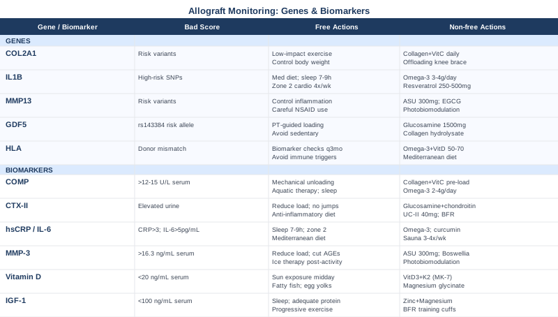
6 Biomarkers That Can Signal Why an Osteochondral Allograft Fails
Monitoring tissue behavior after a graft procedure requires more precision than a periodic MRI. Blood and urine biomarkers give you a window into cartilage metabolism and inflammatory activity that imaging cannot provide — and they become informative months before structural changes are visible. The six tests below cover the most relevant dimensions of graft biology: cartilage matrix integrity, collagen degradation, systemic inflammation, enzymatic destruction, nutritional sufficiency, and anabolic capacity.
1. COMP — Cartilage Oligomeric Matrix Protein
Cartilage Oligomeric Matrix Protein (COMP) is a structural glycoprotein embedded in the extracellular matrix of hyaline and fibrocartilage. When cartilage tissue is mechanically stressed, inflamed, or actively degrading, COMP is released into synovial fluid and subsequently into the bloodstream. Serum COMP is one of the most validated circulating markers of cartilage matrix breakdown, with consistent evidence across both post-traumatic and degenerative joint disease contexts.
In the context of an osteochondral allograft, elevated COMP levels suggest that the donor cartilage — or the native tissue surrounding it — is under stress that exceeds repair capacity. The value of tracking COMP serially is that it gives you a biological trend before imaging shows structural deterioration. Research on serum COMP and cartilage loss has shown correlations with radiographic joint space narrowing and with MRI-confirmed cartilage volume loss in multiple knee disease populations.
How to Measure It
COMP is measured via a serum blood draw. It's available through specialty laboratories including Quest Diagnostics and through some rheumatology practices. Synovial fluid COMP provides a more accurate picture of local joint status but requires aspiration. Cost for serum COMP: $80–$200 through specialty labs; not universally covered by standard insurance. Most labs flag values above 12–15 U/L as elevated in a post-surgical context, though standardization varies. Serial readings taken at consistent time points (morning, similar activity levels) are more informative than isolated values.
If the Score Is High, the Plan Without Supplements
The immediate non-supplement response to elevated COMP is mechanical unloading. Reduce high-impact activities and transition to aquatic therapy, cycling, or elliptical training, which maintain joint motion and muscle activation without cartilage compression spikes. Work with a physical therapist to introduce partial weight-bearing protocols appropriate to your healing stage. Prioritize sleep quality and duration — growth hormone secretion during slow-wave sleep is the primary endogenous driver of tissue repair, and it's frequently disrupted in post-surgical patients. Graded, progressive return to loading should be guided by your biomarker trend, not a calendar alone.
If the Score Is High, the Plan With Supplements or Equipment
The best-supported supplement protocol for elevated COMP is hydrolyzed collagen peptides (10–15 g/day) combined with vitamin C (200–500 mg), consumed 30–60 minutes before a brief session of light mechanical activity. This timing protocol is supported by Dr. Keith Baar's connective tissue synthesis research — discussed in detail later in this article — and maximizes the collagen synthesis window that follows mechanical loading. Omega-3 fatty acids (EPA+DHA, 2–4 g/day in triglyceride form) reduce cartilage degradation markers in joint disease studies. Cycle collagen for 3 months, retest COMP, then reassess. Side effects at standard doses are minimal; exceeding 4 g/day of EPA+DHA long-term may modestly increase bleeding time.
2. CTX-II — The Most Direct Window Into Cartilage Collagen Breakdown
C-terminal crosslinking telopeptide of type II collagen (CTX-II) is a degradation fragment released into urine when type II collagen — the dominant structural collagen in hyaline cartilage — is enzymatically cleaved. Unlike CTX-I, which reflects bone collagen turnover, CTX-II is essentially cartilage-specific. This makes it one of the most direct non-invasive indicators of active cartilage matrix destruction available.
After an osteochondral allograft, the donor tissue is rich in type II collagen. Elevated urine CTX-II levels indicate that this matrix is being broken down — whether by matrix metalloproteinases activated by local inflammation, by immune-driven mechanisms, or by excessive mechanical stress. CTX-II research in joint disease populations has shown that elevated levels at 3–6 months after joint injury predict significantly worse long-term cartilage outcomes, making this a genuinely prognostic test.
How to Measure It
CTX-II is measured from a second morning urine sample (first morning urine reflects an overnight fasted-state peak; second morning provides a more stable baseline), normalized to urinary creatinine to adjust for hydration. Available through specialty and research-oriented labs. Cost: $100–$250, rarely covered by routine insurance. Collect samples at consistent times and with consistent pre-test activity levels for reliable trending. Testing every 3–6 months provides the most useful picture during the first two years post-procedure.
If the Score Is High, the Plan Without Supplements
High CTX-II is a signal that the cartilage degradation rate is elevated and that the mechanical and inflammatory environment of the joint needs to change. Reduce high-force joint loading — particularly compressive activities like running, jumping, or heavy squatting — and substitute with swimming, stationary cycling, or aquatic resistance training. Adopt a whole-foods, anti-inflammatory dietary pattern: minimize ultra-processed foods, refined carbohydrates, and industrial seed oils, which are associated with higher systemic inflammatory burden and accelerated cartilage catabolism. Neuromuscular electrical stimulation (NMES) can preserve periarticular muscle function and joint stability without direct cartilage loading.
If the Score Is High, the Plan With Supplements or Equipment
Glucosamine sulfate (1500 mg/day) and chondroitin sulfate (1200 mg/day) have shown measurable reductions in urinary CTX-II in clinical trials when used consistently for 3–6 months. This combination is well-tolerated; cycle on for 6 months, retest CTX-II, and decide whether to continue. Undenatured type II collagen (UC-II, 40 mg/day) works via a distinct mechanism — oral immune tolerance to type II collagen — and is particularly relevant for allograft contexts where immune activity may be driving breakdown. Blood flow restriction training equipment ($100–$300 for consumer-grade cuffs) allows meaningful musculoskeletal stimulus with substantially lower peak cartilage forces than standard resistance exercise; proper technique instruction is essential before using it.
3. hsCRP and IL-6 — The Systemic Inflammation Checkpoint
High-sensitivity C-reactive protein (hsCRP) and interleukin-6 (IL-6) are the two most practical markers of systemic inflammatory load to track after an allograft. IL-6 is a primary cytokine in the inflammatory cascade; hsCRP is a downstream hepatic response protein that reflects IL-6 and TNF-α activity over the preceding 12–24 hours. Together, they give you a reliable read on whether your systemic inflammatory environment is conducive to graft survival.
Persistent low-grade systemic inflammation — even at levels that wouldn't raise concern on a routine blood panel — is increasingly understood as a driver of post-surgical cartilage failure. When IL-6 is chronically elevated, it upregulates matrix metalloproteinase expression inside the joint, suppresses IGF-1 receptor signaling in chondrocytes, and can directly induce donor chondrocyte apoptosis. For an allograft, the donor cells are already under metabolic stress from storage conditions; placing them in a high-cytokine environment reduces their viability significantly. Research on IL-6 and chondrocyte viability supports inflammation control as a primary protective strategy for cartilage tissue.
How to Measure It
Both markers are available through standard laboratory panels at nearly any facility. hsCRP: approximately $15–$40, typically covered by insurance when ordered for cardiovascular or inflammatory assessment. Serum IL-6: $40–$100, less routinely ordered but available on request. Target thresholds for graft-protection context: hsCRP below 1.0 mg/L is optimal; 1–3 mg/L indicates moderate, manageable inflammatory load; above 3 mg/L warrants direct intervention. IL-6 below 2 pg/mL is normal; values above 5 pg/mL are associated with accelerated joint tissue degradation in published research.
If the Score Is High, the Plan Without Supplements
Sleep duration and quality is the most underrated anti-inflammatory intervention available. Consistently sleeping fewer than 7 hours elevates both CRP and IL-6 significantly in controlled studies; this is a direct biological relationship, not a correlation. Targeting 7–9 hours with sleep hygiene improvements (cool dark room, consistent bedtime, limiting blue light exposure) can meaningfully lower inflammatory markers within 2–3 weeks. Zone 2 aerobic exercise (sustained low-intensity, 30–45 minutes, 4x/week) is one of the best-validated lifestyle interventions for chronic hsCRP reduction. A Mediterranean dietary pattern, adopted over 8–12 weeks, consistently lowers CRP across populations. Address periodontal health — gum disease is a frequently overlooked but potent systemic IL-6 driver.
If the Score Is High, the Plan With Supplements or Equipment
Omega-3 fatty acids (EPA+DHA, 2–4 g/day in triglyceride or re-esterified triglyceride form, taken with meals) lower both hsCRP and IL-6 in peer-reviewed trials, with meaningful effects at 8–12 weeks. Choose a high-purity product with third-party testing for oxidation. Curcumin with piperine (500–1000 mg/day of BCM-95 or Longvida formulation, which improve bioavailability significantly versus standard curcumin) has demonstrated anti-inflammatory effects comparable to low-dose NSAIDs in some trials, without the potential chondrocyte or bone integration concerns associated with prolonged NSAID use. Cycle for 3–6 months, reassess inflammatory markers. Regular sauna use (3–4x/week, 15–20 minutes at 170–180°F) reduces hsCRP and IL-6 in multiple published studies and is a practical, accessible adjunct if your post-surgical condition allows.
4. MMP-3 — The Active Cartilage Matrix Destruction Enzyme
Matrix metalloproteinase 3 (MMP-3), or stromelysin-1, is an enzyme that degrades proteoglycans, fibronectin, and multiple collagen subtypes (II, III, IV, IX, X) present in cartilage. It plays a dual destructive role: it degrades matrix proteins directly and acts as a potent upstream activator of other MMPs, including the critical MMP-13 collagenase. Elevated MMP-3 in serum therefore signals not just current degradation, but an amplified degradation cascade in the joint environment.
In the context of an allograft, high circulating MMP-3 reflects an enzymatic milieu that will progressively destroy both donor tissue and the surrounding native cartilage if left unaddressed. MMP-3 is upregulated by IL-1β and TNF-α — which means it's directly downstream of the inflammatory markers discussed above — but measuring it separately confirms that the inflammatory signaling has translated into active enzymatic destruction. Research on MMP-3 as a joint disease biomarker validates its utility for assessing ongoing cartilage destruction activity.
How to Measure It
Serum MMP-3 is available through specialty labs and some rheumatology panels. Cost: $80–$200. The standard adult reference range is approximately 3.3–16.3 ng/mL; values in the upper quartile of this range in a post-surgical patient with joint symptoms warrant attention. This test is more commonly ordered in rheumatoid arthritis contexts; you may need to specifically request it from your physician. As with all biomarkers, serial trending is more valuable than a single isolated reading.
If the Score Is High, the Plan Without Supplements
Because MMP-3 is downstream of inflammatory cytokine signaling, the primary leverage point is upstream: controlling IL-1β and TNF-α through the interventions described in the hsCRP/IL-6 section. Locally, ice therapy applied to the joint (10–15 minutes post-activity) can transiently reduce local inflammatory mediators that trigger MMP-3 upregulation. Reducing dietary advanced glycation end-products (AGEs) — concentrated in high-heat processed foods like fried items, charred meats, and packaged snacks — lowers MMP expression in tissue and epidemiological studies. Avoid mechanical overload during periods of high MMP-3; the enzyme is already active, and continued high-force loading compounds matrix damage.
If the Score Is High, the Plan With Supplements or Equipment
Avocado-soybean unsaponifiables (ASU, 300 mg/day) have demonstrated MMP-inhibiting properties in European clinical trials and are one of the better-supported nutraceuticals in joint disease research. Boswellia serrata extract (350–500 mg/day standardized to 65% boswellic acids, specifically containing AKBA) reduces metalloproteinase activity and has been studied in knee joint contexts with positive outcomes. Cycle Boswellia for 2–3 months on, then 4 weeks off; it's generally well-tolerated at these doses with occasional mild gastrointestinal effects. Photobiomodulation at 808–830 nm applied periarticular (discussed in more detail in the complementary section) has evidence for reducing MMP-3 expression at the tissue level and can be used as a practical adjunct.
5. 25-Hydroxyvitamin D — The Immune and Bone Integration Factor
25-hydroxyvitamin D (25-OH vitamin D) is commonly framed as a bone health marker, but its relevance to osteochondral allograft recovery extends well beyond calcium metabolism. Vitamin D is a potent immune modulator — it downregulates T-helper 1 and T-helper 17 responses, promotes regulatory T-cell activity, and reduces the production of pro-inflammatory cytokines including IL-6 and TNF-α. For an allograft, this immune-regulatory role is particularly relevant, since immune activation targeting the donor tissue is one of the key failure mechanisms.
The subchondral bone component of an osteochondral allograft must integrate with the recipient's native bone — a process directly dependent on adequate vitamin D for calcium-phosphate metabolism and osteoblast activity. Additionally, vitamin D receptor signaling in chondrocytes influences their differentiation, metabolic activity, and survival. Deficiency (below 20 ng/mL) is associated with more aggressive cartilage degradation, higher inflammatory marker levels, and impaired bone healing in multiple populations. Research on vitamin D and cartilage biology consistently positions it as a modifiable baseline factor with direct biological relevance.
How to Measure It
A standard serum 25-OH vitamin D test is available at any primary care clinic or through direct-to-consumer labs (Everlywell, LabCorp, Quest). Cost: $30–$80, frequently covered by insurance as a preventive test. Test at least twice per year — once in late winter and once in late summer — to capture seasonal variation. For post-surgical optimization, target 40–70 ng/mL; most experts in musculoskeletal medicine suggest this range is meaningfully superior to the basic "sufficiency" threshold of 20 ng/mL.
If the Score Is Low, the Plan Without Supplements
Midday sun exposure (15–30 minutes on arms and legs without sunscreen) is the most physiological route to vitamin D synthesis, but its practical impact is highly variable based on skin tone, season, latitude, and cloud cover. For lighter-skinned individuals in sunny climates during summer months, this may be meaningful; for most post-surgical patients recovering indoors in winter months, it is insufficient. Dietary sources — fatty fish (salmon, sardines, mackerel), egg yolks, organ meats — contribute modestly but cannot correct a deficiency to therapeutic range without supplementation.
If the Score Is Low, the Plan With Supplements or Equipment
Vitamin D3 supplementation (2,000–5,000 IU/day) paired with vitamin K2 in MK-7 form (90–180 mcg/day) is the standard protocol — K2 directs calcium to skeletal tissue rather than arteries and soft tissue, which is particularly relevant post-surgically when bone integration is occurring. For deficiency below 20 ng/mL, a loading approach of 10,000 IU/day for 8–12 weeks followed by maintenance dosing is used in many clinical settings. Magnesium glycinate (300–400 mg/day) must be included as an essential cofactor for vitamin D conversion and activation; magnesium depletion is common and blunts the response to supplementation. Retest at 8–12 weeks to calibrate the maintenance dose. Toxicity risk is negligible at doses under 10,000 IU/day in individuals with normal kidney function.
6. IGF-1 — The Anabolic Signal That Cartilage Repair Depends On
Insulin-like growth factor 1 (IGF-1) is the primary systemic growth factor governing anabolic activity in cartilage tissue. It stimulates chondrocyte proliferation, drives proteoglycan and type II collagen synthesis, and — critically — directly counteracts the catabolic effects of IL-1β and TNF-α. When IGF-1 levels are adequate, chondrocytes have both the survival signal and the synthetic capacity to maintain and rebuild the cartilage matrix. When levels are low — which becomes increasingly common with age, sedentary behavior, chronic stress, and poor sleep — this anabolic protection disappears.
For a freshly implanted osteochondral allograft, the donor chondrocytes are already under significant metabolic challenge. They've been preserved under cold storage conditions that diminish their viability over time, and they must now re-establish themselves in a new host environment. If that environment is low in IGF-1, the cells lack the molecular signal to increase their synthetic activity and rebuild the matrix around them. IGF-1 in chondrocyte biology is extensively documented as central to cartilage repair signaling, making it one of the most actionable systemic factors in post-allograft recovery.
How to Measure It
Serum IGF-1 is a standard blood test, often ordered as part of a growth hormone or longevity assessment panel. Cost: $50–$150, available at most major labs. Reference ranges are age-dependent; for adults aged 30–50, optimal values are generally 150–300 ng/mL. Values below 100 ng/mL in a post-surgical patient with slow recovery warrant investigation of contributing factors. Measure in the morning after a normal night's sleep and without heavy exercise the prior day for the most stable reading.
If the Score Is Low, the Plan Without Supplements
The three most powerful non-pharmacological drivers of IGF-1 are sleep quality (growth hormone — which drives hepatic IGF-1 production — peaks during deep slow-wave sleep), progressive resistance exercise (even low-load resistance training with BFR significantly elevates GH and IGF-1), and adequate dietary protein (1.6–2.2 g/kg body weight per day, distributed across meals). Prolonged fasting beyond 18–20 hours suppresses IGF-1 and is counterproductive in post-surgical recovery contexts. Chronic psychological stress elevates cortisol, which directly antagonizes growth hormone signaling — making stress management a legitimate biological intervention, not a soft recommendation.
If the Score Is Low, the Plan With Supplements or Equipment
Zinc (15–25 mg/day with food) and magnesium glycinate (300–400 mg/day) are essential cofactors for GH and IGF-1 signaling and are commonly depleted in post-surgical patients. Blood flow restriction training generates acute GH spikes and sustained IGF-1 elevation using very low mechanical joint loads — this is one of the most practical tools for patients who cannot tolerate conventional resistance exercise post-operatively. Consumer-grade BFR cuffs ($100–$300) are available, but proper training in technique and pressure settings is important before self-directed use. Retest IGF-1 after 8–12 weeks of sleep optimization and low-load resistance training before considering additional interventions.
The Genetic Factors That Can Shape Graft Outcomes
Biomarkers tell you what is happening in your biology right now. Genetic data tells you about your individual tendencies — how aggressively you degrade cartilage collagen, how strongly you mount an inflammatory response to joint injury, and how well your immune system may tolerate foreign tissue over time. Genetic testing for joint health isn't yet standard practice, but it's increasingly accessible through commercial platforms (including 23andMe raw data interpreted through third-party tools) and through clinical genetic panels. For patients with recurring joint failures or strong family histories of cartilage disease, understanding these five factors can change the framing of an entire recovery strategy.
COL2A1 — The Structural Blueprint of Cartilage Collagen
COL2A1 encodes the alpha-1 chain of type II collagen, the molecular backbone of hyaline cartilage. Severe pathogenic variants in this gene cause skeletal dysplasias, but subclinical risk variants and polymorphisms are associated with reduced cartilage matrix quality at a structural level — making the tissue mechanically less resilient and more vulnerable to enzymatic degradation. For someone with a COL2A1 risk profile, both native and grafted cartilage may function in a structurally disadvantaged environment. COL2A1 variants in cartilage and joint disease have been documented in multiple genome-wide association studies.
If This Gene Has Risk Variants, the Plan Without Supplements
COL2A1 risk variants argue strongly for long-term joint load management as a non-negotiable strategy. High-impact sports, repetitive compressive loading, and activities with high peak knee forces are significantly more damaging to structurally compromised cartilage than to normal tissue. Body composition management is particularly important — each kilogram of additional body weight adds approximately 4 kilograms of compressive force to the knee during level walking. Prioritize low-impact exercise (swimming, cycling, elliptical) as the backbone of a long-term fitness strategy, and maintain strong periarticular musculature as a primary mechanical shock absorber. An unloading brace during high-demand activities provides additional mechanical protection.
If This Gene Has Risk Variants, the Plan With Supplements or Equipment
Supporting collagen synthesis continuously — not cycling on and off — is the most rational approach given that the structural demand is ongoing. Hydrolyzed collagen peptides (10–15 g/day with vitamin C, before mechanical loading) and supplemental lysine and proline (the key amino acids in the collagen triple helix) provide structural building blocks. Track COMP and CTX-II every 6 months as an ongoing monitoring strategy, not just in the acute post-surgical period. An offloading knee brace (custom or over-the-counter for the appropriate compartment) is worth discussing with your orthopedist for daily wear during higher-demand activities.
IL1B — The Inflammatory Response Amplifier
IL1B encodes interleukin-1 beta, one of the most potent pro-inflammatory cytokines in joint biology. Common SNPs in this gene — including rs16944 and rs1143634 — are associated with higher basal IL-1β production and an exaggerated inflammatory response to tissue injury. The biological implications for allograft survival are substantial: IL-1β directly induces chondrocyte apoptosis, upregulates MMP-3 and MMP-13 expression, and suppresses IGF-1 receptor activity in cartilage. Individuals with high-risk IL1B variants essentially have an immune system that reacts more destructively to joint injury. IL1B variants in joint disease risk have been replicated across multiple ethnic populations.
If This Gene Has Risk Variants, the Plan Without Supplements
Carrying a high-risk IL1B variant means that any significant joint insult — mechanical overload, infection, systemic illness, or even overtraining — will produce a more destructive local joint environment than in people with low-risk alleles. Lifestyle inflammation control becomes a primary biological imperative, not a general wellness suggestion: Mediterranean dietary pattern, consistent zone 2 aerobic exercise (4x/week), 7–9 hours of sleep, and active stress management. Avoid smoking and excessive alcohol, which both directly upregulate IL-1β production via NF-κB pathway activation. Monitor hsCRP and IL-6 every 3–4 months rather than annually.
If This Gene Has Risk Variants, the Plan With Supplements or Equipment
Omega-3 fatty acids (EPA+DHA, 3–4 g/day) and resveratrol (250–500 mg/day with food, Trans-resveratrol form for bioavailability) show IL-1β modulating activity through NF-κB suppression in published studies. Berberine (500 mg twice daily with meals) has anti-inflammatory properties with evidence for reducing inflammatory cytokine expression. Cycle omega-3 continuously at therapeutic dose; cycle resveratrol for 3 months on, 1 month off. Diacerhein (50 mg twice daily) is a prescription compound with specific anti-IL-1 mechanism evidence in joint disease and is available in Europe — worth discussing with a physician in relevant countries.
MMP13 — The Gene That Controls the Primary Cartilage Collagen Shredder
MMP13 encodes matrix metalloproteinase 13 (collagenase-3), the primary enzyme responsible for degrading the type II collagen triple helix in hyaline cartilage. Unlike MMP-3, which targets the broader matrix scaffold, MMP-13 directly attacks the structural collagen that gives cartilage its load-bearing properties. Variants in the MMP13 gene and its regulatory regions are associated with upregulated MMP-13 expression in response to inflammatory stimuli, leading to faster collagen degradation rates. In an allograft context, a high-risk MMP13 profile means the enzymatic machinery for type II collagen destruction is particularly active. MMP13 in cartilage pathology research positions it as one of the central drivers of rapid-onset cartilage loss.
If This Gene Has Risk Variants, the Plan Without Supplements
MMP-13 is tightly regulated by IL-1β and TNF-α; keeping the cytokine environment controlled through the interventions described in the IL1B and hsCRP sections is the most upstream defensive strategy. In addition, the timing and duration of anti-inflammatory medications post-surgery warrants specific discussion with your orthopedist: while NSAIDs reduce inflammation, prolonged NSAID use may unfavorably affect chondrocyte metabolism and subchondral bone remodeling — a nuanced trade-off particularly relevant for high-MMP13 expressors who may be tempted to use them chronically.
If This Gene Has Risk Variants, the Plan With Supplements or Equipment
Avocado-soybean unsaponifiables (ASU, 300 mg/day) have shown MMP-13 inhibiting properties in European clinical trials and represent a relatively low-risk continuous supplement option. Green tea extract (EGCG, 300–400 mg/day standardized) inhibits MMP activity in cartilage tissue studies; cycle for 2–3 months on, then 4 weeks off. Photobiomodulation (808–830 nm, periarticular application) has tissue-level evidence for reducing MMP-13 expression and is a practical adjunct with minimal side effects and home-use options at $150–$400.
GDF5 — The Joint Architecture and Repair Signaling Gene
Growth differentiation factor 5 (GDF5), a bone morphogenetic protein (BMP-14), is the single most consistently replicated osteoarthritis susceptibility gene across genome-wide association studies. The risk allele of SNP rs143384 is associated with reduced GDF5 expression in joint tissues across multiple populations. GDF5 plays critical roles in articular cartilage formation, joint tissue homeostasis, and the regenerative response to injury — making this gene directly relevant to how effectively an osteochondral graft environment can sustain and repair itself. GDF5 and joint susceptibility research has identified it in European, Asian, and other ethnic cohort studies.
If This Gene Has Risk Variants, the Plan Without Supplements
Reduced GDF5 expression means the joint's default repair signaling is structurally weaker. This places a higher premium on precise, PT-guided loading protocols — enough mechanical stimulus to activate the residual GDF5 pathway, but not enough to overwhelm a reduced repair capacity. Avoiding prolonged sedentary periods is important: GDF5 signaling in joint tissue is mechanically stimulated, and disuse suppresses its already-limited activity. PRP (platelet-rich plasma) injections deliver concentrated growth factors locally and have shown benefit in cartilage conditions; this is worth discussing with your orthopedic surgeon as an adjunct rather than a standalone intervention.
If This Gene Has Risk Variants, the Plan With Supplements or Equipment
Glucosamine sulfate (1500 mg/day) has some evidence for supporting growth factor signaling in cartilage; given GDF5's role in joint tissue homeostasis, this is a rational supplement choice. Collagen hydrolysate paired with glycine-rich dietary sources (bone broth, gelatin-based foods) supports BMP/GDF-dependent repair pathways by providing substrate and co-factor availability. Low-load progressive resistance training stimulates BMP and GDF signaling in musculoskeletal tissues even when GDF5 expression is reduced — mechanical input remains the most direct known activator of this pathway.
HLA Typing — The Immune Compatibility Factor
The human leukocyte antigen (HLA) system is the molecular identity system by which the immune system distinguishes self from non-self. In solid organ transplantation, HLA matching between donor and recipient is central to preventing rejection. Osteochondral allografts are considered immunologically privileged to a degree because they are avascular — without blood vessels, immune surveillance of the tissue is limited. However, emerging evidence suggests that HLA mismatch and the development of donor-specific antibodies may contribute to a subset of allograft failures that are otherwise labeled unexplained. Research on immune factors in osteochondral allograft failure is still evolving, but the hypothesis is gaining scientific traction.
If HLA Mismatch Is Identified, the Plan Without Supplements
HLA mismatch cannot be reversed after implantation, but it informs monitoring strategy. Known HLA incompatibility between donor and recipient warrants more frequent biomarker surveillance — hsCRP, COMP, and CTX-II every 3 months rather than every 6. Discuss with your surgeon whether periodic donor-specific antibody screening is appropriate for your case. Minimize immune system triggers during the critical graft integration window: avoid overtraining, manage intercurrent illness carefully, and be particularly attentive to systemic inflammatory drivers.
If HLA Mismatch Is Identified, the Plan With Supplements or Equipment
Immune-modulating nutrition — high omega-3, adequate vitamin D at the upper-normal range (50–70 ng/mL), and a Mediterranean dietary pattern — reduces the systemic inflammatory backdrop against which immune activation occurs. Vitamin D3 at therapeutic levels has documented T-regulatory cell-promoting effects that are directly relevant to donor tissue tolerance. Experimental approaches using topical immunosuppressants around allograft sites exist in research settings but should not be self-administered; this is a clinical discussion for patients with evidence of immune-mediated graft failure.
What Dr. Keith Baar's Research and the Huberman Lab Teach About Joint Repair
Most post-surgical recovery advice is built around safety constraints — what not to do, when to progress, which movements to avoid. What it rarely addresses is the molecular timing and nutritional environment that determine whether connective tissue repair actually happens at a biological level. Dr. Keith Baar, professor of molecular exercise physiology at UC Davis, has spent years studying exactly this — and his findings, discussed extensively in the Huberman Lab context and published in peer-reviewed journals, challenge several assumptions embedded in standard rehab protocols. Here are ten of the most important things from that body of work.
1. There Is a Synthesis Window, and Timing Changes Everything
Connective tissue synthesizes new collagen in a window that begins shortly after mechanical loading and peaks in the 4–6 hours that follow. Providing the molecular precursors — amino acids from collagen or gelatin, combined with vitamin C — in the 30–60 minutes before that loading session ensures the tissue has the substrates available when synthesis peaks. This is not a marginal effect: Shaw et al. (2017) in the American Journal of Clinical Nutrition demonstrated roughly double the collagen synthesis rate in subjects who consumed vitamin C-enriched gelatin before intermittent loading compared to placebo. Timing the intervention before loading — not after — is the key distinction that most patients miss.
2. Gelatin and Hydrolyzed Collagen Are Not the Same as Whey Protein for This Purpose
For connective tissue specifically, glycine and proline — the most abundant amino acids in the collagen triple helix — are the rate-limiting substrates for synthesis. Whey protein is rich in leucine and branched-chain amino acids that drive muscle protein synthesis but contains relatively little glycine or proline. For cartilage and tendon repair, 10–15 grams of gelatin or hydrolyzed collagen (not whey, not plant protein isolate) is the biologically appropriate source to take pre-loading.
3. Brief Intermittent Loading Outperforms Extended Rest for Connective Tissue
Unlike muscle, which can grow with progressive overload under extended loading, cartilage and tendons are avascular and rely on intermittent mechanical compression-decompression cycles for nutrient diffusion and synthesis signal generation. Complete rest after surgery is catabolic for connective tissue — it doesn't trigger repair, it triggers atrophy. Brief, graded mechanical loading — 5–15 minutes of appropriate activity — done within clinical healing windows generates the synthesis signals that rest cannot provide.
4. Poor Sleep Undermines Cartilage Repair at a Hormonal Level
Growth hormone secretion peaks during deep slow-wave sleep. GH drives IGF-1 production both systemically (liver) and locally in tissues including cartilage. A single night of shortened or fragmented sleep can significantly suppress GH pulse amplitude. This means sleep disruption directly reduces cartilage anabolic capacity — an issue particularly common in post-surgical patients dealing with pain, anxiety, and medication side effects. Sleep hygiene is a biological repair intervention in this context, not a wellness platitude.
5. Blood Flow Restriction Training Enables High-Stimulus Rehab With Low Joint Load
Blood flow restriction training uses partial venous occlusion to create a high-metabolic-stress stimulus in muscle using very low mechanical loads (20–30% of 1RM). The result is significant GH and IGF-1 elevation, muscle hypertrophy, and connective tissue synthesis stimulus — all with dramatically lower peak joint forces than conventional resistance training. For post-allograft patients who cannot tolerate heavy loading, BFR is among the most research-backed tools for progressive rehabilitation without mechanical compromise of healing tissue.
6. Under-Loading Is as Problematic as Over-Loading for Cartilage Health
Articular cartilage has no direct blood supply. It receives oxygen and nutrients through compression-driven fluid diffusion — a process that depends on regular joint movement. Prolonged immobilization deprives cartilage of nutrient exchange and triggers catabolic remodeling. Daily movement appropriate to the healing stage is not optional for graft viability — it's a metabolic requirement. The dose, type, and timing should be guided by a qualified physical therapist, but the fundamental principle is that controlled motion is protective.
7. Vitamin C Is a Non-Negotiable Enzymatic Co-Factor for Collagen Formation
Collagen synthesis requires vitamin C as a co-factor for prolyl hydroxylase and lysyl hydroxylase, the enzymes that convert proline and lysine into the hydroxylated amino acids that crosslink and stabilize the collagen triple helix. Without adequate vitamin C, newly synthesized collagen cannot form properly. The therapeutic dose for maximal collagen synthesis support — approximately 200–500 mg taken together with the collagen/gelatin before loading — is above basic dietary adequacy requirements but well within safety margins.
8. Chronic Stress and Cortisol Suppress the Entire Repair Cascade
Psychological stress, overtraining, caloric restriction, and sleep deprivation all elevate cortisol chronically. Elevated cortisol directly antagonizes GH pulse amplitude, downregulates IGF-1 receptor expression in chondrocytes, and upregulates inflammatory cytokine production. This creates a biochemical environment in which even optimal loading and nutrition cannot fully compensate. Stress management — through whatever approaches are sustainable for a given individual — is a biologically active recovery intervention.
9. Heat Shock Proteins Offer Chondrocyte Cytoprotection
Brief heat exposure via sauna or hot baths upregulates heat shock proteins (HSPs), particularly HSP70, which protect cells from proteotoxic stress and inflammatory damage. For chondrocytes in a post-surgical inflammatory environment, HSP upregulation may provide a degree of cytoprotection that complements the anti-inflammatory interventions discussed throughout this article. Regular sauna use (2–4x/week, 15–20 minutes at 170–180°F) is a practical application; cardiovascular status should be assessed before regular sauna use post-surgery.
10. The Repair Mechanisms for Cartilage and Tendon Share Core Biology
Both tissues are predominantly collagen-based, largely avascular, and governed by overlapping molecular pathways including GDF5, TGF-β, and BMP signaling for repair. This means that interventions validated in high-quality tendon repair research — the vitamin C + collagen timing protocol, BFR training, mechanical loading periodization — carry strong mechanistic rationale for cartilage contexts, even when direct cartilage-specific human data is limited. The biological logic transfers.
Complementary Modalities With Genuine Evidence for Joint Health
The interventions discussed so far are primarily systemic — biomarker tracking, nutritional protocols, exercise strategy. A handful of complementary modalities have meaningful human evidence that is relevant to the specific biology of osteochondral allograft failure: reducing local inflammation, suppressing cytokine-driven degradation, and improving the systemic conditions under which cartilage repair occurs. The three below were selected for condition-specific relevance and quality of evidence.
Low-Level Laser Therapy and Photobiomodulation
Photobiomodulation (PBM) uses specific wavelengths of red and near-infrared light (typically 630–1000 nm) to stimulate mitochondrial energy production, reduce oxidative stress, and modulate inflammatory signaling in target tissues. For osteochondral allograft recovery, its two most relevant mechanisms are: reduction of chondrocyte apoptosis under inflammatory conditions, and downregulation of pro-inflammatory cytokines including IL-1β, TNF-α, and the MMPs they activate. Both of these are direct drivers of graft failure described earlier in this article, making PBM a biologically plausible adjunct rather than a generic pain relief modality.
A randomized trial using 830 nm PBM applied to the knee for 10 minutes per session, 3x/week over 8 weeks, demonstrated significant reductions in pain, inflammatory markers, and cartilage degradation indices compared to sham treatment in knee osteoarthritis patients. Wavelength and dose matter: 808–830 nm in the near-infrared range penetrates to joint depth more effectively than shorter visible wavelengths, and consistent application over 8–12 weeks is required before meaningful changes in tissue-level markers would be expected.
Consumer-grade near-infrared panels and targeted devices (FDA-cleared, 808–850 nm, 100–500 mW output) are available in the $150–$500 range for home use; clinical-grade devices are also used in physical therapy clinics. Apply to the periarticular knee tissue for 8–12 minutes per session, 3x/week. PBM is generally safe with no known adverse effects at standard doses. Avoid direct eye exposure from emitting elements. As with all adjunctive interventions, consistent use over an adequate time period is necessary for the results seen in clinical trials to be relevant.
Mindfulness-Based Stress Reduction
Mindfulness-Based Stress Reduction (MBSR) is an 8-week structured program combining mindfulness meditation, body scan practices, and gentle yoga. Its relevance to osteochondral allograft recovery is indirect but biologically grounded: MBSR is one of the most consistently validated non-pharmacological interventions for reducing systemic inflammation markers, including the hsCRP and IL-6 that this article has identified as central biomarkers of graft failure risk. Chronic psychological stress drives cortisol elevation, which upregulates NF-κB — the transcription factor directly controlling IL-1β and TNF-α gene expression. MBSR interrupts this pathway at a regulatory level.
A meta-analysis of mindfulness-based interventions found significant reductions in serum CRP and IL-6 across multiple randomized controlled trials, with effects becoming measurable after 6–8 weeks of consistent practice. The mechanism involves HPA axis down-regulation and reduced cortisol reactivity to stressors — both of which directly attenuate the downstream inflammatory cascade. This makes MBSR a genuine biological intervention for the inflammatory biomarkers discussed in this article, not merely a psychological coping tool.
An 8-week MBSR course — available in-person through medical centers or online through established programs (including the University of Massachusetts MBSR Online and various certified instructors) — is the evidence-based entry point. Daily practice of 20–45 minutes produces the effects documented in trials. Post-surgical patients who find sitting practice uncomfortable can use body scan and supine awareness practices with equal effectiveness. Consistency matters more than session length: 15 minutes daily every day produces better inflammatory marker outcomes than 45 minutes twice weekly.
Microbiome-Directed Approaches
The gut-joint axis — the bidirectional relationship between intestinal microbiome composition and systemic inflammatory state — is an emerging area in musculoskeletal medicine with increasing research support. Gut dysbiosis (reduced bacterial diversity, overgrowth of pro-inflammatory species, increased intestinal permeability) is associated with elevated circulating IL-6, TNF-α, and lipopolysaccharides — all of which worsen the cartilage-degrading inflammatory environment in joints. Since systemic inflammation is one of the primary modifiable drivers of osteochondral allograft failure, addressing microbiome composition is a meaningful upstream intervention.
Research connecting gut microbiome to joint inflammation has found measurably different microbiome profiles in osteoarthritis patients compared to controls, with reduced populations of butyrate-producing bacteria (including Faecalibacterium prausnitzii and Akkermansia muciniphila) and elevated intestinal permeability markers. Interventions targeting these imbalances — including high-fiber dietary patterns and fermented food intake — have shown reductions in systemic CRP and IL-6 in clinical trial populations.
A microbiome-directed strategy for joint support involves three pillars: increasing dietary fiber intake to 30–40 grams per day from diverse plant sources (the primary driver of microbial diversity), adding daily fermented foods (yogurt, kefir, kimchi, sauerkraut, or kombucha — all supported by a 2021 Stanford randomized trial for increasing microbial diversity), and considering targeted probiotic supplementation — particularly strains with anti-inflammatory evidence like Lactobacillus acidophilus and Bifidobacterium longum at 10–20 billion CFU/day. Avoid unnecessary antibiotics and proton pump inhibitors during the recovery period; both significantly disrupt microbiome composition. Expect 3–6 months before meaningful changes in systemic inflammatory markers would be expected from this approach alone.
Conclusion
Osteochondral allograft failure is rarely a single-cause event. It reflects a convergence of modifiable biological factors — systemic inflammation, cartilage matrix catabolism, immune compatibility, nutritional status, anabolic signaling capacity, and genetic susceptibility — each of which can be assessed, tracked, and addressed to some meaningful degree. The six biomarkers discussed in this article — COMP, CTX-II, hsCRP/IL-6, MMP-3, vitamin D, and IGF-1 — collectively represent a practical starting panel that provides real-time biological feedback unavailable through imaging alone. The five genetic factors add context for why some patients face structural disadvantages that require specifically adapted strategies.
The most actionable next step for most people is to run the biomarker panel, share the results with an orthopedic physician or sports medicine specialist, and use the findings to have more targeted conversations about loading protocols, nutritional interventions, and monitoring frequency. Genetic data, if accessible, adds a longer-term strategic layer. The complementary modalities and connective tissue repair framework provide additional tools that fit within a broader recovery strategy without replacing clinical care.
Better information, applied with appropriate clinical guidance, consistently produces better decisions. That holds here as much as anywhere in medicine.
Musculoskeletal: Bone Conditions Joint Conditions Sports Injuries
Autoimmune: Inflammatory Conditions Connective Tissue Conditions