This article contains AI generated content.
· UpdatedOsteogenesis Imperfecta: 8 Genes And 6 Biomarkers To Track
Introduction
Living with osteogenesis imperfecta — or watching a child navigate it — means living with a kind of uncertainty that most people never have to face. The bones break. Sometimes from something trivial. Sometimes from nothing at all. And the standard medical response, while necessary, often feels incomplete: bisphosphonates prescribed, fracture counts tracked, and follow-up scheduled for six months later. What happens in between is largely up to you, with very little actionable information.
The frustrating part is that OI is not one disease. It is a family of at least 20 clinically distinct subtypes, driven by different genetic mutations, affecting collagen production and bone quality in different ways. A protocol designed for someone with a mild COL1A1 variant may be entirely wrong for someone with a CRTAP mutation. Generic bone health advice — calcium, vitamin D, weight-bearing exercise — captures only a fraction of what is actually happening at the molecular level.
What has changed in the last decade is the depth of available information. Genetic sequencing can now identify the specific mutation responsible, and that identification changes what interventions are reasonable. At the same time, specific bone turnover biomarkers, once reserved for research labs, are now accessible through standard clinical labs. Together, these tools make it possible to track how bone remodeling is actually behaving in a specific individual — not just how it behaves on average across the population.
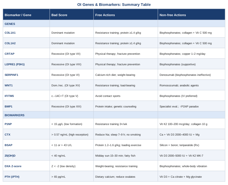
This article takes two complementary approaches. The first, and most actionable for most readers, focuses on the six biomarkers most worth tracking — what they reveal, how to measure them affordably, and what interventions can move them in the right direction. The second examines the key genes implicated in OI — what each one does, and what can realistically be done when a mutation is identified. Neither approach promises a cure. But better information, consistently acted upon, leads to better decisions.
6 Biomarkers Worth Tracking If You Have Osteogenesis Imperfecta
Most OI management focuses on fracture events and DXA scans every one to two years. That leaves enormous gaps. Bone remodeling is a continuous, dynamic process — and in OI, the underlying imbalance between bone formation and resorption is present even between fractures. Tracking biomarkers regularly gives you a real-time window into that process.
The following six markers have the strongest evidence base for clinical relevance in OI and related bone fragility conditions. Several are recommended by specialists like Peter Attia for general bone health monitoring; others are more specific to conditions where collagen synthesis is impaired.
Biomarker 1: P1NP — Procollagen Type I N-Terminal Propeptide
Why it matters: P1NP is the most sensitive marker of bone formation currently available. When osteoblasts synthesize new collagen, they cleave propeptides from both ends of the procollagen molecule. The N-terminal fragment — P1NP — enters the bloodstream in measurable concentrations. In OI, where the defect lies in collagen production itself, P1NP provides a direct readout of how much type I collagen the body is actually making. Low P1NP suggests suppressed bone formation, which is common in patients on long-term bisphosphonate therapy. Elevated P1NP can indicate high turnover states.
How to measure it: P1NP is measured from a fasting morning blood draw. It is part of most standard bone metabolism panels. Cost ranges from $40–$120 depending on laboratory and insurance. The International Osteoporosis Foundation recommends P1NP as the reference bone formation marker for clinical trials and monitoring. Reference intervals vary by lab, but adults typically fall between 15–80 µg/L.
If the score is low: the plan without supplements
Low P1NP (below 15 µg/L in adults) often reflects over-suppression of bone turnover, commonly seen after years of bisphosphonate use. Without supplements, the core non-pharmacological approach includes progressive resistance training — specifically weight-bearing mechanical loading, which stimulates osteoblast activity through the Wnt signaling pathway. Even in OI, carefully supervised resistance exercise has been shown to improve bone formation markers. Aquatic resistance training offers a safer mechanical stimulus for more severe phenotypes. Aim for 3 sessions per week, 20–30 minutes, adapted to fracture risk and current functional capacity.
If the score is low: the plan with supplements or equipment
Vitamin K2 (MK-7 form, 100–200 mcg/day) has evidence for improving osteocalcin carboxylation and supporting bone matrix formation without increasing fracture risk. A 2013 randomized trial showed MK-7 supplementation significantly improved bone strength indices in postmenopausal women. Collagen peptides (10g/day of hydrolyzed type I collagen) have emerging evidence for stimulating P1NP elevation and supporting bone mineral density in combination with resistance training. Vibration platforms (whole-body vibration at 25–50 Hz, 10–20 minutes 3x/week) have been specifically studied in OI populations as a way to stimulate bone formation without impact loading — with mixed but encouraging results. Side effects of K2 are minimal; collagen peptides are generally well tolerated.
Biomarker 2: CTX — C-Terminal Telopeptide of Type I Collagen
Why it matters: CTX (also called beta-CrossLaps) is the primary clinical marker of bone resorption. It reflects osteoclast activity — the cells that break down bone tissue. In OI, the abnormal collagen structure can drive abnormal remodeling dynamics, with some subtypes showing elevated resorption relative to formation. CTX is also the key monitoring marker when bisphosphonate therapy is initiated, since bisphosphonates work by suppressing osteoclast activity. Understanding the ratio of P1NP (formation) to CTX (resorption) gives a far more complete picture than either marker alone.
How to measure it: CTX requires a fasting, morning blood sample — ideally before 10am, as values fluctuate with food intake and circadian rhythm. Cost ranges from $40–$100. Normal adult range is approximately 0.10–0.57 ng/mL for women and 0.10–0.63 ng/mL for men, though labs vary.
If the score is high: the plan without supplements
Elevated CTX signals excessive bone breakdown. Non-pharmacological interventions include eliminating factors that accelerate resorption: reducing sodium intake (high sodium increases urinary calcium loss, which secondarily elevates PTH and thus CTX), eliminating tobacco completely, and moderating alcohol. Sleep optimization is underappreciated — cortisol, which rises sharply with sleep deprivation, directly stimulates osteoclast activity. Targeting 7–9 hours per night with consistent sleep timing has measurable effects on bone turnover markers within weeks.
If the score is high: the plan with supplements or equipment
Calcium (500–600 mg in two divided doses from food or supplement, not exceeding 1000–1200 mg/day total) combined with vitamin D3 (2000–4000 IU/day, titrated to serum 25(OH)D between 40–60 ng/mL) reduces secondary hyperparathyroidism and dampens resorption signaling. Magnesium (200–400 mg/day as glycinate or malate) supports PTH regulation and is frequently depleted in individuals taking proton pump inhibitors. Cycling: run baseline CTX after 3 months to assess response. For persistent elevation, a clinical conversation about bisphosphonate therapy initiation or adjustment is warranted.
Biomarker 3: Bone-Specific Alkaline Phosphatase (BSAP)
Why it matters: Bone-specific alkaline phosphatase is an enzyme produced by osteoblasts during bone matrix mineralization. Unlike total alkaline phosphatase, BSAP is liver-independent, making it a cleaner signal of osteoblast activity. In OI, abnormal collagen scaffolding can impair normal mineralization even when osteoblasts are functionally active — and BSAP helps distinguish between low bone formation and poor mineralization quality. It is particularly relevant in children with OI, where growth-related bone turnover is naturally high.
How to measure it: BSAP requires a specific assay (not just total ALP). It can be ordered through most reference labs. Cost ranges from $50–$130. In adults, normal BSAP is typically 11–43 U/L. Pediatric ranges are considerably higher due to active growth.
If the score is abnormal: the plan without supplements
Disproportionately low BSAP alongside normal or high CTX suggests a coupling defect — where resorption outpaces formation. This pattern argues strongly for maximizing anabolic signals through mechanical loading and adequate dietary protein (1.2–1.6g/kg/day), since protein provides the amino acid substrate for collagen synthesis. Intermittent fasting beyond 16 hours may suppress mTORC1 signaling needed for bone formation — short eating windows are not recommended for individuals with OI or active bone healing.
If the score is abnormal: the plan with supplements or equipment
Silicon (as orthosilicic acid, 6–10 mg/day) has early evidence for supporting collagen cross-linking and osteoblast differentiation. Boron (3–6 mg/day) appears to support vitamin D metabolism and has shown effects on BSAP in small studies. These are low-risk adjuncts. Teriparatide (PTH 1-34), a prescription anabolic agent, has been used off-label in severe OI to improve bone formation — it directly elevates BSAP and P1NP. This requires specialist management and is typically reserved for adults with severe disease not responding to antiresorptive therapy.
Biomarker 4: 25-Hydroxyvitamin D
Why it matters: Vitamin D deficiency is prevalent in OI populations for several reasons: reduced outdoor mobility, avoidance of activities that carry fracture risk, and in some cases, impaired vitamin D metabolism. 25(OH)D is the storage form and the clinically measured marker of vitamin D status. At levels below 30 ng/mL, secondary hyperparathyroidism begins to develop, which accelerates bone resorption — a significant problem in a condition where bone quality is already compromised. Thomas Dayspring and Peter Attia both emphasize targeting 40–60 ng/mL as an optimal functional range, not merely the laboratory "normal" threshold of 20 ng/mL.
How to measure it: Standard serum 25(OH)D test via blood draw. Cost is $30–$80, frequently covered by insurance when bone conditions are documented. Test in late winter (when levels are lowest) and again in late summer to understand seasonal range. Retest 3 months after any change in supplementation.
If the score is below 40 ng/mL: the plan without supplements
Deliberate sun exposure — 15–30 minutes of midday sun to large skin surface areas (arms, legs, torso) — can generate 10,000–20,000 IU of vitamin D3 in lighter-skinned individuals. Darker skin requires longer exposure. This is free and has no overdose risk at normal exposures. Fatty fish (salmon, sardines, mackerel) 3–4 times per week contributes meaningful dietary D3.
If the score is below 40 ng/mL: the plan with supplements
Vitamin D3 supplementation at 2,000–5,000 IU/day is the standard approach. Take with the largest meal of the day (D3 is fat-soluble). Co-administer vitamin K2 (MK-7, 100–200 mcg/day) to ensure calcium is directed toward bone rather than arterial tissue — this pairing is increasingly emphasized by functional medicine practitioners including Attia. Retest at 3 months and adjust. Do not exceed 10,000 IU/day without monitoring serum calcium and 25(OH)D, as toxicity — though uncommon — occurs at very high doses.
Biomarker 5: DXA Bone Mineral Density (Z-Score)
Why it matters: DXA (dual-energy X-ray absorptiometry) measures bone mineral density at the lumbar spine and hip. In OI, the Z-score (which compares to age-matched peers rather than the T-score used in osteoporosis) is the relevant metric. It is the most direct structural measure of whether interventions are working. While DXA doesn't capture bone quality — particularly important in OI where the matrix itself is defective — it remains the standard clinical monitoring tool and directly predicts fracture risk at the population level.
How to measure it: DXA scan at a bone density center or hospital radiology department. Cost: $100–$300 without insurance; often covered for diagnosed OI. Repeat every 1–2 years while monitoring treatment. HR-pQCT (high-resolution peripheral quantitative CT) is an emerging superior option that captures bone microarchitecture — available at specialized centers for $200–$500.
If the Z-score is low: the plan without supplements
Mechanical loading is the most powerful non-pharmacological stimulus for DXA improvement. Resistance training at 70–85% of 1RM (adapted for OI severity) has the strongest evidence. Swimming and aquatic therapy preserve cardiovascular fitness without fracture risk but are less effective for DXA improvement due to the absence of gravitational loading. If ambulatory, short bouts of weight-bearing throughout the day (accumulated step counts) show dose-dependent benefits for BMD in longitudinal studies.
If the Z-score is very low: the plan with supplements or equipment
Bisphosphonates (pamidronate, zoledronic acid) remain the most evidence-supported pharmacological intervention in OI and directly improve DXA scores — though their effect on fracture reduction in OI is more modest than in osteoporosis. Whole-body vibration platforms (Galileo or similar, 20–35 Hz, 10–15 minutes 3x/week) have been specifically studied in OI cohorts, including children, with improvement in cortical bone parameters. A controlled trial by Semler et al. showed positive trends in motor function and bone density with vibration therapy in children with OI.
Biomarker 6: PTH — Parathyroid Hormone
Why it matters: PTH is the primary regulator of calcium homeostasis. When serum calcium drops — due to inadequate intake, poor absorption, or vitamin D deficiency — the parathyroid glands secrete PTH, which mobilizes calcium from bone by stimulating osteoclast activity. Chronically elevated PTH (secondary hyperparathyroidism) accelerates the bone resorption that OI patients are already vulnerable to. Conversely, very low PTH may indicate vitamin D toxicity or primary hyperparathyroidism. PTH is the key regulator connecting vitamin D status, calcium intake, and bone resorption — making it a critical connector biomarker.
How to measure it: Intact PTH (iPTH) measured from a morning blood draw, ideally alongside 25(OH)D and calcium. Cost: $40–$100. Optimal range per functional medicine standards: 15–55 pg/mL. Secondary hyperparathyroidism is typically defined as iPTH above 65 pg/mL in the presence of normal calcium.
If PTH is elevated: the plan without supplements
Eliminate factors driving low calcium availability: reduce high-oxalate foods (spinach, almonds in excess) that bind calcium and reduce absorption, increase dietary calcium from dairy or sardines with bones, and optimize magnesium intake from whole foods (legumes, seeds, dark chocolate), since magnesium deficiency impairs PTH regulation. Assess for celiac disease or gut malabsorption if PTH remains elevated despite adequate dietary intake — this is underdiagnosed in OI populations.
If PTH is elevated: the plan with supplements
The PTH-lowering protocol centers on the vitamin D/calcium/magnesium triad. Vitamin D3 (2000–5000 IU/day) is the cornerstone intervention. Calcium citrate (not carbonate) at 500 mg twice daily improves absorption in individuals with low stomach acid. Magnesium glycinate (200–400 mg/day in the evening) supports PTH suppression. Reassess PTH and 25(OH)D at 3 months. If PTH remains elevated despite optimized levels of these nutrients, evaluation by an endocrinologist is warranted to rule out autonomous parathyroid function.
Building on the biomarker picture, understanding the genetic drivers adds the final layer — particularly for families evaluating reproductive decisions or for clinicians trying to match treatment to mechanism.
8 Key Genes in Osteogenesis Imperfecta
COL1A1 — The Core Collagen Gene
COL1A1 encodes the alpha-1 chain of type I collagen, the structural backbone of bone, tendons, and skin. The majority of OI cases (approximately 85–90%) arise from dominant mutations in COL1A1 or COL1A2. Quantitative mutations reduce collagen output (milder disease, OI type I); qualitative mutations produce structurally abnormal collagen that disrupts the entire fibril (more severe disease, types II, III, IV).
If the COL1A1 gene is affected: plan without supplements
Because COL1A1 mutations cannot currently be corrected at the somatic level outside clinical trials, management focuses on maximizing the functional output of normal alleles. Mechanical loading — particularly resistance training — upregulates collagen synthesis at the transcriptional level. Adequate dietary protein (1.4–1.6g/kg/day) provides glycine and proline, the rate-limiting amino acids for collagen biosynthesis.
If the COL1A1 gene is affected: plan with supplements
Hydrolyzed collagen peptides (10g/day) may support collagen production by providing bioavailable proline and glycine as precursors. Vitamin C (500–1000 mg/day) is a required cofactor for prolyl hydroxylase, the enzyme that stabilizes the collagen triple helix — deficiency directly impairs collagen cross-linking. Cycling: ongoing use of both is reasonable with no significant side effects at these doses.
COL1A2 — The Partner Collagen Chain
COL1A2 encodes the alpha-2 chain of type I collagen. Two alpha-1 chains and one alpha-2 chain form the collagen heterotrimer. Mutations here follow similar patterns to COL1A1 and produce similar phenotypic outcomes — the same intervention logic applies. A specific subset of recessive COL1A2 mutations has been associated with OI combined with Ehlers-Danlos syndrome features, suggesting overlap in connective tissue fragility beyond bone.
CRTAP — The Collagen Modification Gene
CRTAP (cartilage-associated protein) is essential for hydroxylation of a specific proline residue (Pro986) in the collagen alpha-1 chain. This modification is required for proper collagen folding. Recessive mutations in CRTAP produce OI type VII — typically severe to lethal. Because CRTAP functions in post-translational modification of collagen rather than collagen synthesis itself, the resulting collagen is overmodified (hyperglycosylated), which impairs fibril formation. Family genetic counseling is critical — the autosomal recessive pattern means siblings of affected individuals carry a 25% recurrence risk.
If the CRTAP gene is affected: plan without supplements
No targeted therapy for CRTAP loss currently exists outside clinical trials. Management is primarily supportive: fracture prevention, bisphosphonate therapy to reduce resorption, and physical therapy adapted to severity. Gene therapy trials targeting recessive OI subtypes are in early stages.
If the CRTAP gene is affected: plan with supplements
Supporting collagen processing indirectly through copper (1–2 mg/day, as lysyl oxidase — the key collagen cross-linking enzyme — is copper-dependent) may have minor supportive value. This is not curative but ensures at least one cross-linking pathway is not further impaired by nutrient deficiency. Avoid high-dose zinc supplementation (above 40 mg/day) without concurrent copper, as zinc and copper compete for absorption.
LEPRE1 (P3H1) — The Prolyl Hydroxylase Gene
LEPRE1 encodes prolyl 3-hydroxylase 1, the enzyme that modifies Pro986 — working in a complex with CRTAP and PPIB. Recessive mutations produce OI type VIII, clinically similar to type VII. This subtype is notably more common in individuals of West African ancestry. The clinical presentation is severe, with white sclerae (differentiating it from the blue sclerae of classic OI), and the fracture burden is high. Understanding this genetic distinction matters because it changes prognostic expectations and signals that the primary defect is in collagen chaperoning, not collagen synthesis.
SERPINF1 — The Vascularization and Bone Density Gene
SERPINF1 encodes pigment epithelium-derived factor (PEDF), a secreted glycoprotein with antiangiogenic and osteogenic functions. Recessive mutations cause OI type VI — characterized by a distinctive "fish-scale" pattern of bone lamellation on histology, extremely low bone density, and the absence of the typical elevated alkaline phosphatase response to bisphosphonates. SERPINF1-related OI is one of the subtypes where standard bisphosphonate therapy shows poor response — a critical genetic distinction. Anti-RANKL therapy (denosumab) has shown more promise in this subtype.
WNT1 — The Signaling Gene
WNT1 mutations — both dominant (causing OI type XV) and recessive (causing severe early-onset osteoporosis) — disrupt Wnt/β-catenin signaling, one of the primary pathways driving osteoblast differentiation and bone formation. Individuals with WNT1 mutations tend to have very low bone formation markers (low P1NP, low BSAP) with relatively preserved bone resorption — a decoupling that limits the effectiveness of antiresorptive monotherapy. Anabolic agents targeting the Wnt pathway, including romosozumab (anti-sclerostin antibody), may be particularly relevant in this subtype, though human trial data in WNT1-specific OI remains limited.
IFITM5 — The Type V OI Gene
IFITM5 mutations are responsible for OI type V — dominantly inherited, and unusual because the same recurrent mutation (c.-14C>T in the 5'UTR) accounts for essentially all cases. Type V OI is characterized by calcification of the forearm interosseous membrane, a hyperplastic callus formation following fractures, and radio-opaque metaphyseal bands — features not seen in collagen-mutation OI. Bisphosphonates remain first-line. The recurrent nature of the mutation means that if one family member is confirmed to have type V OI, the diagnostic workup for relatives is straightforward.
BMP1 — The Collagen Processing Gene
BMP1 encodes bone morphogenetic protein 1, a metalloprotease responsible for cleaving the C-propeptide of procollagen — an essential step in converting procollagen to mature collagen. Recessive mutations cause OI type XIII, often associated with elevated P1NP (because the uncleaved propeptide accumulates) — a paradox where high formation markers coexist with poor bone quality. This highlights why interpretation of biomarkers always requires genetic context. BMP1 mutations are rare but clinically recognizable by the unique P1NP pattern.
A Book That Reframes How to Think About Bone Fragility
Outlive: The Science and Art of Longevity by Peter Attia dedicates substantial attention to musculoskeletal health and bone density as pillars of what Attia calls "the centenarian decathlon" — the ability to perform the physical tasks that matter in later decades. While not specifically about OI, the framework is highly applicable.
The 10 Most Relevant Insights for OI from Attia's Framework
1. Bone density peaks in your early 20s and the investment made before then determines the floor for the rest of life. In OI, this window is compressed by fracture risk — but the principle of maximizing peak bone density through every available safe means during adolescence applies directly.
2. The P1NP to CTX ratio matters more than either marker alone. Attia repeatedly emphasizes that uncoupled bone turnover — where resorption outpaces formation — is far more dangerous than symmetrically elevated turnover. In OI, monitoring both markers at the same time provides a ratio that captures this dynamic.
3. Grip strength is a proxy for systemic musculoskeletal health and predicts fracture risk independently of BMD. Grip strength measurement (dynamometer, $30–$50 device) is a cheap, repeatable functional test. For OI patients with upper extremity involvement, grip strength trends over time reveal whether the functional component of bone-muscle interaction is improving.
4. Zone 2 cardio (conversational pace, 45–60 minutes, 4x/week) reduces systemic inflammation and improves insulin sensitivity — both of which affect bone remodeling through IGF-1 and cortisol pathways. Even aquatic Zone 2 exercise achieves this metabolic benefit.
5. Protein intake is chronically underestimated in bone health protocols. Attia targets 1.6g/kg/day minimum. For OI, where collagen is the primary defect, ensuring adequate protein substrate is foundational — yet most bone health guidelines emphasize minerals and vitamins while leaving protein targets vague.
6. Sleep is anabolic. Growth hormone — which directly stimulates collagen synthesis and bone formation — is secreted almost entirely during slow-wave sleep. Prioritizing sleep duration and quality is not optional for anyone trying to optimize bone formation.
7. Muscle mass is the largest reservoir of amino acids that feed collagen synthesis during periods of dietary inadequacy. Building and maintaining lean mass creates metabolic buffer. In OI patients who are frequently immobilized after fractures, muscle mass preservation during recovery directly supports bone healing speed.
8. The relationship between statins and musculoskeletal health is nuanced. Some statins have been associated with myopathy and impaired CoQ10 production — relevant for OI patients who may also be managing cardiovascular risk. Attia recommends CoQ10 (100–200 mg/day) if statins are prescribed alongside any bone or muscle-health protocol.
9. Omega-3 fatty acids (EPA/DHA, 2–4g/day combined) reduce inflammatory cytokines — including IL-6 and TNF-alpha — that stimulate RANKL expression and osteoclast activation. In OI, chronic micro-inflammation from repeated fracture healing may chronically elevate these pathways. Fish oil at therapeutic doses is one of the lowest-risk, highest-value adjuncts.
10. Continuous glucose monitoring (CGM) reveals post-meal glucose spikes that chronically elevate cortisol and advanced glycation end-products (AGEs) — both of which impair collagen cross-linking. A two-week CGM trial ($50–$100) can identify dietary patterns that are quietly degrading bone matrix quality.
Complementary Approaches Worth Considering
Mindfulness Meditation and MBSR
Chronic pain is a near-universal feature of OI across all subtypes — stemming from fractures, skeletal deformity, and the psychological anticipation of injury. Mindfulness-Based Stress Reduction (MBSR), developed by Jon Kabat-Zinn, involves 8 weeks of structured training in present-moment attention, body scanning, and non-reactive awareness of pain signals. Its relevance in OI lies partly in direct pain modulation (through endogenous opioid pathways and altered pain perception in the anterior cingulate cortex) and partly in reducing the cortisol load of chronic anxiety — cortisol being a direct driver of bone resorption.
A 2016 meta-analysis published in JAMA Internal Medicine confirmed that mindfulness meditation programs produced significant improvements in chronic pain, depression, and anxiety compared to control conditions. Evidence specifically in OI is limited, but the chronic pain mechanism is shared with other musculoskeletal conditions where MBSR shows consistent benefit.
For practical application in OI: begin with a guided 8-week MBSR program (available via apps such as Insight Timer or formalized programs through hospital-based integrative medicine departments). Body scan practice for 20–45 minutes daily is the core technique. The focus for OI should be distinguishing between pain that signals acute fracture risk versus habitual pain that does not — developing this discrimination reduces protective restriction while maintaining genuine safety awareness.
Biofeedback
Biofeedback uses real-time physiological monitoring — surface electromyography, heart rate variability, skin conductance, or temperature sensors — to help individuals learn voluntary control over bodily states. In OI, it is most relevant as a pain management and muscle tension tool. Fracture events and chronic pain create compensatory muscle guarding patterns that increase biomechanical load on already fragile bone segments. Learning to consciously reduce muscle tension in affected areas reduces this secondary loading — a genuinely specific mechanism for OI.
A 2011 review in Applied Psychophysiology and Biofeedback confirmed efficacy of biofeedback for chronic musculoskeletal pain, with effects on both pain intensity and functional disability. EMG biofeedback in particular has shown benefit for guarding-related pain patterns.
Practical protocol: 10 sessions with a certified biofeedback practitioner (BCIA-certified), followed by home practice with a consumer HRV biofeedback device (Polar H10 or similar, $80–$120). HRV biofeedback specifically — slow breathing at approximately 6 breaths/minute — enhances parasympathetic tone and reduces circulating cortisol, providing the secondary bone benefit of blunting chronic resorption signals.
Music Therapy
Music therapy involves structured, clinician-guided use of music as a therapeutic tool — distinct from passive music listening. In OI, which disproportionately affects children and carries significant psychological burden (fear of fracture, social isolation, limited physical participation), music therapy addresses the psychological dimension that standard medical care frequently neglects. Active music making — drumming, singing, instrument play — also provides gentle upper-body motor engagement and a sense of physical competence that fracture avoidance often erodes.
A 2016 Cochrane systematic review of music interventions for pain found significant reductions in pain intensity and analgesic requirements across clinical populations. Evidence specifically in OI is absent, but the pain reduction and psychological wellbeing mechanisms are well-established in pediatric hospital settings, where OI patients are frequently encountered.
Practical approach: board-certified music therapists (MT-BC credential) offer individual or group sessions. For children with OI, rhythm-based activities such as adaptive drumming allow physical expression without significant fracture risk. Sessions of 30–45 minutes twice weekly represent the standard therapeutic dose in pediatric pain programs. This is low-risk with no adverse effects.
Breathing-Based Therapies
Dysregulated breathing patterns — particularly chronic hyperventilation — are common in individuals with chronic pain and anxiety, including OI. Habitual overbreathing reduces arterial CO2, which paradoxically increases pain sensitivity and impairs sleep quality. Breathing retraining based on the Buteyko method or coherence breathing (approximately 6 breaths/minute) normalizes CO2 levels, reduces sympathetic activation, and improves HRV — all relevant to the cortisol-bone axis. There is also a direct musculoskeletal dimension: accessory respiratory muscle overuse from chronic hyperventilation creates chronic tension in cervical and thoracic musculature that is particularly problematic in OI patients with vertebral compression.
A study published in the journal Chest demonstrated that breathing retraining reduced dysfunctional breathing symptoms and improved functional status in patients with chronic respiratory conditions. For OI, the application is primarily analgesic and anxiolytic rather than respiratory. Slow-breathing protocols (4-7-8 breathing, box breathing at 4 seconds per phase) practiced for 10 minutes twice daily show measurable HRV improvements within 4 weeks.
Low-Level Laser Therapy (Photobiomodulation)
Low-level laser therapy (LLLT), also called photobiomodulation (PBM), applies near-infrared or red light to tissue at non-thermal power levels. The proposed mechanism involves absorption by cytochrome c oxidase in mitochondria, increasing ATP production, reducing oxidative stress, and modulating inflammatory signaling. For OI, the most relevant application is post-fracture healing acceleration — pre-clinical studies consistently show faster bone callus formation and improved mechanical properties with LLLT, and early human data supports accelerated fracture healing.
A 2014 systematic review of LLLT for bone repair found positive effects on bone healing parameters, though study quality was variable. No trials in OI specifically exist, but the healing acceleration mechanism is not OI-specific — it operates at the cellular level regardless of collagen genotype.
Protocol: 830–980 nm near-infrared at 50–100 mW/cm², applied for 60–90 seconds per point over a fracture site or adjacent bone, 3–5 times per week during active healing. Devices such as the Joovv Solo or similar clinical panels ($400–$1200 for home devices) can be used after clearance from the treating orthopedic team. Adverse effects are minimal — avoid direct eye exposure. This should be considered adjunctive to, not a replacement for, standard fracture management.
Conclusion
Osteogenesis imperfecta is a condition where the difference between passive management and active monitoring is significant. The biomarkers covered in this article — P1NP, CTX, BSAP, 25(OH)D, DXA Z-score, and PTH — form a practical, measurable framework that reveals what is actually happening with bone turnover between clinic visits. The genetic picture, while less immediately actionable, determines which interventions are likely to work and which may not — information worth having before committing to years of a particular treatment.
The most useful next step is straightforward: if you have not had a bone turnover panel in the last six months, request one that includes P1NP and CTX alongside 25(OH)D and PTH. If you have not had genetic testing, discuss it with a medical geneticist or a specialized OI center — the OI Foundation maintains a directory of specialized clinics. And if the complementary strategies in this article — breathing work, biofeedback, or photobiomodulation — seem relevant to your situation, bring them to your care team as questions, not replacements. The goal is not to replace medical management but to fill the gaps that medical management consistently leaves open.
Musculoskeletal Endocrine & Metabolic
Musculoskeletal: Bone Conditions
Autoimmune: Connective Tissue Conditions