This article contains AI generated content.
· UpdatedOsteopoikilosis Genes Biomarkers – 3 Genes And 7 Biomarkers To Track
When a Rare Bone Condition Leaves You With More Questions Than Answers
Most people with osteopoikilosis discover it by accident. An X-ray taken for a sprained ankle or an unrelated scan comes back with a radiologist's note about "multiple sclerotic foci" or "bone islands" scattered throughout the skeleton. The orthopedist or GP then delivers what has become the default script: it is a benign finding, probably hereditary, nothing to worry about. Go home. Live your life.
For many people, that answer lands badly — not because it is technically wrong, but because it offers nothing useful. If you have joint discomfort that appeared around the time of diagnosis, or a family member who carries the same finding, or if you simply feel unsettled by a condition most doctors have never seen in a living patient, "benign and rare" is not especially helpful guidance.
The biology of osteopoikilosis is more specific than that reassurance implies. The condition is driven by a disruption in how bone-forming signals — particularly those traveling through the BMP (bone morphogenetic protein) and TGF-β (transforming growth factor beta) pathways — are regulated inside osteoblasts. That disruption has a precise genetic origin in most cases, and it produces downstream effects that are measurable through blood markers. None of this makes osteopoikilosis dangerous. But it does mean there is considerably more to understand, and more to track, than a single X-ray report conveys.
This article approaches osteopoikilosis through two complementary lenses. The primary focus is on seven biomarkers that can give a clearer, actionable picture of bone metabolism and systemic inflammation — markers that researchers like Peter Attia and Thomas Dayspring have helped bring into meaningful clinical practice. The second lens covers the genetic architecture of the condition: what the key genes do, what happens when they go wrong, and what can be done about it. Between these two frameworks, most people with osteopoikilosis will find something more useful than a reassuring shrug.
Summary
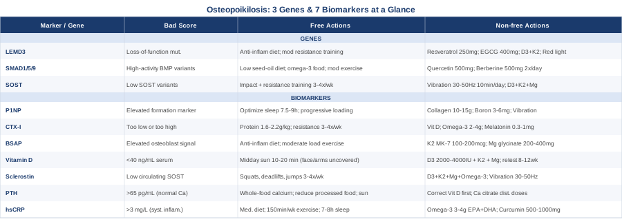
This article covers seven measurable biomarkers — including why P1NP and CTX-I are the gold standard starting point, what sclerostin reveals that standard panels entirely miss, and why tracking hsCRP matters even in a condition not classified as inflammatory. It examines three key genes behind osteopoikilosis — including the primary LEMD3/MAN1 gene, how its loss directly drives the excessive bone formation visible on X-ray, and what BMP pathway variants mean for the severity of the condition. Each marker and gene comes with a practical protocol: what to do when results are suboptimal, with and without supplementation, including frequency, cycling guidance, and side effects. Beyond the core biomarker and genetics material, the article synthesizes the most relevant bone health insights from Peter Attia's work in Outlive — reframed for someone with this specific condition — and examines three complementary modalities with real human clinical evidence behind them.
7 Biomarkers Worth Tracking If You Have Osteopoikilosis
Tracking biomarkers in osteopoikilosis is not standard practice — most clinicians will not order a bone marker panel for what they have labeled a benign incidental finding. But the right frame here is not diagnostic. These markers do not tell you whether you have OPK. They reveal the current functional state of your bone metabolism, the degree of systemic inflammation, and how your body is balancing bone formation against resorption. For someone with a known BMP pathway disruption in their biology, that information is genuinely valuable.
The markers below range from inexpensive and widely available — vitamin D, hsCRP, alkaline phosphatase — to more specialized and harder to access, like sclerostin. Start with what your clinician can order and what your budget allows, then expand the panel over time.
1. Bone-Specific Alkaline Phosphatase (BSAP)
Why it matters: Alkaline phosphatase (ALP) is a byproduct of osteoblast activity. When bone-forming cells are working, ALP levels rise. Standard blood panels include total ALP, which also captures liver-derived ALP and is therefore too noisy for bone-specific interpretation. Bone-specific ALP (BSAP) isolates the osteoblast signal, making it a cleaner indicator of bone formation activity.
In osteopoikilosis, the BMP pathway is overactive in affected bone regions. BSAP gives a rough systemic sense of whether that overactivity is reflected in the broader bone formation environment. In most OPK patients, BSAP falls within normal range — but establishing a personal baseline is what makes future readings interpretable. A single number without a trend is limited; two numbers separated by six months is the beginning of actual information.
How to measure it: BSAP can be ordered as a standalone test or as part of a bone marker panel. Out-of-pocket cost typically ranges from $40–$120 depending on the lab and coverage. Quest Diagnostics and LabCorp both offer it. Some clinicians use total ALP from a comprehensive metabolic panel as a rough proxy, though BSAP is meaningfully more precise. Fasting is not required. Retest every 6–12 months once a baseline is established.
If BSAP is elevated — the plan without supplements: Elevated BSAP suggests active bone formation is running above expected levels. Focus on reducing systemic stressors that amplify osteoblast activity: eliminate ultra-processed foods, prioritize an anti-inflammatory dietary pattern (Mediterranean, whole-food-based), and reduce alcohol. Increase load-bearing exercise — but moderate intensity over excessive volume, since extreme endurance training can paradoxically spike bone turnover markers without proportional functional bone quality gains.
If BSAP is elevated — the plan with supplements or equipment: Vitamin K2 (as MK-7, 100–200 mcg daily) directs calcium into the bone matrix and has been shown in human trials to support appropriate bone mineralization without promoting pathological calcification. Magnesium glycinate (200–400 mg nightly) supports ALP enzyme function and overall bone matrix quality. Frequency: ongoing, no cycling required. Side effects: vitamin K2 can theoretically interfere with warfarin — confirm with a clinician if on anticoagulants.
2. P1NP (Procollagen Type 1 N-terminal Propeptide)
Why it matters: P1NP is considered by most bone health specialists — including Peter Attia and the International Osteoporosis Foundation (IOF) — as the gold standard biomarker for bone formation. It is released when type I collagen, the main structural protein of bone, is being synthesized. That makes it a highly sensitive and specific indicator of how actively new bone is being laid down right now.
For someone with osteopoikilosis, P1NP provides more granular information than BSAP alone. Because OPK involves localized overactivation of bone formation, P1NP helps distinguish between someone whose overall systemic bone turnover is normal and someone whose formation rate is genuinely elevated beyond the sclerotic zones. It is also the best single marker to track when implementing any dietary, supplemental, or exercise-based intervention — it tells you clearly whether what you are doing is working.
How to measure it: P1NP is available through most major reference labs. Expect $60–$150 out-of-pocket. The IOF and ISCD both recommend it as the reference bone formation marker in clinical protocols. Most clinicians who work with bone markers order P1NP alongside CTX-I (see below) to read the full remodeling picture. Collect a morning, fasting sample for the lowest variability and the most meaningful longitudinal comparisons.
If P1NP is elevated — the plan without supplements: Prioritize sleep quality as the first intervention — growth hormone is secreted primarily during slow-wave sleep, and it is one of the strongest natural regulators of bone formation activity. Target 7.5–9 hours with consistent timing. Resistance training with progressive overload — not excessive volume — is the most evidence-backed non-pharmacological way to direct elevated formation activity toward functional bone quality rather than disordered accumulation.
If P1NP is elevated — the plan with supplements or equipment: Collagen peptides (10–15 g/day with vitamin C) support structured bone matrix formation by providing the amino acid substrate for type I collagen synthesis. Boron (3–6 mg/day from food or supplementation) has shown effects on bone turnover markers in small human trials and supports estrogen and testosterone metabolism, both of which directly influence P1NP levels. Whole-body vibration platforms (30–60 Hz range, 10 minutes daily) have demonstrated effects on bone marker profiles in postmenopausal women in randomized trials; evidence in OPK specifically is absent, but the mechanobiological rationale is sound. Frequency: no mandatory cycling for these supplements. Side effects: collagen peptides are well tolerated; boron at very high doses (above 20 mg/day) can cause nausea — stay within the 3–6 mg range.
3. CTX-I (C-terminal Telopeptide of Type 1 Collagen)
Why it matters: CTX-I (also called Beta-CrossLaps) is the direct complement to P1NP. While P1NP reflects bone formation, CTX-I reflects bone resorption — the process by which osteoclasts break down old bone tissue. Healthy bone remodeling requires balance between these two processes. Neither marker tells a complete story alone.
In osteopoikilosis, where the predominant issue is overactive formation, understanding resorption is equally important. A low CTX-I alongside an elevated P1NP means net bone accumulation is occurring — consistent with OPK. If both markers are elevated together, systemic turnover has accelerated for an additional reason: inflammation, vitamin D deficiency, hormonal change, or something else worth investigating. Reading the two markers as a pair is where the real diagnostic value lives.
How to measure it: CTX-I is typically ordered alongside P1NP as a bone turnover pair. Cost is similar: $50–$130 out-of-pocket. Critical collection detail: CTX-I shows pronounced diurnal variation — it is highest in the early morning and lowest in the afternoon. Always use a fasting, pre-9 AM sample for consistent, comparable results. A single afternoon result is not reliably usable for longitudinal tracking and should be disregarded or repeated under proper conditions.
If CTX-I is too low — the plan without supplements: Very low CTX-I alongside elevated P1NP confirms net bone accumulation consistent with OPK. Ensure adequate dietary protein (1.6–2.2 g/kg bodyweight daily), which is essential for osteoclast health and balanced remodeling. Avoid excessive supplemental calcium without vitamin D3 and K2 pairing, since high isolated calcium may further blunt resorption signals.
If CTX-I is too high — the plan without supplements: Elevated resorption signals can be driven by chronic stress, poor sleep, low estrogen or testosterone, or vitamin D deficiency — identify which applies. Eliminate excess caffeine (above 400 mg/day) and alcohol, both of which reliably elevate CTX. Resistance training remains the most evidence-based non-pharmacological way to shift the P1NP/CTX ratio toward formation. Aim for 3–4 sessions per week with compound movements.
If CTX-I is elevated — the plan with supplements: Optimize vitamin D to 40–60 ng/mL serum (see below) — this alone corrects CTX elevation in many deficient individuals. Omega-3 fatty acids at 2–4 g EPA+DHA per day have shown modest anti-resorptive effects in human trials. Low-dose melatonin (0.3–1 mg at bedtime) has been shown in small human studies to reduce bone resorption markers by supporting osteoblast over osteoclast activity. Frequency: omega-3s can be taken continuously; melatonin can be cycled at 5 days on, 2 days off if used long-term for this purpose. Side effects: low-dose melatonin is well tolerated; doses above 3 mg may impair natural melatonin production over time.
4. 25-OH Vitamin D (Serum Vitamin D)
Why it matters: Vitamin D deficiency impairs bone mineralization and drives compensatory increases in PTH (parathyroid hormone), which in turn accelerates bone resorption. Even in a condition where bone formation is already overactive — as in OPK — adequate vitamin D is essential for ensuring that the matrix being formed is well-mineralized rather than structurally compromised.
Beyond bone mechanics, vitamin D receptors are present on immune cells throughout the body, meaning vitamin D status measurably influences systemic inflammation. This matters for OPK patients who experience joint symptoms, where an inflammatory driver may be contributing to discomfort that gets attributed solely to the structural bone finding.
How to measure it: A 25-OH vitamin D blood test is widely available and costs $30–$70, often covered by insurance. Target serum level: most integrative clinicians — including Peter Attia — recommend 40–60 ng/mL as the functional optimum, compared to many labs' lower "sufficient" threshold of 30 ng/mL. Re-test every 3–6 months while actively optimizing; annually once stable.
If the score is below 40 ng/mL — the plan without supplements: Midday sun exposure (10–20 minutes, face and arms uncovered) produces meaningful vitamin D3 synthesis in fair-to-medium skin tones; longer exposure is needed for darker complexions or high-latitude residents. This approach costs nothing and carries additional benefit through circadian signaling and mild photobiomodulation to skin and bone-adjacent tissue.
If the score is below 40 ng/mL — the plan with supplements: Vitamin D3 supplementation at 2,000–4,000 IU daily with a fatty meal (fat-soluble absorption) is among the most evidence-backed bone interventions available. Pair with vitamin K2 (MK-7, 100–200 mcg) to minimize soft-tissue calcification risk and magnesium (200–400 mg) to support activation of the 25-OH to 1,25-OH conversion. Re-test after 8–12 weeks. Some individuals with variants in the VDR gene or the CYP27B1 conversion enzyme show blunted response and may need higher doses under clinical supervision. Side effects: toxicity requires sustained intake well above 10,000 IU/day; never exceed 100 ng/mL serum without medical guidance.
5. Sclerostin (SOST Protein)
Why it matters: Sclerostin is a protein produced primarily by osteocytes — mature bone cells embedded within the mineralized matrix. Its biological role is to act as a brake on bone formation by inhibiting Wnt signaling in osteoblasts. The pharmaceutical world recognized this mechanism clearly enough to develop romosozumab (Evenity), an anti-sclerostin antibody now approved for osteoporosis that works by blocking this brake to allow bone formation to proceed.
In osteopoikilosis, where BMP pathway signaling is overactive — a pathway that overlaps with and interacts with Wnt signaling — measuring sclerostin adds a layer of bone biology information that standard panels entirely miss. Low circulating sclerostin, whether from genetic predisposition or reduced osteocyte function, could contribute to the persistence of overactive bone formation signals in OPK-affected areas. This is still an emerging area of research for this specific condition, but the mechanistic relevance is real.
How to measure it: Serum sclerostin assays are more specialized than the markers above. ARUP Laboratories and some research-affiliated labs offer it in the US. Cost ranges from $80–$200 and insurance coverage is unlikely. It is not part of any standard clinical bone panel at present, so access typically requires a functional or integrative medicine physician willing to work with reference labs. Interpret alongside P1NP and CTX-I rather than in isolation.
If sclerostin is low — the plan without supplements: Mechanical loading is the primary physiological activator of sclerostin production. Resistance training and high-impact exercise stimulate osteocytes to secrete sclerostin, which then feeds back to limit excessive bone formation — this is part of the normal mechanosensing regulatory loop. For OPK patients, this is an important exercise-specific application: compound, load-bearing movements (squats, deadlifts, weighted carries, jumping when appropriate to the individual) support the body's own bone formation braking system. Aim for 3–4 sessions per week.
If sclerostin is low — the plan with supplements or equipment: No supplement has been shown to reliably raise sclerostin levels in humans. Optimizing the broader bone metabolism environment — vitamin D, K2, magnesium, omega-3s, protein — supports appropriate BMP/Wnt interaction within its intended parameters. For equipment: vibration platforms at 30–50 Hz for 10–15 minutes daily have been shown in human studies to activate osteocyte mechanosensing responses, which includes SOST regulation. This is an active area of bone biology research and early evidence is promising. Side effects: whole-body vibration at appropriate frequencies is safe for most adults; avoid frequencies above 60 Hz without clinical guidance.
6. PTH (Parathyroid Hormone)
Why it matters: PTH is the body's primary regulator of calcium homeostasis. When calcium or vitamin D is insufficient, PTH rises to mobilize calcium from bone, accelerating resorption in the process. Secondary hyperparathyroidism — PTH elevated because of low vitamin D rather than a primary gland problem — is common and frequently goes undetected.
For someone with osteopoikilosis, elevated PTH is particularly relevant because it adds a resorption stimulus on top of the dysregulated formation environment that already exists. Tracking PTH alongside vitamin D gives a complete picture of the calcium-bone axis. A vitamin D level of 35 ng/mL with a normal PTH looks categorically different from the same vitamin D level paired with elevated PTH — the second scenario means the body is already compensating for perceived insufficiency, and the bone is paying the cost.
How to measure it: Intact PTH (iPTH) is the standard assay, often included in comprehensive metabolic panels or ordered alongside vitamin D. Cost: $30–$80. Target: ideally in the lower half of the reference range, approximately 15–45 pg/mL by most lab standards. PTH consistently above 65–70 pg/mL in the context of normal serum calcium warrants investigation into vitamin D status and dietary calcium patterns before any further workup.
If PTH is elevated — the plan without supplements: The most common correctable drivers are vitamin D insufficiency, dietary calcium intake below physiological requirements (1,000–1,200 mg/day for most adults), and excess dietary phosphorus from ultra-processed food suppressing calcium absorption. Prioritize whole-food calcium sources: sardines with bones, full-fat dairy, fortified plant foods. Increase sun exposure. Reduce soft drinks and highly processed food.
If PTH is elevated — the plan with supplements: Correct vitamin D first (see above) — this is the most common and fully reversible driver of elevated PTH. If calcium intake from food is genuinely insufficient, calcium citrate (better absorbed than calcium carbonate, especially without a heavy meal) is the preferred supplemental form. In the context of OPK's overactive bone formation signal, avoid large single-dose calcium supplements (above 500 mg at once) and instead distribute intake across meals. Side effects: calcium supplementation at high doses (above 2,500 mg/day total from all sources) has been associated with cardiovascular risk in some observational data — stay within physiological ranges and prioritize food sources.
7. hsCRP (High-Sensitivity C-Reactive Protein)
Why it matters: Osteopoikilosis is not classified as an inflammatory condition, but that classification does not make inflammation irrelevant for people who have it. Chronic low-grade systemic inflammation — reflected by elevated hsCRP — accelerates bone resorption, impairs bone mineralization, and is independently associated with joint pain and stiffness. A meaningful subset of OPK patients reports musculoskeletal symptoms, and elevated hsCRP may be the missing link between a genetic predisposition and functional, daily complaints.
Peter Attia has written extensively about hsCRP as a foundational health marker — not a disease-specific test but a systemic risk indicator that cuts across cardiovascular, metabolic, and musculoskeletal health. For OPK patients, it serves as an important contextualizer for everything else in the bone marker panel. An elevated hsCRP changes how you interpret elevated CTX-I. It changes what dietary and lifestyle interventions to prioritize.
How to measure it: hsCRP — specifically high-sensitivity CRP, not standard CRP, which is too coarse to detect meaningful low-grade inflammation — is widely available and costs $15–$40. Target: below 1 mg/L for low systemic inflammatory burden; above 3 mg/L indicates elevation worth addressing. Always retest if results are high during or shortly after an illness, which can transiently spike the marker independent of chronic status.
If hsCRP is elevated — the plan without supplements: The lifestyle levers for lowering hsCRP are among the most studied in medicine: a Mediterranean or anti-inflammatory dietary pattern, elimination of trans fats and refined carbohydrates, at least 150 minutes of moderate exercise per week, 7–8 hours of quality sleep, and consistent stress reduction practices. Each of these independently lowers hsCRP; in combination, the effect is substantial. Prolonged sitting elevates hsCRP even in people who exercise — brief activity breaks throughout the day matter.
If hsCRP is elevated — the plan with supplements: Omega-3 fatty acids at therapeutic doses (3–4 g EPA+DHA/day) consistently reduce hsCRP in randomized controlled trials. Bioavailable curcumin (500–1,000 mg/day in forms like BCM-95 or Meriva) has shown significant hsCRP reductions in human trials. Vitamin D and magnesium (already discussed) carry anti-inflammatory effects as secondary benefits. Cycling: omega-3s can be taken continuously; curcumin can be taken continuously or cycled at higher doses (8 weeks on, 4 weeks off) to assess individual response. Side effects: high-dose omega-3s may modestly prolong bleeding time — note if taking NSAIDs or anticoagulants. Curcumin can interact with certain medications; review with a clinician if on chronic prescriptions.
Moving from these seven measurable markers into the genetic layer adds a fundamentally different dimension to understanding osteopoikilosis — not "what is happening right now" but "why your bone biology is wired the way it is, and what that means for everything you do next."
The Genetics Behind Osteopoikilosis: What Your DNA Reveals
Understanding the genetics of osteopoikilosis does not change the diagnosis — that is still a radiological finding. But it changes how you interpret everything else about the condition. The genes involved explain not just the bone islands visible on imaging but the entire signaling environment that produced them. Some of those pathways respond to targeted interventions even when the underlying mutation is not correctable.
Gene 1: LEMD3 (Also Known as MAN1)
What it does: LEMD3 is the primary gene responsible for osteopoikilosis. Located on chromosome 12q14.3, it encodes an inner nuclear membrane protein that serves as a critical negative regulator of both BMP and TGF-β signaling. The mechanism is elegant and specific: LEMD3 binds to phosphorylated SMAD proteins — the intracellular messengers that carry BMP and TGF-β signals from the cell surface to the nucleus — and facilitates their dephosphorylation, effectively switching the signal off before it can drive excessive gene expression.
When LEMD3 carries a loss-of-function mutation, this braking mechanism fails. Phosphorylated SMADs accumulate in the nucleus and remain active longer than they should. BMP and TGF-β target genes stay on, driving osteoblasts to excessive formation activity in specific bone locations. The result, over years, is the sclerotic foci characteristic of OPK — patches of dense, well-mineralized bone where the formation signal ran unchecked. Inheritance is autosomal dominant, meaning a single mutated copy is sufficient. The landmark work identifying LEMD3 mutations as causative for osteopoikilosis, Buschke-Ollendorff syndrome, and melorheostosis was published in Nature Genetics in 2004 by Hellemans and colleagues, establishing the shared molecular origin of these related conditions.
What it affects in practice: The primary consequence is localized bone overformation visible on imaging. Secondary effects may include altered systemic bone remodeling dynamics, joint involvement in some families (particularly in Buschke-Ollendorff presentations), and a variably expressive phenotype even within the same family carrying the same mutation — suggesting that modifier genes or epigenetic factors play a meaningful role in how the condition manifests.
If the gene is bad — the plan without supplements: LEMD3 function cannot be restored with lifestyle interventions. What can be done is modulating the downstream consequences. Since BMP/TGF-β signaling drives excessive osteoblast activity, avoid lifestyle factors that further amplify this pathway: reduce ultra-processed food intake (which increases systemic TGF-β-promoting cytokines), maintain a healthy body weight (adipose tissue secretes signaling molecules that amplify TGF-β), and perform moderate resistance training without extreme volume. Schedule regular radiological follow-up to monitor for any changes in the distribution or density of bone islands over time — particularly if you develop new joint symptoms or the condition has been atypical in presentation.
If the gene is bad — the plan with supplements or equipment: Several natural compounds have documented BMP/TGF-β pathway interactions: - Resveratrol (250–500 mg/day of a high-bioavailability form) has shown SMAD pathway modulation in preclinical bone models, though human bone-specific evidence remains limited. Cycling: 8 weeks on, 4 weeks off. Side effects: generally well tolerated; avoid high doses in pregnancy; may interact with blood thinners. - EGCG from green tea (400–600 mg/day standardized extract) has shown TGF-β modulation in some human studies, particularly in the context of fibrotic and proliferative processes. Cycling: 8–12 weeks on, rest period. Side effects: avoid on an empty stomach — can cause nausea; limit total daily caffeine from all sources. - Vitamin D3 with K2 (as described above): supports appropriate calcium metabolism in the context of overactive bone formation — a foundational ongoing intervention with no cycling required. - For equipment: red and near-infrared light therapy at 660 nm and 850 nm combined has emerging evidence for modulating BMP pathway signaling in bone tissue (discussed further in the complementary section). A home device used 3–5 times per week over affected joint areas represents a low-risk adjunct to the above.
Gene 2: SMAD1 / SMAD5 / SMAD9 (BMP Signaling Cascade)
What they do: SMAD1, SMAD5, and SMAD9 are the three receptor-regulated SMADs that specifically mediate BMP signaling. When a BMP ligand binds to its receptor at the cell surface, the receptor phosphorylates one of these SMADs, which then partners with SMAD4 to translocate into the nucleus and activate target genes — including Runx2, the master transcription factor for osteoblast differentiation, and osteocalcin. LEMD3 normally terminates this cascade by dephosphorylating these SMADs.
When LEMD3 function is lost, SMADs 1, 5, and 9 remain phosphorylated and nuclear for longer than intended. Their persistent activity drives osteoblast gene expression beyond what is physiologically appropriate. The result is both the localized bone accumulation of OPK and, potentially, subtler systemic effects on bone remodeling dynamics throughout the skeleton.
What genetic testing may reveal: Standard clinical genetic panels focused on OPK test for LEMD3 mutations, not SMAD variants. However, whole-genome sequencing or polygenic scoring can identify common variants in SMAD1, SMAD5, and SMAD9 that modify BMP signal amplitude. Individuals carrying higher-activity SMAD variants may experience more pronounced OPK expression — a hypothesis supported by the well-documented variability in clinical presentation among LEMD3 mutation carriers. This remains an active research area rather than established clinical practice.
If the score is bad — the plan without supplements: Dietary interventions that reduce baseline BMP/TGF-β signaling input are the most accessible lifestyle levers. A dietary pattern low in linoleic acid (found in seed oils: corn, sunflower, soybean) and high in omega-3 fats (fatty fish, sardines, mackerel) reduces the cytokine environment that amplifies SMAD activation. Moderate-intensity exercise normalizes the SMAD signaling environment in bone-forming cells; both prolonged inactivity and excessive high-intensity training have been shown to dysregulate it in opposite directions.
If the score is bad — the plan with supplements or equipment: Beyond the BMP-modulating compounds mentioned under LEMD3: - Quercetin (500–1,000 mg/day with bromelain for enhanced absorption) has shown BMP/SMAD interaction in preclinical studies and anti-inflammatory effects in human trials. Cycling: 6–8 weeks on, 2–4 weeks off. Side effects: generally well tolerated at standard doses; may interact with certain antibiotics and immunosuppressants. - Berberine (500 mg 2–3 times/day with meals) activates AMPK and has been shown in animal models to modulate BMP4 expression and osteogenic signaling pathways. Human bone data is limited but directionally consistent with the preclinical work. Cycling: 8 weeks on, 4 weeks off to prevent tolerance. Side effects: GI distress is common if started at full dose — titrate from 250 mg/day upward over 2 weeks; avoid in pregnancy.
Gene 3: SOST (The Sclerostin Gene)
What it does: SOST encodes sclerostin, the protein discussed as biomarker #5 above. The gene is expressed primarily by osteocytes in response to mechanical loading and BMP pathway stimulation. Sclerostin's role is to inhibit Wnt signaling in osteoblasts — it functions as the body's internal limit on how much bone formation is allowed to proceed before a regulatory checkpoint applies.
In osteopoikilosis, SOST is relevant on two levels. First, in areas of active bone island formation, the local SOST response may be insufficient to restrain the overactive BMP signal, contributing to the persistence of bone accumulation. Second, common genetic variants in SOST — including rs851054 and other population-level SNPs — are associated with variation in bone mineral density and potentially in the extent of bone island formation. Individuals with OPK who also carry low-SOST-expression variants may experience more widespread or denser bone islands than those with higher natural sclerostin activity.
If the SOST score suggests low sclerostin activity — the plan without supplements: Mechanical loading is the primary activator of SOST expression — this is not hypothetical but demonstrated in human bone biology research. High-impact and resistance exercise stimulate osteocytes to secrete more sclerostin, which provides feedback inhibition of excessive formation. For OPK patients, this makes exercise specific: the goal is not just cardiovascular health or muscle mass but supporting the body's own brake on the overactive bone formation signal. Jump training, weighted walking, and compound resistance movements (squats, deadlifts, lunges) are the preferred modalities. Frequency: 3–4 sessions per week, progressively loaded.
If the SOST score suggests low sclerostin activity — the plan with supplements or equipment: No supplement reliably increases sclerostin levels in humans. Optimizing the full bone biology environment — vitamin D, K2, magnesium, protein, omega-3s — creates conditions for appropriate BMP/Wnt cross-regulation to function within intended parameters. For equipment: vibration platforms at 30–50 Hz for 10–15 minutes daily have been shown in human studies to activate osteocyte mechanosensing responses in ways that include SOST pathway engagement. This is among the more mechanistically grounded equipment choices for bone biology optimization in this context. Side effects: whole-body vibration at appropriate frequencies is safe for most healthy adults; avoid above 60 Hz without specific clinical guidance.
What Peter Attia's "Outlive" Reveals About Bone Health Monitoring
Peter Attia's Outlive: The Science and Art of Longevity devotes significant attention to bone health as one of the pillars of what Attia calls "Medicine 3.0" — proactive, data-driven health management built around personal baselines and long-horizon tracking. While the book does not address osteopoikilosis specifically, its framework for bone monitoring goes considerably further than the standard "take calcium and get a DEXA scan" advice most patients receive, and it is directly applicable to the OPK context.
1. Bone Density Is Not the Same as Bone Quality
OPK patients often show locally higher bone density in affected areas, but this is not functionally superior bone. Quality — cross-linking, mineralization homogeneity, collagen architecture — matters as much as density, and no DEXA scan measures it. P1NP and CTX-I together give more actionable insight into bone quality than density imaging alone.
2. The Window for Building Bone Capital Closes in Your 30s
Peak bone mass is reached in the late 20s to early 30s. Every decade after involves a gradual net shift toward resorption outpacing formation. For OPK patients, this means the sclerotic foci may remain stable for decades, but the non-affected skeleton follows the same trajectory as everyone else. This is a compelling reason to prioritize bone capital across both affected and unaffected skeletal regions.
3. Protein Is the Most Underappreciated Bone Nutrient
Attia is consistent on this point: dietary protein at 1.6–2.2 g/kg bodyweight is foundational for bone matrix. Type I collagen — the scaffold into which calcium and phosphate are deposited — requires adequate amino acid supply. Most bone health discussions fixate on minerals while ignoring the protein matrix that holds them. This is a significant oversight, particularly for OPK patients whose collagen production and cross-linking quality directly influences the structure of newly formed bone.
4. Zone 2 Cardio Does Less for Bone Than Most People Assume
Walking and cycling — among the most recommended cardiovascular activities — provide minimal bone-loading stimulus. Resistance training and impact activities (running, jumping, weighted carries) drive bone formation signals. Attia explicitly recommends prioritizing these for bone capital, and that recommendation applies with equal or greater force in osteopoikilosis.
5. P1NP and CTX-I Must Be Read as a Pair, Not in Isolation
The ratio and trend of formation to resorption markers tells a story that neither marker tells alone. A flat P1NP with rising CTX is a categorically different clinical picture from elevated P1NP with flat CTX, even if both markers are technically within lab reference range. Attia treats the pair as a single diagnostic unit for bone health assessment.
6. Hormonal Status Is Bone Status
Estrogen and testosterone are major determinants of the P1NP/CTX balance. Perimenopause in women and declining testosterone in men shift the remodeling balance toward net resorption. Attia's framework integrates hormone assessment directly alongside bone markers — they cannot be read independently. For OPK patients who also experience hormonal change, this adds another variable to the bone monitoring picture.
7. Vitamin D Is a Conditional Hormone, Not Just a Supplement
At therapeutic serum levels (40–60 ng/mL), vitamin D functions as a hormone modulating hundreds of genes across multiple tissues, including those in the bone formation pathway. Attia does not recommend low-dose supplementation without measurement — he treats vitamin D status as a core optimization variable requiring titration to a specific functional target.
8. Sleep Deprivation Measurably Spikes Bone Resorption
Growth hormone secretion during slow-wave sleep is one of the strongest natural regulators of bone formation. Even partial sleep restriction acutely shifts bone marker profiles toward resorption. Chronic poor sleep is one of the most underappreciated drivers of accelerated bone loss in adults under 50, independent of diet, exercise, or supplementation.
9. Muscle Mass and Bone Are Coupled Systems
Muscle tissue produces myokines — including irisin and IGF-1 — that directly stimulate osteoblast activity. Sarcopenia and bone loss tend to develop together for biological, not coincidental, reasons. Building and preserving muscle simultaneously protects bone, which is why resistance training is consistently the most high-yield exercise investment for anyone concerned with skeletal health.
10. Annual Tracking Beats a Single Measurement Every Time
A single set of biomarkers is a snapshot. Longitudinal tracking — even just once per year — turns those snapshots into a trend line, which is where meaningful information lives. For OPK patients specifically, this means establishing a personal baseline now rather than waiting until something clinically noticeable changes, when intervention options become narrower.
Complementary Approaches With Relevant Evidence
Standard medical management of osteopoikilosis offers little beyond periodic observation. The following modalities have meaningful human clinical evidence that makes them relevant either to bone health in the BMP pathway context or to the musculoskeletal and psychological symptoms that some OPK patients experience.
Low-Level Laser Therapy / Photobiomodulation
Photobiomodulation (PBM) uses specific wavelengths of red (630–700 nm) and near-infrared (800–1,100 nm) light to stimulate mitochondrial function in target cells — including osteoblasts and osteocytes — through absorption by cytochrome c oxidase. In the context of osteopoikilosis, PBM is mechanistically relevant because BMP pathway dysregulation affects cellular energy metabolism, and PBM has been shown in preclinical bone models to modulate SMAD signaling and osteoblast differentiation in measurable ways.
A review published in Lasers in Medical Science assessed photobiomodulation effects on bone metabolism and found consistent evidence across multiple human and animal studies for enhanced osteoblast activity and reduced inflammatory cytokine profiles in treated tissue. A separate randomized controlled trial in adults with musculoskeletal pain found that near-infrared PBM at 830 nm applied over weight-bearing joints significantly reduced inflammatory markers and subjective pain scores compared to sham treatment over 12 weeks.
For practical OPK application: combination red (660 nm) and near-infrared (850 nm) light devices delivering 40–100 mW/cm² at skin surface, applied to symptomatic joint areas for 10–20 minutes per session, 3–5 times per week. Commercial home devices in the $200–$800 range meeting these specifications are available. Start at lower treatment frequency (3x/week) to assess individual response before increasing. Contraindications: avoid direct application over active malignancy or directly to the eyes. Pregnant women should consult a clinician before use. Evidence specific to OPK is not yet available, but the mechanistic rationale and musculoskeletal safety profile make it one of the more reasonable complementary modalities to explore.
Mindfulness-Based Stress Reduction (MBSR)
MBSR is an 8-week structured program combining meditation, body scan practice, and gentle yoga, developed at the University of Massachusetts by Jon Kabat-Zinn. Its relevance for osteopoikilosis operates through two mechanisms: reducing the chronic stress response, which elevates cortisol — a known driver of bone resorption and elevated CTX-I — and providing a framework for managing the psychological weight of carrying a rare, incidentally discovered diagnosis that most physicians dismiss without adequate explanation.
A randomized controlled trial published in Psychoneuroendocrinology found that MBSR reduced the cortisol awakening response by approximately 25% in healthy adults after 8 weeks — a magnitude associated with meaningful downstream effects on bone turnover markers. A meta-analysis of MBSR in chronic musculoskeletal pain, reviewing 12 randomized trials, found significant reductions in pain intensity and self-reported disability, with effect sizes comparable to low-dose pharmacological interventions without the associated side effects.
For realistic application: MBSR is most accessibly delivered through validated online programs (the UMass Medical School program offers a structured online format), or through local programs frequently offered through hospitals and integrative health centers. Commit to the full 8-week protocol, including home practice of approximately 45 minutes per day during the active training period. The evidence supports continuing a scaled-down version of the practice afterward for sustained cortisol and inflammatory benefit. Caution: a small proportion of individuals find intensive body scan practices emotionally activating — working with a trained instructor is advisable if prior trauma is a factor.
Yoga
Yoga's relevance for osteopoikilosis is centered on two distinct mechanisms: mechanical loading for bone formation signaling and joint mobility maintenance for the stiffness and discomfort that affect a subset of OPK patients. Weight-bearing yoga postures — particularly those involving single-leg balance, isometric muscle engagement, and slow controlled loading — provide a meaningful bone-loading stimulus without the impact risk of running or jumping.
The IAYT-sponsored BONE study led by Dr. Loren Fishman at Columbia University followed participants practicing a 12-pose yoga sequence for 10 minutes daily over a 10-year period and found significant improvements in bone mineral density of the hip and spine as measured by DEXA and quantitative CT. The study included participants with established low bone density, and results showed that consistent weight-bearing yoga practice was associated with meaningful and sustained bone density improvement. Effect sizes were clinically relevant even in older adults, suggesting the stimulus is sufficient to counteract age-related resorption.
For OPK patients: modified yoga styles — Iyengar yoga or therapeutic yoga specifically — that emphasize alignment and controlled weight-bearing are preferable over hot yoga or fast-flow dynamic styles. Recommended postures for bone loading include Warrior I and II, Triangle, Chair pose, Tree pose, and Downward Dog. Frequency: 3–5 sessions per week, 20–30 minutes per session. Work with an experienced instructor initially, particularly if any of your affected skeletal sites are in high-load areas such as the femoral head or lumbar spine. Evidence for OPK specifically is absent; the bone density evidence is extrapolated from low-density populations, but the loading mechanism applies broadly.
Conclusion
Osteopoikilosis is a condition that looks deceptively simple on paper — a benign radiological curiosity. The biology underneath it, involving LEMD3 loss-of-function and dysregulated BMP/TGF-β signaling, is specific enough to warrant a more structured monitoring approach than standard medical practice currently offers.
The clearest next step for most readers is to order a baseline bone marker panel. P1NP and CTX-I together, alongside 25-OH vitamin D and hsCRP, cost less than most people spend on supplements in a month and provide a far more accurate picture of actual bone metabolism than a DEXA scan. Adding PTH and BSAP if your clinician is willing rounds out the panel without significant additional cost. Repeat the panel in 6–12 months and let the trend tell you something.
If you have not yet pursued genetic testing for LEMD3 or related pathway genes, a referral to a clinical geneticist is worth considering — especially if family members are affected, if the presentation has been atypical, or if you want the clearest possible understanding of what is driving the biology. Osteopoikilosis is not something to fear. But it is something to understand with precision. With the right data in hand, the decisions that follow become considerably clearer.
Musculoskeletal: Bone Conditions Joint Conditions
Autoimmune: Inflammatory Conditions Connective Tissue Conditions