This article contains AI generated content.
· UpdatedPachydermoperiostosis Genes and Biomarkers: 2 Genes And 6 Biomarkers To Track
Introduction
Living with pachydermoperiostosis means navigating a condition most physicians have never encountered in clinical practice — one where the facial coarsening, painful joints, clubbed fingers, and bone overgrowth do not fit any common disease category. Many patients spend years moving between specialists before receiving a confirmed diagnosis, and even then, the conversation often stops at symptom management. What makes this especially frustrating is not the rarity of the condition, but the gap between what the science now understands about its biology and what actually reaches the patient.
Generic advice — eat less inflammatory food, manage stress, see a rheumatologist — is not wrong, but it is far too broad to be useful here. Pachydermoperiostosis is a monogenic disorder, driven by mutations in one of two specific genes that control a very precise biological process: the degradation of prostaglandin E2 (PGE2). Without understanding that mechanism, any intervention is largely directional guessing rather than targeted action.
This article takes a more precise approach. It examines the two genes that cause PDP at the root level — what they do, what breaks when they mutate, and what evidence-based strategies (with and without supplements) can support the underlying biology. It also covers six measurable biomarkers that allow you to track disease activity, bone turnover, and inflammatory burden over time, turning follow-up appointments into data-driven conversations rather than subjective symptom reports.
Neither direction promises a reversal of the condition. What they offer is something more durable: a clearer map. Better information leads to better decisions — about what to eat, what to measure, what to discuss with specialists, and when to push for more rigorous management. The genetic and biomarker sections form the backbone of this article, and they are followed by a summary of key insights from inflammation research and a review of complementary approaches with meaningful clinical support.
Summary
Pachydermoperiostosis (PDP) is one of the few chronic conditions where the root cause is biologically specific enough to build a rational intervention strategy around it. Two genes — HPGD and SLCO2A1 — are responsible for nearly all primary cases, and both converge on the same problem: excess prostaglandin E2 (PGE2), an inflammatory lipid mediator that drives skin thickening, bone overgrowth, and joint pain when it cannot be adequately cleared. This article explains what each gene does when it misfires, how to distinguish between the two subtypes, and — crucially — what you can do about it with and without pharmaceutical support. The biomarker section that follows covers six measurable indicators — including urinary PGE2 metabolites, alkaline phosphatase, bone turnover markers, and serum albumin — that let you track biological activity in a concrete, actionable way. Beyond the genetics and labs, you will find key insights drawn from prostaglandin and inflammation research that most clinicians don't discuss, plus complementary approaches including photobiomodulation, mindfulness, microbiome strategies, and massage therapy that have real clinical support for symptom management. If you have been told there is little to do beyond managing symptoms as they arise, the information in this article may shift that conversation significantly.
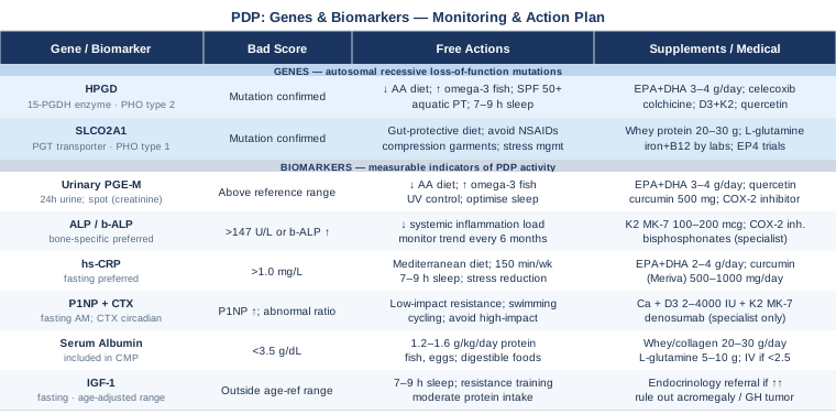
The Two Genes Behind Pachydermoperiostosis — And What You Can Do About Them
Pachydermoperiostosis, also classified as primary hypertrophic osteoarthropathy (PHO), is one of the rare conditions where the genetic architecture is understood in enough detail to build a rational, targeted management strategy. The condition is caused by loss-of-function mutations in one of two genes — HPGD or SLCO2A1 — both of which function as brakes on the prostaglandin E2 pathway. When either brake fails, PGE2 accumulates, and it is that unchecked accumulation that drives virtually every feature of the condition.
Knowing which gene is involved matters clinically. The two subtypes — PHO type 2 (HPGD mutations) and PHO type 1 (SLCO2A1 mutations) — differ in severity, organ involvement, and the specific risks patients face. This distinction shapes which monitoring and intervention strategies are most appropriate. Genetic testing is increasingly accessible and should be considered foundational for anyone with a clinical PDP diagnosis.
Why Prostaglandin E2 Is at the Center of Everything
Prostaglandin E2 is a lipid mediator derived from arachidonic acid — an omega-6 fatty acid abundant in the modern diet — through the action of cyclooxygenase (COX) enzymes. Under normal conditions, PGE2 is produced transiently in response to tissue stress and then rapidly inactivated by a two-step clearance process: cellular uptake via the prostaglandin transporter, followed by enzymatic oxidation by 15-hydroxyprostaglandin dehydrogenase (15-PGDH). In PDP, one of these steps is broken.
PGE2 acts through four receptor subtypes (EP1 through EP4). In bone, sustained EP4 receptor activation drives osteoblast activity and periosteal new bone formation — the source of the characteristic bone overgrowth in PDP. In skin, PGE2 promotes fibroblast proliferation and excessive collagen deposition, producing pachydermia. In joints, chronic PGE2 signaling sustains synovial inflammation. These are not incidental side effects; they are the direct molecular consequence of uncontrolled PGE2 activity. Any meaningful management strategy must, at some level, address this upstream driver.
Gene 1: HPGD — When the Prostaglandin-Degrading Enzyme Is Missing
What the gene does: HPGD encodes 15-hydroxyprostaglandin dehydrogenase (15-PGDH), the primary enzyme responsible for converting active PGE2 into its inactive 15-keto metabolite. This enzyme is expressed broadly across tissues — skin, gut, lung, and bone — and represents the terminal step in PGE2 inactivation. Without functional 15-PGDH, PGE2 accumulates wherever it is produced, with no mechanism for local clearance.
Mutation profile: HPGD mutations in PDP are autosomal recessive — both copies of the gene must carry loss-of-function variants for the condition to manifest. Missense, nonsense, frameshift, and splice-site variants have all been documented. The condition was first linked to HPGD in a landmark 2008 study by Uppal and colleagues, which definitively established PDP as a disorder of prostaglandin catabolism rather than an unexplained bone dysplasia (Uppal et al., 2008, Nature Genetics).
Clinical profile: Patients with HPGD mutations typically present with the complete clinical triad: facial pachydermia with furrowing (cutis verticis gyrata), digital clubbing, and diaphyseal periostosis. Hyperhidrosis, myelofibrosis, and gastric hypertrophy are seen in a subset of patients. Severity is variable and likely modulated by dietary arachidonic acid load and other environmental inputs that influence PGE2 production volume.
HPGD Impaired — Plan Without Supplements
The most direct non-pharmacological approach when HPGD is dysfunctional is to reduce the upstream dietary supply of PGE2 — specifically, to limit arachidonic acid, the fatty acid precursor that COX enzymes convert into prostaglandins.
Reduce dietary arachidonic acid: Arachidonic acid is concentrated in red meat, processed meat, organ meats, and egg yolks from conventionally raised animals. Shifting toward a Mediterranean or plant-forward dietary pattern — where seafood replaces red meat as the primary animal protein — reduces substrate availability for COX enzymes. This does not block PGE2 production, but it meaningfully lowers the chronic production rate. Application: Daily dietary habit, not a temporary restriction. Sustainable implementation is more important than strict elimination. Side effects: None at dietary quantities; extreme restriction of all omega-6 fatty acids would theoretically cause essential fatty acid deficiency over years, but this is not a realistic risk from a food-first approach.
Increase omega-3 fatty acids from food: EPA and DHA from fatty fish — sardines, mackerel, wild salmon, herring — compete with arachidonic acid for COX enzyme access. When EPA is present in higher concentrations, it shifts prostaglandin production toward series-3 prostaglandins (including PGE3), which are substantially less inflammatory than PGE2. Eating fatty fish three to four times per week produces a meaningful shift in the omega-3/omega-6 ratio over weeks to months. Side effects: None at food-level quantities.
Sun protection as a PGE2 modulator: UV-B radiation induces COX-2 expression in keratinocytes within hours of exposure, directly amplifying PGE2 production in skin. For patients with HPGD mutations who cannot clear that PGE2, UV exposure is a controllable and significant amplifier of skin-level prostaglandin burden. Daily SPF 50+ application and minimizing midday sun exposure is a simple, cost-free intervention with a direct biological rationale. Application: Daily habit, year-round.
Low-impact physical therapy for periosteal pain: Periosteal pain and joint swelling in PDP respond modestly to aquatic therapy and gentle range-of-motion exercise. These do not address the gene but preserve joint function without loading inflamed periosteum. High-impact activities (running, jumping) may exacerbate periosteal pain during active phases. Frequency: Three to four sessions per week during symptomatic periods; maintain between flares.
Sleep optimization: Sleep deprivation elevates inflammatory cytokines and increases PGE2 metabolite excretion measurably. For patients with impaired PGE2 clearance, consistently poor sleep compounds the biochemical burden. Consistent sleep timing, a cool dark room, and a target of 7–9 hours represents a low-cost, high-impact intervention across nearly every disease state involving chronic inflammation. Application: Non-negotiable daily practice.
HPGD Impaired — Plan With Supplements or Medical Support
Omega-3 supplementation (EPA + DHA): When dietary intake is insufficient, high-dose fish oil supplements at 2–4 g of combined EPA and DHA per day provide the prostaglandin-competing effect at a therapeutic dose. Choose third-party tested products for purity verification (IFOS or NSF certified). Cycling: Continuous use is generally supported; some practitioners use 8 weeks on, 2–4 weeks off to monitor platelet function, particularly before surgical procedures. Side effects: Fishy aftertaste, GI discomfort at high doses, mild platelet function effects. Discuss with physician if on anticoagulants.
COX-2 selective inhibitors (medical): NSAIDs — particularly celecoxib — reduce PGE2 production at the COX step, upstream of the absent 15-PGDH enzyme. Documented case reports show improvement in periosteal pain and, in some patients, modest changes in skin findings with COX-2 inhibitor use. This is a medical prescription decision, not a self-directed strategy. Frequency: As prescribed; typically continuous or pulsed. Side effects: Cardiovascular risk with long-term COX-2 use, renal effects, GI effects (milder than non-selective NSAIDs). Requires physician monitoring.
Colchicine (medical): Case-level reports document improvement in digital clubbing and periosteal pain with colchicine, an anti-inflammatory agent that modulates cytokine release and neutrophil activity. Evidence is case-level, not trial-level, but the mechanism is plausible given PDP's inflammatory signature. Dose: Typically 0.5–1.5 mg/day as prescribed. Side effects: GI symptoms, myopathy at high doses, drug interactions; physician supervision required.
Vitamin D3 and K2: Vitamin D modulates prostaglandin metabolism and bone remodeling signaling. Optimizing serum 25-OH-D to the 40–60 ng/mL range — not exceeding 80 ng/mL — is a rational adjunct given bone metabolism involvement. K2 as MK-7 (100–200 mcg/day) helps direct calcium toward bone matrix rather than ectopic soft tissue deposits. Cycling: Year-round if sun exposure is limited. Side effects: Hypercalcemia at very high vitamin D doses; generally safe at 2000–4000 IU D3/day with K2 co-administration.
Magnesium glycinate: Magnesium cofactors numerous anti-inflammatory enzyme pathways and is commonly suboptimal in individuals with chronic inflammatory conditions. Supplementing at 300–400 mg elemental magnesium per day (glycinate form for GI tolerance) supports cellular anti-inflammatory pathways without significant risk. Side effects: Loose stools at high doses; glycinate form is better tolerated than oxide.
Polyphenols with COX-2 modulating activity: Quercetin (500 mg/day) and bioavailable curcumin formulations (Meriva or theracurmin, 500–1000 mg/day) have documented partial COX-2 inhibitory effects in both cellular and clinical contexts, without the COX-1 suppression that makes NSAIDs GI-damaging. Direct PDP evidence is absent, but the mechanism is relevant. Cycling: 8–12 weeks on, 3–4 weeks off as a precautionary approach. Side effects: Curcumin may interact with blood thinners; take with food to reduce GI irritation. Quercetin is generally well-tolerated.
Gene 2: SLCO2A1 — When PGE2 Cannot Enter Cells for Degradation
What the gene does: SLCO2A1 encodes the prostaglandin transporter (PGT), a membrane protein responsible for shuttling extracellular PGE2 into cells, where it can then be inactivated by 15-PGDH. When SLCO2A1 is non-functional, PGE2 cannot efficiently enter cells for degradation — even if 15-PGDH is fully intact — and it accumulates in the extracellular space. The transporter step is a prerequisite for the degradation step, and losing it has clinical consequences nearly identical to losing 15-PGDH itself.
Distinguishing PHO type 1 from type 2: SLCO2A1 mutations produce PHO type 1, which carries an important distinguishing feature: a significant proportion of these patients develop chronic enteropathy — intestinal ulcers, protein-losing enteropathy, and GI inflammation — in addition to the classic triad of skin, bone, and digit changes. This gastrointestinal involvement creates a distinct management challenge. Low serum albumin, iron deficiency, B12 depletion, and malabsorption may all compound the systemic inflammatory burden from excess PGE2. HPGD patients (PHO type 2) typically do not develop this GI phenotype.
SLCO2A1 Impaired — Plan Without Supplements
Gut-protective dietary strategy: Given enteropathy risk in SLCO2A1 patients, a gut-supportive diet is foundational. This means strict avoidance of NSAIDs (which exacerbate mucosal damage and may precipitate GI bleeding in an already compromised gut), prioritizing easily digestible proteins (fish, eggs, well-cooked legumes) to support albumin levels, and favoring cooked rather than raw vegetables during active gut inflammation phases. Bone broth and collagen-rich foods support mucosal repair. Application: Daily, as a sustained dietary framework, not a temporary restriction.
NSAID avoidance during gut flares: This is not a general recommendation but a specific caution for SLCO2A1 patients. Even topical NSAIDs can worsen intestinal permeability in enteropathy. This creates a therapeutic tension — NSAIDs reduce PGE2 production but damage the gut that is already vulnerable. Specialist guidance on balancing this tradeoff is essential before initiating any NSAID therapy in PHO type 1 patients.
Compression therapy for limb edema: Periosteal involvement and low albumin together can produce significant dependent edema in the lower limbs. Medical-grade graduated compression garments (20–30 mmHg), worn during daytime hours, reduce fluid accumulation without pharmacological intervention. Application: Daily daytime wear; professionally fitted to ensure appropriate gradient. Side effects: Skin irritation; do not use in peripheral arterial disease without vascular assessment.
Stress management for gut barrier function: Psychological stress amplifies intestinal permeability through glucocorticoid-mediated effects on gut tight junctions. In SLCO2A1 patients with active enteropathy, chronic stress substantially worsens mucosal integrity and systemic inflammatory load. This is one of the more compelling reasons to formalize a daily stress reduction practice — not as an optional wellness add-on, but as a measurable gut-protective intervention.
SLCO2A1 Impaired — Plan With Supplements or Medical Support
Albumin and protein support: For patients with measurably low albumin (below 3.5 g/dL) due to protein-losing enteropathy, targeted protein supplementation supports serum levels without the high arachidonic acid load of red meat. Whey protein isolate or collagen hydrolysate at 20–30 g/day in divided doses provides high-bioavailability protein. Cycling: Continuous as needed based on albumin levels. Side effects: GI tolerance is generally good; monitor renal function if there is concurrent kidney involvement.
Iron and B12 supplementation based on labs: Chronic enteropathy reliably impairs iron and B12 absorption. Deficiencies are common, measurable, and correctable. Iron as ferrous bisglycinate (better GI tolerance than ferrous sulfate in an inflamed gut) at doses guided by serum ferritin and transferrin saturation. B12 as sublingual methylcobalamin (1000 mcg/day) bypasses gut absorption if gastric involvement is present. Recheck every 3 months until stable.
Glutamine for mucosal repair: L-glutamine (5–10 g/day) provides a preferential fuel source for enterocytes and has evidence for supporting intestinal mucosal integrity in enteropathy contexts. Dissolved in water, taken on an empty stomach. Side effects: Generally safe at these doses; avoid in patients with seizure disorders or liver failure.
EP receptor-targeting therapies (emerging): Because both HPGD and SLCO2A1 mutations converge on excess PGE2 signaling through EP2 and EP4 receptors, selective EP receptor antagonists represent a rational pharmacological target under active investigation in rare disease research. These are not yet standard of care but are an area of genuine scientific interest. Patients with genetically confirmed PDP should ask their specialist about clinical trial eligibility through rare disease registries.
Getting Genetic Testing for PDP
Whole-exome sequencing or targeted gene panels covering HPGD and SLCO2A1 are available through academic medical centers and commercial genetics laboratories. A confirmed genetic result does not always change immediate medical management, but it definitively establishes the diagnosis, distinguishes PHO type 1 from type 2 (which matters for gut monitoring), provides information relevant to family members, and opens access to rare disease registries and clinical trials. Genetic counseling before and after testing is strongly recommended, both to interpret results and to address implications for relatives.
6 Biomarkers Worth Tracking in Pachydermoperiostosis
While genetics identifies what is broken at the root, biomarkers reveal how active the condition is right now. In a condition where progression is slow and often subtle, having objective numbers to follow — rather than relying entirely on symptom perception — gives you and your clinicians a far more accurate picture over time. The six biomarkers below are selected for their direct relevance to PDP biology, their practical availability, and their ability to guide specific clinical decisions.
Biomarker 1: Urinary PGE2 Metabolites (PGE-M / Tetranor-PGEM)
Why it matters: This is the most direct biomarker for PDP disease activity. Because HPGD and SLCO2A1 mutations both impair PGE2 clearance, the body compensates partly through alternative catabolism, generating tetranor-PGEM (commonly called PGE-M), the primary urinary metabolite of PGE2. Elevated urinary PGE-M reliably reflects excess systemic PGE2 burden and is measurably elevated in most patients with confirmed PDP. No other single lab value captures the primary pathophysiological mechanism as directly.
How to measure it: 24-hour urine collection, or a spot urine sample normalized to creatinine. Academic centers and commercial reference labs (including Mayo Clinic Laboratories) offer this as part of a prostaglandin metabolite panel. Cost range: $80–$200 depending on the panel and laboratory. Results are reported in ng/mg creatinine; reference ranges vary by laboratory.
If the score is elevated — plan without supplements: Dietary reduction of arachidonic acid (the PGE2 precursor) directly lowers PGE2 production and therefore its urinary metabolite. Tracking PGE-M quarterly while implementing dietary changes allows you to confirm whether the intervention is moving the marker in the right direction — providing a biochemical feedback loop that is rare in rare disease management.
If the score is elevated — plan with supplements or medical support: High-dose EPA/DHA (3–4 g/day as combined omega-3s), COX-2 inhibitors (medical, prescribed), and polyphenol-based COX-2 modulators (quercetin 500 mg/day, bioavailable curcumin 500 mg/day) all have plausible mechanisms for reducing PGE2 synthesis volume. Recheck urinary PGE-M every 3 months when introducing interventions to assess biological response. Cycling for polyphenols: 8–12 weeks on, 3–4 weeks off. Side effects: See supplement notes under the genetics section.
Biomarker 2: Alkaline Phosphatase (ALP) and Bone-Specific ALP
Why it matters: ALP is released by osteoblasts during active bone synthesis and is typically elevated in PDP as a reflection of pathological periosteal bone formation. Tracking ALP longitudinally provides a rough index of periosteal activity. However, total ALP is non-specific — liver, gut, and placental sources also contribute — making bone-specific ALP (b-ALP) a more informative version when ordering targeted monitoring.
How to measure it: Total ALP is included in standard metabolic panels and most routine bloodwork. Bone-specific ALP is an add-on available at larger laboratories. Cost range: Total ALP $0–$20 (typically bundled in panels); bone-specific ALP $30–$80. Normal adult total ALP: 44–147 U/L (lab-dependent); interpret in age and sex context.
If the score is elevated — plan without supplements: Bone-specific ALP in PDP is driven by the gene-level mechanism, not a lifestyle variable that can be directly targeted. However, reducing total-body inflammatory burden (through dietary and lifestyle changes) may reduce the paracrine signals that amplify periosteal osteoblast activity. Monitor ALP every 6 months as a trend indicator rather than a target number for lifestyle intervention.
If the score is elevated — plan with supplements or medical support: COX-2 inhibitors have been associated with measurable ALP reductions in PDP case series, suggesting that reducing PGE2 signaling slows periosteal activity biochemically as well as symptomatically. Vitamin K2 (MK-7, 100–200 mcg/day) may support appropriate bone matrix mineralization and reduce poorly organized periosteal bone deposition, though direct PDP evidence is lacking. Bisphosphonates have been used in severe periosteal overgrowth cases but evidence is inconsistent and specialist management is essential given osteonecrosis risks with long-term use.
Biomarker 3: High-Sensitivity C-Reactive Protein (hs-CRP)
Why it matters: CRP is the most widely used and accessible measure of systemic inflammatory activity. Chronic PGE2 excess in PDP sustains a low-grade inflammatory state that is reflected in hs-CRP. While not specific to PDP, it serves as a useful longitudinal tracker for assessing whether lifestyle and supplemental interventions are reducing the systemic inflammatory burden. The high-sensitivity version detects the lower ranges relevant to chronic disease — below 3 mg/L — rather than only the acute infection range.
How to measure it: Blood test; fasting is preferred but not required. Cost range: $10–$40 at most laboratories. Target for cardiovascular and chronic inflammatory health: below 1.0 mg/L; functional medicine practitioners often target below 0.5 mg/L.
If the score is elevated — plan without supplements: Mediterranean dietary pattern (the most studied dietary anti-inflammatory approach), consistent sleep, moderate aerobic movement (150 minutes per week), and stress reduction each have robust evidence for reducing hs-CRP by 20–40% in chronically elevated populations. These represent foundational changes with cumulative effects when maintained consistently.
If the score is elevated — plan with supplements or medical support: Omega-3 fatty acids (2–4 g EPA+DHA/day) reduce hs-CRP by approximately 0.2–0.5 mg/L in pooled analyses. Bioavailable curcumin (500–1000 mg/day as Meriva or theracurmin) has documented CRP-lowering effects in multiple clinical trials. Cycling: Curcumin: 12 weeks on, 3–4 weeks off; omega-3s are generally continuous. Side effects: Curcumin interacts with anticoagulants; take with food. Monitor ferritin alongside iron supplementation in SLCO2A1 patients, as CRP can artificially elevate ferritin during active inflammation.
Biomarker 4: Bone Turnover Markers — P1NP and CTX
Why it matters: Two complementary markers characterize bone metabolism more precisely than ALP alone: P1NP (procollagen type 1 N-terminal propeptide) reflects bone formation rate, while CTX (C-terminal telopeptide of type 1 collagen) reflects bone resorption. In PDP, periosteal formation is typically elevated out of proportion to resorption, creating an abnormal P1NP-to-CTX ratio. These markers are recommended as the gold-standard bone turnover assessment by the International Osteoporosis Foundation and are used in clinical bone health monitoring contexts by specialists including Peter Attia in his bone-focused patient protocols.
How to measure it: Fasting morning blood draw — CTX is particularly circadian-variable and must be drawn fasting before 10 AM for meaningful interpretation. Cost range: P1NP $50–$100; CTX $50–$100; often ordered together. Establish a personal baseline and track the trend every 6–12 months.
If the score is abnormal — plan without supplements: Low-impact resistance exercise (resistance bands, swimming, cycling) provides the mechanical stimulus that supports normalized bone remodeling without excessive periosteal loading. Weight-bearing resistance training can worsen periosteal pain in high-impact forms; adapt the stimulus to the patient's pain tolerance. Check P1NP and CTX every 6–12 months to monitor trends.
If the score is abnormal — plan with supplements or medical support: The foundational bone metabolic support stack — calcium (from food first, supplement if needed at 500 mg/day split doses), vitamin D3 (2000–4000 IU/day), and K2 as MK-7 (100–200 mcg/day) — supports bone matrix quality even when periosteal formation is pathologically elevated. In cases of severe P1NP elevation with significant periosteal overgrowth, specialist-directed anti-RANKL therapy (denosumab) has been explored. Caution: Denosumab requires specialist management, careful transition planning, and monitoring for hypocalcemia and atypical fractures.
Biomarker 5: Serum Albumin
Why it matters: Serum albumin is the most practical single marker of nutritional status and gut integrity. It is particularly critical for SLCO2A1 mutation carriers who may have protein-losing enteropathy, where gut mucosal damage allows albumin to leak into the intestinal lumen. Low albumin (below 3.5 g/dL) signals either inadequate protein intake, impaired absorption, or active gut losses. Even in HPGD patients without enteropathy, albumin tracks general nutritional resilience and is sensitive to chronic inflammation (which suppresses albumin synthesis as part of the acute phase response).
How to measure it: Included in a comprehensive metabolic panel (CMP). Cost range: $0–$20 (bundled in routine labs). Target: above 4.0 g/dL; values below 3.5 g/dL warrant investigation and dietary or medical intervention.
If the score is low — plan without supplements: Increase dietary protein to 1.2–1.6 g per kilogram body weight per day from high-bioavailability sources: fish, eggs, well-cooked legumes, and collagen-rich preparations. During active enteropathy flares, cooked and easily digestible food preparations are better tolerated than raw or high-fiber options. Oral rehydration if there is significant fluid loss.
If the score is low — plan with supplements or medical support: Whey protein isolate or collagen hydrolysate (20–30 g/day in divided servings) provides a high-bioavailability protein source that is generally well-tolerated even in compromised gut states. L-glutamine (5–10 g/day) supports enterocyte function and mucosal integrity and has evidence for reducing intestinal permeability in chronic enteropathy. Medical: In severe hypoalbuminemia (below 2.5 g/dL), intravenous albumin infusion may be indicated as a bridge while gut integrity is restored. This is a hospital-level decision.
Biomarker 6: IGF-1 (Insulin-Like Growth Factor 1)
Why it matters: IGF-1 is a growth factor that regulates connective tissue, bone, and skin cell proliferation. It has been studied in PDP context because the periosteal overgrowth and dermal thickening share some phenotypic features with IGF-1 excess states (such as acromegaly), and because IGF-1 levels reflect the anabolic activity of the connective tissue compartment. More practically, measuring IGF-1 helps to distinguish primary PDP from secondary hypertrophic osteoarthropathy (caused by lung tumors, cardiac disease, or other underlying conditions) where IGF-1 and growth hormone dynamics differ. An abnormally elevated IGF-1 in a patient presenting with osteoarthropathy features warrants investigation beyond the PDP gene panel.
How to measure it: Fasting blood test. Cost range: $50–$120. Target: age-adjusted ranges (typically 115–307 ng/mL in adults, declining progressively with age). Interpret in the context of age and sex.
If the score is abnormal — plan without supplements: IGF-1 is sensitive to sleep quality, caloric intake, and resistance exercise. Ensuring 7–9 hours of quality sleep, maintaining adequate but not excessive protein intake, and performing regular moderate resistance training normalizes IGF-1 within the appropriate physiological range. Caloric restriction lowers IGF-1 (which theoretically reduces anabolic bone and skin signaling) but simultaneously reduces overall metabolic resilience — balance is more important than absolute reduction.
If the score is significantly elevated — plan with supplements or medical support: Substantially elevated IGF-1 alongside osteoarthropathy features warrants endocrinological evaluation to rule out acromegaly or a GH-secreting tumor. There are no standard supplements that safely lower IGF-1 without also compromising muscle mass and recovery. The main dietary lever is moderating very high protein intake (particularly from animal sources) if IGF-1 is already at the high end of normal. Medical management, if true GH/IGF-1 axis dysregulation is identified, is a specialist decision.
Key Insights From Inflammation and Prostaglandin Research
Dr. Andrew Huberman and the researchers he regularly references on his podcast — including those working on eicosanoid biology, fatty acid metabolism, and chronic inflammation — have collectively mapped a body of knowledge that is directly applicable to the mechanisms driving PDP, even though the condition is too rare for dedicated coverage. The following ten findings from inflammation and prostaglandin research represent the most actionable insights for PDP patients.
1. Arachidonic Acid Is the Raw Material for Every PGE2 Molecule
Every molecule of PGE2 in the human body originates from arachidonic acid (AA), a 20-carbon omega-6 fatty acid. AA is abundant in red meat, processed meat, organ meats, egg yolks, and conventionally raised poultry. Reducing dietary AA is one of the only food-first mechanisms for reducing PGE2 synthesis capacity — not by eliminating the molecule, but by reducing substrate availability for the COX enzymes that manufacture it.
2. Omega-3 and Omega-6 Fatty Acids Compete at the Same Enzyme
EPA (eicosapentaenoic acid, from fish oil) competes directly with AA for COX enzyme binding. When EPA is present at higher concentrations, it preferentially occupies the enzyme and generates series-3 prostaglandins — including PGE3 — which are dramatically less inflammatory than PGE2. This competitive inhibition is not incidental; it is the primary mechanism by which dietary omega-3 increases shift the prostaglandin profile toward less inflammatory outcomes.
3. The Omega-6 to Omega-3 Ratio Matters More Than Absolute Quantities
Most modern Western diets carry an omega-6 to omega-3 ratio of 15:1 to 20:1. Evolutionary estimates suggest a ratio closer to 4:1 or lower. For PDP patients where PGE2 clearance is already impaired, a high dietary omega-6 load dramatically amplifies the accumulation problem. Shifting toward a 4:1–6:1 ratio through food changes alone has a measurable biochemical impact on prostaglandin production over weeks to months.
4. COX-2 Is Inducible — and Many of Its Triggers Are Modifiable
COX-2, the primary enzyme responsible for inflammatory PGE2 production, is not constitutively active. It is induced by specific biological signals: IL-1β and TNF-α (inflammatory cytokines), lipopolysaccharide from gut bacteria, UV radiation, and psychological stress. This means that managing lifestyle stressors is not peripheral to PDP management — it directly modulates one of the primary prostaglandin production enzymes, upstream of both HPGD and SLCO2A1.
5. Sleep Restriction Elevates Prostaglandin Output
Experimental studies have shown that sleep restriction (below 6 hours per night) significantly increases inflammatory cytokines, COX-2 expression, and prostaglandin metabolite excretion. For PDP patients who already have impaired PGE2 clearance, consistently poor sleep may substantially worsen the biochemical burden. Sleep is not passive recovery — in this context, it is an active regulator of prostaglandin metabolism.
6. UV Radiation Directly Amplifies Skin PGE2
UV-B exposure induces COX-2 expression in keratinocytes within hours, producing a local surge in PGE2 production in the skin. For PDP patients with impaired PGE2 degradation in dermal tissue, this represents a direct, controllable amplifier of skin-level prostaglandin accumulation. The rationale for daily broad-spectrum sun protection is stronger in PDP than in the general population.
7. Gut Bacteria Continuously Drive COX-2 Expression
Gram-negative gut bacteria produce lipopolysaccharide (LPS), which activates TLR4 receptors on immune cells and epithelial surfaces, inducing COX-2 systemically. A dysbiotic gut microbiome with high LPS-producing bacteria generates a continuous, low-grade COX-2 induction signal that adds to any dietary AA load. Prebiotic fiber, fermented foods, and reduced ultra-processed food intake reduce this bacterial inflammatory signal at its source.
8. Polyphenols Inhibit COX-2 Without Blocking COX-1
Plant polyphenols — quercetin, luteolin, resveratrol, and curcuminoids — partially inhibit COX-2 expression and activity through multiple molecular mechanisms, without suppressing the constitutive COX-1 enzyme that protects the gastric mucosa. This distinguishes them from NSAIDs mechanistically and makes them particularly interesting for PDP patients who also have GI vulnerability (especially SLCO2A1 patients). The evidence base is stronger in cellular and animal models than in human trials for most polyphenols, but the mechanistic rationale is sound.
9. Visceral Adiposity Is a Sustained Prostaglandin Generator
Visceral adipocytes constitutively express COX-2 and produce PGE2, TNF-α, and IL-6 as part of their inflammatory secretome. Excess visceral fat represents a chronic, ambient source of PGE2 production that is independent of dietary inputs and accumulates on top of the genetic clearance deficit. Reducing visceral adiposity through caloric moderation and regular movement is one of the few lifestyle interventions that durably reduces background systemic PGE2 production volume.
10. EP4 Is the Receptor That Drives Periosteal Bone Overgrowth
Among the four EP receptor subtypes, EP4 is primarily responsible for mediating periosteal osteoblast activation and the new bone formation response to PGE2. Research across multiple bone biology groups has identified EP4 as a potentially druggable target in conditions of PGE2-driven periosteal expansion. EP4 antagonists are under investigation for bone-related conditions. Understanding this receptor specificity reinforces the mechanistic rationale for COX-2 inhibitors (which reduce PGE2 production upstream) over non-specific anti-inflammatory approaches in PDP.
Complementary Approaches With Clinical Evidence
The approaches below have meaningful human clinical evidence for managing specific components of PDP — particularly musculoskeletal pain, inflammation, gut integrity, and quality of life. None address the underlying gene mutation, but each has a plausible biological rationale in the context of PGE2-driven disease, and each adds value beyond what pharmacological management alone provides.
Low-Level Laser Therapy / Photobiomodulation
Photobiomodulation (PBM) delivers red and near-infrared light wavelengths (typically 630–1070 nm) to tissue, where it is absorbed by mitochondrial chromophores and modulates cellular inflammatory signaling. The proposed mechanism of particular relevance in PDP is PBM's documented capacity to reduce COX-2 expression and local PGE2 production in treated tissue — directly targeting one element of the prostaglandin pathway that is relevant to this condition.
Systematic reviews and meta-analyses support PBM for musculoskeletal pain, including joint and periosteal pain syndromes, with significant reductions in inflammatory mediators in treated tissue (Hamblin, 2017, Photobiomodulation in musculoskeletal conditions). Multiple randomized trials in chronic joint pain and inflammatory arthritis show significant pain reduction with clinical-grade PBM devices compared to sham treatment.
A practical protocol involves a clinical-grade device (class 3B or class 4 laser, or a high-irradiance LED panel at 810–850 nm) applied to the most symptomatic joints and long bones at 30–60 J/cm² per session, three sessions per week for 8–12 weeks. Home near-infrared panels are available at $300–$1000; clinical sessions typically cost $50–$150 each. Side effects: Minimal; avoid direct eye exposure; clinical supervision recommended for first sessions. Direct PDP evidence is absent — this is a mechanistically plausible extrapolation from joint pain and inflammation data.
Mindfulness Meditation / MBSR
Mindfulness-Based Stress Reduction (MBSR) is an 8-week structured program combining formal meditation, body scan practice, and mindful movement. Its relevance in PDP operates on two levels: as a well-validated chronic pain management approach, and as a physiological mechanism for reducing the psychological stress that drives COX-2 expression. Both rationales are directly relevant to PDP.
A landmark randomized controlled trial published in JAMA Internal Medicine (Cherkin et al., 2016) demonstrated that MBSR significantly reduced chronic musculoskeletal pain and improved function compared to usual care, with effects maintained at 26-week follow-up (Cherkin et al., 2016). Additional research has shown that consistent mindfulness practice reduces cortisol, IL-6, and CRP — all measurable markers relevant to the inflammatory burden in PDP.
Practically, the MBSR protocol begins with 10–20 minutes of daily body scan meditation and builds toward 40–45 minutes over 8 weeks. Free programs (Palouse Mindfulness online) follow the validated Jon Kabat-Zinn structure. Certified in-person or virtual MBSR programs are available through hospitals and academic centers at $200–$600 for the full 8-week course. Side effects: Minimal; a minority of individuals experience transient emotional discomfort when beginning formal practice — this is normal and typically resolves.
Massage Therapy
Therapeutic massage addresses two of the most functionally limiting features of PDP: periosteal joint pain and the psychological burden of living with a chronic rare condition. Mechanically, soft-tissue manipulation improves local circulation, reduces muscle guarding around painful and swollen joints, and has documented anti-inflammatory effects in chronic musculoskeletal conditions.
A systematic review in Pain Medicine (Bervoets et al., 2015) found that massage therapy significantly reduced pain intensity and improved function in patients with chronic musculoskeletal conditions compared to active controls (Bervoets et al., 2015). Lymphatic drainage massage — a specific technique that facilitates fluid movement — is particularly relevant for managing limb edema secondary to periosteal disease or low albumin states.
A realistic protocol involves weekly 60-minute sessions with a therapist experienced in working with inflammatory or hypersensitive musculoskeletal conditions. Direct pressure over affected periosteal sites should be avoided; work should focus on soft tissue overlying bones and surrounding joint musculature. During active flares, lighter pressure and lymphatic drainage techniques are better tolerated. Cost: $60–$130 per session. Communicate the diagnosis clearly to the therapist, as standard deep-tissue protocols may be inappropriate.
Microbiome-Directed Therapies
The gut microbiome's role in regulating systemic inflammation through the LPS-TLR4-COX-2 axis is directly relevant to PDP, where any additional driver of COX-2 induction amplifies an already impaired PGE2 clearance system. For SLCO2A1 patients with enteropathy, gut dysbiosis may be a secondary but significant contributor to both GI and systemic inflammatory burden.
Clinical trials examining prebiotic fiber supplementation in chronic inflammatory conditions have documented reductions in LPS, intestinal permeability markers (including zonulin), and inflammatory cytokines. Probiotic supplementation with Lactobacillus acidophilus NCFM and Bifidobacterium lactis Bi-07 has demonstrated modest but consistent reductions in CRP and inflammatory markers in randomized trials. The combination of prebiotics and probiotics (synbiotic approach) shows stronger effects than either alone in most meta-analyses.
A practical microbiome-directed strategy includes increasing dietary prebiotic fiber (inulin from chicory and garlic; resistant starch from cooled cooked potatoes or green bananas), adding a multi-strain probiotic containing the above strains, reducing ultra-processed food intake that selectively enriches pro-inflammatory Gram-negative bacteria, and including fermented foods (kefir, kimchi, sauerkraut) if GI tolerance allows. Cost: Probiotics $20–$50/month. Side effects: Prebiotic fiber can cause initial bloating — start with small amounts and titrate up over 2–4 weeks. In active SLCO2A1 enteropathy, introduce changes slowly and track GI symptoms carefully.
Chinese Herbal Medicine
Several compounds from botanical and traditional Chinese medicine practice have documented COX-2 modulating and anti-inflammatory effects that are mechanistically relevant to PDP. Boswellia serrata (standardized to AKBA content) inhibits 5-lipoxygenase and modulates inflammatory cytokine production in musculoskeletal conditions. It has a more favorable safety profile than stronger botanicals and a modest but consistent evidence base in chronic joint inflammation contexts.
A systematic review of Boswellia extract in chronic joint conditions found significant reductions in pain and inflammatory markers compared to placebo in multiple randomized trials (Siddiqui, 2011, International Journal of Biological Sciences). For musculoskeletal pain in rare inflammatory conditions like PDP, the lack of direct clinical trial evidence is a genuine limitation — but the COX pathway relevance is mechanistically sound.
AKBA-standardized Boswellia extract at 200–400 mg/day represents the safer starting point for self-directed botanical use. Cycling: 12 weeks on, 4 weeks off. Tripterygium wilfordii (Thunder God Vine) has stronger anti-inflammatory evidence in rheumatological conditions but carries significant hepatotoxic and reproductive side effect risk — it requires experienced practitioner supervision and should not be used without formal monitoring. Side effects of Boswellia: GI effects at high doses; avoid if pregnancy is planned; disclose to physician before combining with NSAIDs or anticoagulants.
A Practical Way Forward
Pachydermoperiostosis is a condition where having precise information meaningfully changes what is possible in managing it. Two genes — HPGD and SLCO2A1 — account for essentially all primary cases, both pointing to the same core problem: prostaglandin E2 that builds up because the clearance machinery is broken. That mechanism is not yet fully correctable, but it is workable. Reducing dietary PGE2 precursors, tracking key biomarkers, controlling the lifestyle factors that amplify COX-2 expression, and thoughtfully incorporating complementary strategies creates a more active, data-informed management approach than symptom management alone.
The practical next step is a three-part foundation: confirm the genetic subtype if not already done (PHO type 1 vs. type 2 changes the monitoring and intervention priorities); establish a baseline biomarker panel (urinary PGE-M, ALP with bone-specific fractionation, hs-CRP, P1NP, CTX, albumin, and IGF-1); and commit to the dietary and lifestyle changes with the clearest biological rationale for this specific condition. Bring these measurements and frameworks to a rheumatologist, internist, or specialist familiar with rare bone disorders. That conversation is considerably more productive when you arrive with data rather than symptoms alone.
Musculoskeletal: Bone Conditions Joint Conditions
Skin: Inflammatory Skin Conditions
Autoimmune: Inflammatory Conditions