This article contains AI generated content.
· UpdatedPanniculitis: 6 Genes and 7 Biomarkers to Track
Introduction
Panniculitis is one of those conditions where getting a confirmed diagnosis already feels like a win — and yet, the real complexity begins right there. The inflammation in the subcutaneous fat can have a dozen different causes, and the painful nodules, redness, and swelling that characterize it can look almost identical regardless of the underlying driver. Many people spend years cycling through corticosteroids and NSAIDs, finding temporary relief but never a lasting answer.
The problem with generic anti-inflammatory treatment is that it addresses the fire without asking what is feeding it. Panniculitis can be triggered by autoimmune dysregulation, hereditary enzyme deficiencies, innate immune overactivation, or occult infection — and each of these pathways responds to completely different interventions. A treatment plan built around a label rather than a mechanism will, at best, suppress symptoms and, at worst, miss something entirely correctable.
This is where biomarker tracking and genetic context start to become genuinely useful tools. Certain blood markers can reveal whether your panniculitis is being driven by complement activation, autoantibody production, macrophage overactivation, or a missing protease inhibitor. Certain genetic variants — including mutations in SERPINA1 or MEFV — can explain why some people develop panniculitis spontaneously, repeatedly, or at unusually young ages. Understanding which of these factors applies to you changes the conversation with your physician from "manage the flare" to "address the root."
This article covers two complementary perspectives: a deep look at the seven most clinically useful biomarkers for tracking and contextualizing panniculitis activity, and a focused review of six genes with meaningful associations to the condition. Beyond that, you will find a summary of a widely-cited functional medicine framework on inflammation, evidence-backed complementary approaches, and practical plans for each marker and gene — both with and without supplementation. The goal is not a cure. It is better information so that better decisions can follow.
Summary
- 7 biomarkers — Alpha-1 Antitrypsin, high-sensitivity CRP, ESR, Ferritin, IL-6, Complement C3/C4, and the ANA/autoantibody panel — can reveal which specific inflammatory mechanism is driving your panniculitis and guide targeted intervention - 6 genes — SERPINA1, MEFV, TNF (-308A), HLA-B8/DRB1*03, NLRP3, PTPN22 — explain why some individuals develop panniculitis recurrently and why flares can be disproportionately severe - For each biomarker and gene, the article includes concrete action plans: what to do before turning to supplements, and what to consider with targeted supplementation or equipment — with dosing, cycling, and side effects - A structured summary of Will Cole's The Inflammation Spectrum distills 10 clinically actionable insights about personalizing inflammation reduction - Five complementary approaches — including the Autoimmune Protocol from Sarah Ballantyne, photobiomodulation, MBSR, microbiome-directed therapy, and breathwork — are reviewed with condition-relevant protocols and study references - All strategies are designed to be used alongside standard medical care, not as alternatives to it
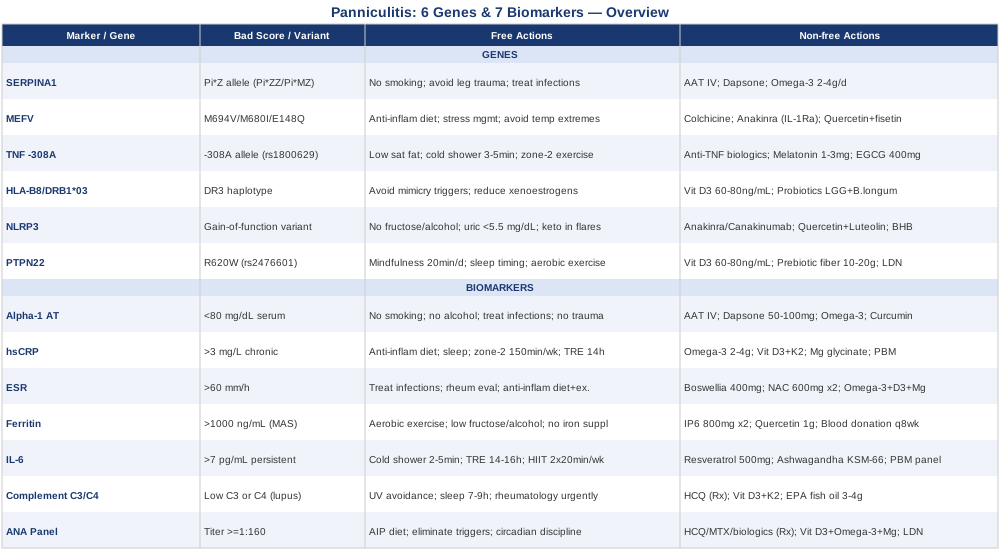
7 Biomarkers That Reveal What Is Actually Driving Your Panniculitis
Tracking biomarkers in panniculitis is not only about monitoring disease activity — it is about identifying which inflammatory pathway is dominant so that interventions can be far more precisely targeted. The seven markers below are clinically actionable, widely available, and together paint a picture that no single CRP test can provide on its own.
Biomarker 1: Alpha-1 Antitrypsin (AAT)
Why it matters: Alpha-1 antitrypsin is a serine protease inhibitor produced primarily by the liver, whose key role in the skin and subcutaneous tissue is to protect against neutrophil elastase — an enzyme that, when unregulated, degrades fat cells and triggers the lobular panniculitis pattern. AAT deficiency is one of the most well-established causes of recurring panniculitis, particularly in adults without an obvious infectious or autoimmune trigger. The association is strong enough that AAT levels should be among the first tests ordered when the etiology remains unclear.
What it may reveal: Low AAT levels (below 80–100 mg/dL) suggest a functional deficiency, which can be confirmed by genotyping the SERPINA1 gene. Even heterozygous carriers (Pi*MZ) can experience clinically relevant deficiency during acute inflammatory stress. Importantly, AAT is an acute-phase reactant — levels can appear artificially normal during a flare even when underlying production capacity is limited. A follow-up measurement during clinical remission is more accurate for detecting true deficiency.
How to measure it: Standard serum AAT test, available at most hospital laboratories. Cost ranges from $30 to $80 USD. Genotyping for Pi*ZZ, Pi*MZ, and Pi*SS phenotypes adds $100–$300 and is typically a one-time test. The published literature on AAT panniculitis supports testing in all cases of lobular panniculitis without a clearly identified cause.
If the score is low — the plan without supplements: - Eliminate smoking entirely: cigarette smoke oxidizes and inactivates AAT, accelerating neutrophil elastase activity at the tissue level - Minimize alcohol consumption: heavy use impairs hepatic AAT synthesis - Treat infections promptly: systemic infections increase neutrophil burden and deplete functional AAT faster than the liver can compensate - Avoid vigorous physical trauma to the lower extremities, where panniculitis in AAT-deficient individuals concentrates; deep-pressure massage over active nodules can trigger Köbner-like responses - Request periodic liver function monitoring: Pi*ZZ homozygotes carry hepatic AAT accumulation risk independent of panniculitis severity
If the score is low — the plan with supplements or equipment: - Intravenous AAT augmentation therapy (Prolastin-C, Zemaira — 60 mg/kg IV weekly): the most direct replacement approach; strongest evidence base in pulmonary disease, but case series support benefit in severe recurring panniculitis; insurance coverage depends on documented deficiency - Dapsone (50–100 mg/day, prescription): reduces neutrophil infiltration directly and is the most commonly used pharmacological approach in confirmed AAT panniculitis; monitor CBC regularly for hemolytic anemia; contraindicated in G6PD deficiency; use continuously during active disease - Omega-3 fatty acids (EPA+DHA 2–4 g/day): modulate neutrophil elastase activity through lipid mediator signaling; take daily with food; effects visible at 8–12 weeks; fish oil or algal oil both valid; no cycling required - Curcumin with piperine (500–1000 mg curcumin/day): suppresses NF-kB-driven neutrophil recruitment; bioavailability requires piperine or a lipid-based formulation; cycle 8 weeks on, 4 weeks off
Biomarker 2: High-Sensitivity CRP (hsCRP)
Why it matters: C-reactive protein is synthesized by the liver in response to IL-6 signaling and remains the most practical marker of active systemic inflammation. In panniculitis, elevated hsCRP reflects ongoing inflammatory burden in the subcutaneous tissue and is the clearest tool for tracking treatment response between flares. Peter Attia consistently includes hsCRP in his baseline inflammatory panel, noting that the high-sensitivity version captures chronic low-grade inflammation that standard tests report as normal.
What it may reveal: An hsCRP above 3 mg/L in the absence of obvious infection warrants investigation of chronic inflammatory drivers. In panniculitis, persistently elevated hsCRP between visible flares often signals ongoing subclinical fat inflammation. Values above 100 mg/L suggest bacterial infection or a severe systemic flare and require urgent evaluation — they do not simply reflect a bad chronic disease day.
How to measure it: Standard blood test; cost $10–$40 USD. Always specify "high-sensitivity CRP" — this is analytically different from standard CRP and detects low-level chronic inflammation below 10 mg/L. Measure every 3 months during active disease or during treatment trials to capture directional change.
If hsCRP is elevated — the plan without supplements: - Transition to an anti-inflammatory dietary pattern: eliminate refined seed oils, reduce ultra-processed foods, increase fatty fish, leafy greens, and berries daily - Improve sleep quality: a single night of under 6 hours measurably raises next-morning CRP; maintain a fixed wake time to protect circadian rhythm - Add zone-2 aerobic exercise: 150 minutes per week reduces CRP chronically through IL-6-mediated anti-inflammatory myokine pathways; walking, cycling, and swimming all qualify; consistency over intensity matters here - Reduce visceral fat if present: visceral adipose tissue is a major independent CRP driver; a 14–16 hour overnight eating window shows consistent CRP lowering in trials
If hsCRP is elevated — the plan with supplements or equipment: - Omega-3 fatty acids (2–4 g EPA+DHA/day): among the best-studied CRP-reducing interventions; take daily with food; meta-analyses confirm consistent CRP lowering at 8–12 weeks; triglyceride form has superior absorption - Vitamin D3 + K2: deficiency is independently associated with elevated CRP; target serum 25(OH)D of 50–80 ng/mL; typical starting dose 2000–5000 IU D3 daily with 100–200 mcg K2 MK-7; adjust after retesting; no cycling needed - Magnesium glycinate (300–400 mg at night): low magnesium correlates with elevated CRP; this form is well-tolerated for chronic daily use; take continuously - Photobiomodulation (red/near-infrared light) at 630–850 nm, 10–20 minutes 3–5x per week over affected areas: reduces local IL-6 and CRP production through mitochondrial activation; consumer-grade devices range $150–$500; no known systemic adverse effects at recommended exposures
Biomarker 3: Erythrocyte Sedimentation Rate (ESR)
Why it matters: ESR measures how quickly red blood cells settle in a test tube, which is proportional to circulating protein levels — particularly fibrinogen and immunoglobulins. In panniculitis, ESR often rises alongside CRP but is additionally sensitive to immunoglobulin elevation, making it especially useful when an autoimmune-mediated subtype is suspected: lupus panniculitis, dermatomyositis-associated panniculitis, or lymphoma-associated panniculitis. Thomas Dayspring has emphasized ESR as a useful complement to CRP precisely because it captures a partially different dimension of the inflammatory protein landscape.
What it may reveal: An elevated ESR with a normal or only mildly elevated CRP is an unusual pattern that should prompt investigation for hypergammaglobulinemia, chronic infection such as tuberculosis, or lymphoproliferative disease. ESR above 60–80 mm/h in panniculitis typically correlates with more systemic disease activity. Because ESR is also sensitive to anemia and age, it is best interpreted alongside CRP rather than in isolation.
How to measure it: Inexpensive ($5–$20 USD), universally available. Order simultaneously with CRP for the most interpretable picture. Retest every 3 months alongside CRP when elevated.
If ESR is elevated — the plan without supplements: - Investigate the underlying driver with a rheumatologist: persistently elevated ESR with depressed CRP warrants specific autoimmune, infectious, and hematological workup before any lifestyle change can be optimally targeted - Treat chronic infections: latent tuberculosis, dental infections, chronic sinusitis, and occult urinary infections can sustain ESR elevation independently of panniculitis activity - Apply the anti-inflammatory dietary and exercise pattern described under hsCRP above — these reduce fibrinogen and immunoglobulin-driven ESR through the same mechanisms
If ESR is elevated — the plan with supplements or equipment: - Boswellia serrata extract (200–400 mg AKBA-standardized extract twice daily): reduces leukotriene and 5-LOX-mediated inflammation; cycle 12 weeks on, 4 weeks off; well-tolerated; reduces ESR in several autoimmune condition trials - N-acetylcysteine (NAC) (600 mg twice daily): supports glutathione production and reduces fibrinogen-driven ESR elevation; cycle 8 weeks on, 4 weeks off; avoid in active bleeding; check for interaction with nitroglycerin if relevant - Omega-3, vitamin D3/K2, and magnesium stack as listed under hsCRP; these three together form a consistent ESR-lowering baseline
Biomarker 4: Serum Ferritin
Why it matters: Ferritin is both an iron storage protein and a powerful acute-phase reactant. In panniculitis, markedly elevated ferritin — above 1000 ng/mL and especially above 10,000 ng/mL — is a critical warning sign for macrophage activation syndrome (MAS), a potentially life-threatening hyperinflammatory state that can accompany severe panniculitis, particularly in Still's disease or lupus. Even moderately elevated ferritin (above 500 ng/mL) in panniculitis should prompt evaluation for MAS or hemophagocytic lymphohistiocytosis (HLH) before attributing it to metabolic causes.
What it may reveal: Ferritin below 30 ng/mL signals iron deficiency, which modulates immune function and worsens fatigue — both meaningful in someone already dealing with panniculitis. Ferritin in the high-normal to mildly elevated range (150–500 ng/mL) is often associated with metabolic inflammation and visceral fat accumulation. In this range, reducing metabolic inflammation is the appropriate response rather than iron supplementation. A glycosylated ferritin fraction — when available — can help distinguish reactive elevation from iron overload or MAS-pattern activation.
How to measure it: Included in most comprehensive metabolic panels or ordered separately for $15–$50 USD. Glycosylated ferritin is a specialized test at larger reference labs; a low glycosylated fraction (below 20%) in the context of suspected Still's disease or lupus raises diagnostic specificity for MAS meaningfully.
If ferritin is markedly elevated — the plan without supplements: - Urgent medical evaluation if ferritin exceeds 5,000 ng/mL: this is a potential medical emergency requiring prompt assessment, not a home-management situation - For moderately elevated ferritin (200–1,000 ng/mL): focus on reducing visceral fat through consistent aerobic exercise and modest caloric reduction; this is more effective than trying to lower ferritin through supplements - Avoid supplemental iron unless confirmed deficient on a full iron panel - Reduce fructose and alcohol: both activate NLRP3 inflammasome in liver cells, accelerating ferritin production through pathways that are distinct from actual iron storage needs
If ferritin is elevated — the plan with supplements or equipment: - IP6 (inositol hexaphosphate) (800 mg on an empty stomach twice daily): chelates excess stored iron and reduces ferritin over 8–12 weeks; cycle 8 weeks on, 4 weeks off; take well away from meals to avoid mineral binding - Quercetin (500–1000 mg/day with food): mild iron chelator and NLRP3 inflammasome inhibitor; cycle 8 weeks on, 2 weeks off; also reduces macrophage activation markers that drive ferritin synthesis - Regular blood donation (every 8–12 weeks): effectively and cost-freely lowers stored iron and ferritin; avoids pharmacological approaches; not appropriate during active panniculitis flares or while on certain medications - Heart rate monitor or smartwatch for zone-2 quantification: sustained aerobic exercise in zone 2 (60–70% max HR) for 150–180 minutes per week consistently lowers metabolic ferritin elevation; the device turns imprecise exercise into a measurable anti-inflammatory intervention
Biomarker 5: Interleukin-6 (IL-6)
Why it matters: IL-6 is the upstream orchestrator that drives both CRP production and ferritin elevation. In panniculitis, IL-6 can be produced directly by activated macrophages within the inflamed fat tissue, making it a more proximal readout of disease activity than the secondary markers it drives. Studies on subcutaneous adipose inflammation show that locally produced IL-6 from fat tissue macrophages can sustain systemic inflammatory patterns even in the absence of visible nodules. IL-6 also suppresses adiponectin, which creates a self-amplifying pro-inflammatory loop in adipose tissue.
What it may reveal: Persistently elevated IL-6 (above 7 pg/mL in most reference ranges) between flares indicates ongoing inflammatory activity in fat depots that may not be clinically visible yet. Very high IL-6 (above 50 pg/mL) in panniculitis raises concern for cytokine storm risk, particularly in individuals with confirmed NLRP3 mutations or an underlying autoimmune diagnosis. When CRP is chronically elevated but a specific cause has not been identified, measuring IL-6 directly narrows the differential.
How to measure it: Not a routine order — requires a specific cytokine panel, available at specialty and large reference laboratories. Cost: $60–$150 USD. Less commonly included in standard rheumatology panels but increasingly ordered by functional medicine physicians and immunologists. Most useful when hsCRP is persistently elevated without a clear explanation.
If IL-6 is elevated — the plan without supplements: - Cold exposure (2–5 minutes of cold water at the end of daily shower, 4–5x/week): durably downregulates IL-6 signaling through norepinephrine-mediated pathways; evidence from cryotherapy and Wim Hof protocol studies supports this as a genuine anti-inflammatory tool, not only a recovery aid - Time-restricted eating (14–16 hour overnight fast, 5+ days per week): reduces adipose tissue macrophage activity and the consequent adipose-derived IL-6 secretion; effects visible at 6–8 weeks of consistent practice - High-intensity interval training (2 brief sessions of 20 minutes per week): transiently elevates IL-6 as a myokine but durably lowers baseline IL-6 through adipose remodeling; avoid during active flares; begin gently and progress
If IL-6 is elevated — the plan with supplements or equipment: - Resveratrol (500 mg/day with food): inhibits NF-kB and STAT3, reducing IL-6 transcription; cycle 8 weeks on, 4 weeks off; lipid-based or micronized forms have meaningfully higher bioavailability than standard powder - Ashwagandha KSM-66 or Sensoril (300–600 mg/day): reduces cortisol-driven IL-6 elevation through HPA axis modulation; cycle 8 weeks on, 4 weeks off; avoid during pregnancy; check for nightshade sensitivity - Full-body or panel photobiomodulation device (630–850 nm, 10–20 minutes over affected areas 4–5x/week): reduces NF-kB activation in adipose tissue directly; consumer-grade panels range $300–$1,500; a useful hardware investment for people with frequent or bilateral panniculitis
Biomarker 6: Complement C3 and C4
Why it matters: The complement system is a cascade of proteins that amplifies immune responses. In panniculitis, complement activation is particularly relevant in lupus panniculitis (also called lupus profundus), where complement deposition in fat tissue is a recognized pathological hallmark. Low C3 and C4 reflect ongoing complement consumption — the system is being activated faster than it is being replenished, signaling immune complex-mediated tissue destruction in the subcutaneous layer. Normal complement with elevated CRP and ESR points more toward autoinflammatory or infectious panniculitis rather than autoimmune disease, which is a diagnostically meaningful distinction.
What it may reveal: Depressed C3 and C4 in a panniculitis patient strongly raise suspicion for lupus panniculitis, mixed connective tissue disease, or cryoglobulinemia-associated panniculitis — all of which require specific immunosuppressive management. Isolated low C4 with normal C3 may reflect inheritance of a C4A null allele, which is common in lupus-prone individuals and can appear even outside a flare. Studies on lupus panniculitis show complement depression correlates with local tissue damage severity, not just systemic disease activity.
How to measure it: Standard serum complement assay (C3, C4, and optionally CH50), $30–$80 USD. Order alongside ANA when autoimmune panniculitis is suspected. Repeat every 3 months when low values are documented to track treatment response.
If complement is low — the plan without supplements: - UV avoidance: sunlight can trigger complement-mediated flares in lupus panniculitis; physical sun protection (UPF clothing, midday sun avoidance) is among the most effective non-pharmacological tools available for this subtype - Collaborate urgently with a rheumatologist: low-complement panniculitis typically requires prescription hydroxychloroquine or immunosuppressants; lifestyle measures alone are insufficient - Prioritize sleep duration (7–9 hours): complement proteins are synthesized by the liver in a circadian-dependent pattern; sleep deprivation measurably reduces complement levels and blunts recovery between flares
If complement is low — the plan with supplements or equipment: - Hydroxychloroquine (prescription, 200–400 mg/day): first-line pharmacological treatment for lupus panniculitis; reduces complement activation indirectly through toll-like receptor interference; requires annual ophthalmologic monitoring for retinopathy - Vitamin D3 + K2 to 50–80 ng/mL 25(OH)D: vitamin D deficiency is associated with dysregulated complement activity in autoimmune conditions; supplementation to the upper optimal range consistently reduces complement consumption markers in lupus cohort studies - EPA-dominant fish oil (3–4 g/day, triglyceride form): reduces immune complex formation by modulating B-cell signaling; take continuously with food; choose an independently tested product for oxidation levels
Biomarker 7: ANA Panel and Specific Autoantibodies
Why it matters: Antinuclear antibodies are the gateway test for autoimmune panniculitis. A positive ANA at a titer of 1:160 or higher should trigger a full autoantibody panel to identify the specific autoimmune condition driving the subcutaneous inflammation. Published literature on lupus panniculitis demonstrates that anti-dsDNA and anti-Ro/SSA antibodies are present in a significant proportion of panniculitis cases even when systemic lupus symptoms are entirely absent — meaning the skin and fat can be the first and only targets of the autoimmune process for years.
What it may reveal: Anti-dsDNA elevation correlates with lupus panniculitis and predicts flare severity. Anti-Jo-1 or anti-MDA5 antibodies implicate dermatomyositis-associated panniculitis. RF and anti-CCP positivity alongside panniculitis may indicate rheumatoid fasciitis-panniculitis overlap. A negative ANA makes autoimmune-mediated panniculitis substantially less likely, though it does not exclude seronegative autoinflammatory presentations.
How to measure it: ANA screen with reflex panel: $100–$200 USD. An extended panel (anti-dsDNA, anti-Sm, anti-Ro/SSA, anti-La/SSB, anti-Jo1, anti-Scl-70, anti-CCP) adds $100–$300 depending on the laboratory. This is a one-time test to start, with repeat testing if clinical status changes significantly.
If ANA is positive and autoimmune panniculitis is confirmed — the plan without supplements: - Eliminate known autoimmune triggers through a structured elimination trial: gluten (in HLA-DQ2/DQ8 carriers), casein, excessive sun exposure, and hormonal fluctuations are among the most relevant; keep a detailed symptom journal linked to diet, stress, sleep, and menstrual cycle phases if applicable - Implement the Autoimmune Protocol (AIP) dietary approach: a 30–90 day elimination of grains, legumes, nightshades, eggs, dairy, nuts, seeds, and all processed foods followed by systematic reintroduction to identify personal triggers - Maintain consistent circadian timing: autoimmune activity follows clock gene-regulated rhythms; a stable sleep-wake schedule reduces early-morning inflammatory spiking that is characteristic of many autoimmune conditions
If ANA is positive — the plan with supplements or equipment: - Prescription immunomodulators as directed by a rheumatologist: hydroxychloroquine, low-dose methotrexate, or biologics (belimumab for lupus spectrum) depending on the specific autoantibody profile; these are non-negotiable in confirmed autoimmune panniculitis and no lifestyle tool substitutes for them - Vitamin D3/K2, omega-3 fatty acids, magnesium glycinate as described above — all three support immune regulation and reduce autoimmune trigger thresholds as an adjunct layer - Low-dose naltrexone (LDN) (1.5–4.5 mg at bedtime, prescription required): off-label but with growing evidence in autoimmune conditions; modulates TLR4 and microglial overactivation; requires a prescribing physician familiar with LDN protocols; no cycling; monitor liver enzymes at initiation
The picture that these seven biomarkers create, when read together, is far more actionable than any single inflammatory marker examined alone. The next step is understanding what genetic background might be shaping that inflammatory picture from the very beginning.
6 Genes That Shape Your Panniculitis Risk and Pattern
Genetics does not determine fate in panniculitis, but it does set the conditions under which inflammation is more likely to occur, more severe when it does, and harder to resolve with standard treatment. Understanding which variants are present allows for preemptive intervention — addressing downstream effects before flares reveal the problem.
Gene 1: SERPINA1
What it may affect: SERPINA1 encodes alpha-1 antitrypsin. Mutations — particularly the Pi*Z allele — result in misfolded AAT that accumulates in hepatocytes rather than entering circulation. Without sufficient circulating AAT, neutrophil elastase activity in fat tissue goes unchecked, producing the proteolytic destruction of adipocytes that characterizes lobular panniculitis. Pi*ZZ homozygotes carry the highest risk, but Pi*MZ heterozygotes can also develop clinically significant panniculitis under sufficient inflammatory stress. Genetic testing is a one-time investment that explains recurrence and guides treatment choices permanently.
If the gene is bad — the plan without supplements: - Eliminate smoking entirely: oxidized AAT is functionally inactive; smoke exposure can negate residual AAT activity and accelerate tissue destruction during every flare - Update vaccinations and treat infections promptly: bacterial and viral infections increase neutrophil burden, depleting functional AAT rapidly - Avoid mechanical trauma to lower extremities: panniculitis in SERPINA1 carriers concentrates in the legs, and deep pressure or trauma can initiate new nodules through mechanical fat injury - Monitor liver function regularly: Pi*ZZ homozygotes accumulate misfolded AAT in hepatocytes with independent hepatic risk; this requires monitoring separate from panniculitis management
If the gene is bad — the plan with supplements or equipment: - AAT augmentation therapy (Prolastin-C, Zemaira — 60 mg/kg IV weekly): the most direct replacement option; evidence is strongest in pulmonary disease but case series in panniculitis show benefit; insurance approval requires documented AAT deficiency - Dapsone (50–100 mg/day, prescription): inhibits neutrophil infiltration directly; most commonly used pharmacological approach in confirmed AAT panniculitis; monitor CBC for hemolytic anemia; contraindicated in G6PD deficiency; used continuously during active disease - Doxycycline (100 mg twice daily for 2–4 week courses during flares): inhibits matrix metalloproteinases and neutrophil elastase activity by a mechanism distinct from its antibiotic properties; intermittent use with antibiotic stewardship awareness
Gene 2: MEFV
What it may affect: MEFV encodes pyrin, a protein that regulates the NLRP3 inflammasome and IL-1β production. Mutations — including M694V, M680I, and E148Q — destabilize pyrin's inhibitory function, allowing spontaneous inflammasome activation and recurrent bouts of systemic inflammation. Published case series describe lobular panniculitis as a presenting feature of MEFV-related autoinflammatory disease even in the absence of classic familial Mediterranean fever (FMF) criteria. For individuals with unexplained, recurrent panniculitis and Mediterranean ancestry — or those with FMF symptoms that have gone unconnected to their skin condition — MEFV genotyping is informative.
If the gene is bad — the plan without supplements: - Low-starch, anti-inflammatory diet: restricting dietary starch reduces the bacterial fermentation load that activates inflammasome-adjacent gut pathways; Mediterranean and whole-food diets fit this naturally - Strict stress management: psychological stress activates the HPA axis and IL-1β signaling; structured breathing, consistent sleep timing, and reduced social stress load are non-negotiable for MEFV carriers - Avoid rapid temperature extremes during known high-risk periods: extreme cold and heat can trigger autoinflammatory episodes in MEFV carriers; plan temperature-regulated environments during travel or seasonal transitions
If the gene is bad — the plan with supplements or equipment: - Colchicine (0.6–1.8 mg/day, prescription): the gold standard for MEFV-related FMF and autoinflammatory panniculitis; used continuously; dose-adjust for renal function; GI side effects common at higher doses; dramatically reduces flare frequency in most carriers - Anakinra (IL-1 receptor antagonist): for colchicine-resistant cases; daily subcutaneous injection; highly effective in MEFV/NLRP3-mediated panniculitis; requires periodic immunological monitoring - Quercetin + fisetin combination (500 mg quercetin + 100 mg fisetin daily): both inhibit NLRP3 inflammasome activation in preclinical studies; cycle 8 weeks on, 2 weeks off; take with food; a useful non-prescription complement to colchicine management, not a replacement for it
Gene 3: TNF Promoter Variant (-308A)
What it may affect: The TNF gene encodes tumor necrosis factor-alpha, a master inflammatory cytokine. The -308A promoter variant (rs1800629) increases TNF-α transcription in response to inflammatory stimuli, producing higher tissue TNF-α levels and faster NF-kB activation. In panniculitis, this translates to more aggressive macrophage recruitment into fat tissue and nodules that are harder to resolve. Human studies link the -308A allele to greater severity in multiple inflammatory conditions including IBD, lupus, and rheumatoid arthritis — all conditions with recognized panniculitis manifestations.
If the gene is bad — the plan without supplements: - Reduce saturated fat intake and eliminate trans fats: both are direct NF-kB activators; in -308A carriers, even modest dietary inflammatory load produces disproportionate TNF-α responses compared to individuals without the variant - Consistent cold exposure (3–5 minutes cold water ending the shower, 5x/week): norepinephrine release durably suppresses TNF-α signaling through alpha-2 adrenergic receptors on macrophages; this is one of the strongest non-pharmacological TNF-lowering tools available - Sustained zone-2 aerobic exercise: reduces TNF-α through adiponectin upregulation and adipose tissue macrophage phenotype remodeling; 150–200 minutes per week; consistency over 3 months is needed for durable effect
If the gene is bad — the plan with supplements or equipment: - Anti-TNF biologics (adalimumab, etanercept — prescription only): considered in severe treatment-resistant panniculitis with documented -308A variant; significant immunosuppression requiring TB screening, infection monitoring, and specialist oversight - Melatonin (1–3 mg at bedtime): suppresses TNF-α transcription via MT1 receptor signaling on macrophages; start at 1 mg; use the minimum effective dose; cycle continuously at low dose; avoid exceeding 5 mg without physician guidance - Green tea extract (EGCG) (400–800 mg/day of standardized extract): inhibits NF-kB activation and downstream TNF-α expression; cycle 8 weeks on, 4 weeks off; take with food; avoid at high doses in liver disease
Gene 4: HLA-B8 and HLA-DRB1*03
What it may affect: The HLA-B8/DRB1*03 (HLA-DR3) haplotype is one of the most studied genetic risk architectures for autoimmune conditions involving skin and connective tissue — including lupus, Sjögren's syndrome, and autoimmune hepatitis, all of which can present with panniculitis. These MHC class I and II alleles affect how the immune system presents self-peptides to T cells, creating a background where tolerance breakdown is more likely when exposed to molecular mimicry triggers such as certain infections or dietary proteins.
If the gene is bad — the plan without supplements: - Address molecular mimicry triggers: certain infections (EBV, Klebsiella) and dietary proteins (gliadin in wheat, casein in dairy) share peptide sequences with human connective tissue; in HLA-DR3 carriers, this mimicry more readily drives autoantibody production; systematic elimination trials are a justified investment - Reduce xenoestrogen exposure: endocrine disruptors increase autoimmune risk in this haplotype; reduce plastic food container use, filter tap water, and prioritize organic produce for the Environmental Working Group's dirty dozen list - Begin monitoring for Sjögren's overlap: HLA-DRB1*03 carriers with panniculitis have elevated risk for concurrent Sjögren's; check anti-Ro/SSA and anti-La/SSB antibodies and discuss symptoms of dry eyes or dry mouth with your physician
If the gene is bad — the plan with supplements or equipment: - Vitamin D3 to 60–80 ng/mL: HLA-DR3 carriers tend toward impaired vitamin D signaling; reaching the upper optimal range helps dampen autoreactive T-cell activation specifically through VDR pathways on dendritic cells; test and titrate rather than assuming a fixed dose is sufficient - Probiotic strains (Lactobacillus rhamnosus GG + Bifidobacterium longum): reduce intestinal permeability and the molecular mimicry-driven immune activation most relevant to this haplotype; take daily, 10–20 billion CFU minimum; refrigerated strains preferred; cycle 12 weeks on, 4 weeks off
Gene 5: NLRP3 Inflammasome Variants
What it may affect: NLRP3 encodes a central component of the inflammasome — the molecular platform that activates IL-1β and IL-18 in response to danger signals including uric acid crystals, cholesterol crystals, fatty acids, and bacterial toxins. Gain-of-function variants cause Cryopyrin-Associated Periodic Syndromes (CAPS), a spectrum where panniculitis is a recognized feature. Even milder polymorphisms are associated with exaggerated IL-1β responses, faster macrophage activation in fat tissue, and impaired immune self-resolution once a trigger is removed. Research on NLRP3 in subcutaneous inflammation continues to grow, with fat tissue increasingly recognized as a site of autonomous inflammasome activity that may sustain systemic inflammation independently of its triggering event.
If the gene is bad — the plan without supplements: - Eliminate fructose and alcohol completely during active disease: both are potent direct NLRP3 activators via uric acid and acetaldehyde pathways; reducing these is among the highest-impact dietary interventions for NLRP3 carriers - Maintain uric acid below 5.5 mg/dL: hyperuricemia is one of the strongest NLRP3 activators; check uric acid on any standard blood panel; the dietary targets are reduced red meat, organ meat, shellfish, and beer - Therapeutic ketogenic diet during flares: beta-hydroxybutyrate, the primary ketone body, directly inhibits NLRP3 inflammasome activation through a well-characterized mechanism; a strict ketogenic diet during flare periods has direct mechanistic support for NLRP3 carriers
If the gene is bad — the plan with supplements or equipment: - Anakinra or canakinumab (prescription IL-1 blockers): highly effective in NLRP3-related panniculitis; canakinumab requires only monthly injections, which improves compliance substantially; both reduce inflammasome-driven IL-1β with focused rather than broad immunosuppression - Quercetin + luteolin combination (500 mg quercetin + 100 mg luteolin/day): among the most studied natural NLRP3 inhibitors, acting through NLRP3 ATPase inhibition; cycle 8 weeks on, 2 weeks off; take with food to improve absorption - Exogenous ketone supplement (beta-hydroxybutyrate) (10–15 g/day in divided doses): directly inhibits NLRP3 activation; useful during periods when a strict ketogenic diet is not feasible; no significant adverse effects at this dose; cycling not required
Gene 6: PTPN22
What it may affect: PTPN22 encodes lymphoid tyrosine phosphatase (Lyp), which regulates T-cell and B-cell receptor signaling thresholds. The R620W variant (rs2476601) paradoxically creates both hyperactivated autoreactive lymphocytes and impaired regulatory T-cell function — a combination that makes immune tolerance substantially harder to maintain. This variant is one of the most consistently replicated autoimmune risk alleles, associated with rheumatoid arthritis, lupus, type 1 diabetes, and autoimmune thyroid disease — conditions that all carry panniculitis as a possible complication or concurrent finding.
If the gene is bad — the plan without supplements: - Maximize regulatory T-cell function through lifestyle interventions: 20 minutes of daily mindfulness practice, consistent sleep timing (same wake time 7 days per week), and moderate aerobic exercise all measurably increase circulating Treg frequency; these are not soft recommendations — they have immunological endpoints - Practice strict infection hygiene during any immunomodulation treatment: PTPN22 R620W carriers have altered responses to certain infections; air filtration (HEPA) at home and standard hand hygiene during respiratory virus season reduce immunological burden on an already-stressed regulatory system
If the gene is bad — the plan with supplements or equipment: - Vitamin D3 to 60–80 ng/mL: specifically promotes regulatory T-cell expansion through VDR signaling on thymic precursors; among the most evidence-backed single-nutrient interventions for autoimmune genetic risk; test and titrate - Short-chain fatty acid support via prebiotic fiber (10–20 g/day of mixed inulin, FOS, arabinogalactan): SCFAs produced by gut bacteria from prebiotic fermentation directly expand Tregs in gut-associated lymphoid tissue; build the dose gradually to avoid gas; continue indefinitely as a maintenance habit - Low-dose naltrexone (LDN) (3–4.5 mg at bedtime, prescription required): expanding evidence in PTPN22-linked autoimmune disease; expands Treg populations, reduces TLR-mediated inflammation, and modulates microglial overactivation; requires a physician familiar with LDN; no cycling; monitor mood during the first 4–6 weeks
With these six genetic lenses in place alongside the biomarker framework, the next question becomes how to organize all of this information into a coherent lifestyle strategy. The following section distills one of the most practically useful frameworks for doing exactly that.
The Inflammation Spectrum: 10 Things Worth Knowing From Will Cole's Framework
Will Cole's book The Inflammation Spectrum (2019) offers a functional medicine framework for understanding how inflammation manifests differently in different people — and why the same diagnosis requires completely different approaches depending on the individual's root drivers. While it does not address panniculitis specifically, its principles map directly onto the personalization challenge that sits at the center of managing inflammatory subcutaneous disease. The book challenges the conventional one-size-fits-all model of anti-inflammatory treatment and replaces it with a testing-first, layered approach.
1. Inflammation Exists on a Spectrum, Not as an On/Off State
Cole's foundational argument is that inflammation ranges continuously from cellular stress and subthreshold immune activation to full systemic autoimmune attack. Many panniculitis patients live in the chronic middle zone — not ill enough to qualify for aggressive immunosuppressants, not well enough to function normally. Recognizing this middle zone changes the strategy: the goal is moving down the spectrum consistently, not just suppressing the acute event.
2. Food Is Either Feeding Inflammation or Fighting It — Not a Neutral Input
Cole identifies specific food categories as consistently pro-inflammatory across his clinical population: refined seed oils high in omega-6, refined sugar and fructose, gluten in genetically susceptible individuals, and industrial dairy. His consistent clinical finding is that removing these before adding any supplement or intervention is the necessary precondition for lasting improvement — not an optional preliminary.
3. The Gut Is the Primary Control Center of Immune Regulation
Roughly 70% of the immune system resides in gut-associated lymphoid tissue. Cole's core thesis is that intestinal permeability — allowing bacterial lipopolysaccharide (LPS) into circulation — persistently activates systemic TLR4-mediated inflammation in tissues far from the gut, including subcutaneous fat. Healing the intestinal barrier through dietary change, gut-supportive supplementation, and microbiome restoration is his foundational step before any other intervention.
4. Anti-Inflammatory Foods Are a Daily Medicine, Not an Occasional Addition
Cole emphasizes building meals around wild-caught fatty fish, leafy greens, berries, beets, ginger, turmeric, green tea, extra-virgin olive oil, and fermented foods rather than treating them as adjuncts. The dose required for clinical anti-inflammatory benefit is daily and consistent — weekly "healthy meals" do not move the needle.
5. Your Stress Response Is a Direct Inflammatory Input
Chronically elevated cortisol upregulates NF-kB and suppresses immune tolerance through multiple converging pathways. Cole argues that psychological and social stress is as real an inflammatory input as dietary sugar — and that practices which structurally lower cortisol (breathwork, deliberate nature exposure, structured social connection) produce measurable reductions in CRP and IL-6 independently of diet within 8–12 weeks.
6. Thyroid Function and Immune Health Are Deeply Interdependent
Cole notes that subclinical hypothyroidism — low free T3 or elevated TSH — reduces NK cell activity and impairs inflammatory clearance, creating conditions where immune dysregulation sustains itself without resolution. For women with panniculitis specifically, undiagnosed Hashimoto's thyroiditis is more common than is generally recognized; adding an anti-TPO antibody test and a full thyroid panel to the biomarker list outlined in this article is a worthwhile step.
7. Mitochondrial Dysfunction Raises the Inflammatory Ceiling
When mitochondria are impaired — through toxin exposure, nutrient deficiencies, or chronic stress — cells leak reactive oxygen species that activate NF-kB independently of any external trigger. Cole identifies coenzyme Q10, B vitamins, alpha-lipoic acid, and red light therapy as his primary mitochondrial support tools; all are relevant to panniculitis given the metabolic demands on inflamed adipocytes.
8. No Single Anti-Inflammatory Protocol Works for Every Person
This is arguably Cole's most important argument: he uses detailed symptom questionnaires and laboratory testing to identify each patient's dominant "inflammation type" — whether gut-driven, hormone-driven, blood-sugar-driven, toxin-driven, or immune-driven. For panniculitis, mapping your biomarker and genetic profile onto these categories makes any lifestyle intervention dramatically more targeted than a general anti-inflammatory approach could be.
9. Testing Before Treating Saves Years of Trial and Error
A recurring theme in the book: most patients begin supplements or elimination diets without baseline testing, making it impossible to attribute improvements to specific interventions. Cole argues that a baseline panel — stool microbiome analysis, comprehensive metabolic panel, inflammatory cytokine panel, hormone panel — provides the leverage that turns scattered lifestyle experiments into a coherent, measurable plan. This approach directly parallels the biomarker-first strategy in this article.
10. Lasting Improvement Requires Depth, Not Just Different Supplements
Cole closes by observing that superficial interventions produce superficial results. Deep, durable improvement in chronic inflammatory disease requires simultaneously addressing gut integrity, hormonal balance, toxic burden, mitochondrial function, and psychological regulation. For panniculitis, this means no single supplement or dietary tweak will change the course of the disease — but a systematic, layered approach informed by actual measurements consistently can.
The complementary approaches below offer specific, evidence-backed tools that align well with this kind of layered strategy.
Complementary Approaches With Clinical Evidence for Panniculitis and Related Inflammatory Conditions
The following five modalities have meaningful clinical evidence in inflammatory or autoimmune conditions relevant to panniculitis. None replace standard medical treatment. Each should be adapted to the specific subtype and current disease activity.
The Autoimmune Protocol (AIP) — Sarah Ballantyne
The Autoimmune Protocol, developed by Dr. Sarah Ballantyne, is a structured dietary and lifestyle elimination-reintroduction program designed to reduce autoimmune inflammation through gut healing and immunological recalibration. For panniculitis with confirmed or suspected autoimmune origin — lupus panniculitis, dermatomyositis-associated panniculitis, or panniculitis arising alongside a positive ANA — AIP represents one of the most practically grounded and systematically designed non-pharmacological interventions available. It deserves mention for any panniculitis patient in whom autoimmune mechanisms are present or suspected.
The protocol begins with a strict 30–90 day elimination of grains, legumes, dairy, nightshades, eggs, nuts, seeds, alcohol, and all processed foods. It then systematically reintroduces these categories one at a time to identify specific individual triggers. A pilot trial published in Inflammatory Bowel Diseases demonstrated significant clinical remission rates in IBD patients following the AIP protocol, with measurable reductions in CRP and fecal calprotectin — a shared autoimmune inflammatory mechanism. Panniculitis-specific trials have not been published, but the mechanistic overlap with other autoimmune conditions with skin and connective tissue involvement makes the evidence applicable.
To apply it for panniculitis: begin the elimination phase during a stable remission period rather than during an active flare, when additional caloric restriction would stress an already burdened system. Use a detailed food journal to track symptom changes throughout both phases. Pair the dietary protocol with Ballantyne's lifestyle pillars: 8+ hours of quality sleep, structured stress management, daily gentle movement, and intentional social connection. Most patients who respond to AIP report noticeable change within 6–12 weeks of consistent adherence.
Low-Level Laser Therapy (Photobiomodulation)
Photobiomodulation (PBM) uses low-power red and near-infrared light (typically 630–850 nm) to modulate cellular energy metabolism and reduce local inflammation without heat or tissue damage. It reduces NF-kB activation in target tissue, lowers local IL-6 and TNF-α production, and promotes mitochondrial ATP generation in stressed cells — a set of mechanisms directly relevant to the inflamed subcutaneous fat compartment in panniculitis. Because PBM is non-invasive, targeted to a specific tissue depth, and accumulates benefit with consistent use, it is a meaningful adjunct for skin and soft tissue inflammatory conditions without significant interaction risk.
Systematic reviews on PBM in dermatology and soft tissue conditions show consistent anti-inflammatory and tissue-repair effects, including reductions in inflammatory cytokines and promotion of tissue regeneration. Direct panniculitis-specific trials have not been published, but several case series describe benefit in lobular inflammatory conditions of comparable pathology.
Practically: use a panel or handheld device delivering at least 50 mW/cm² at the target tissue, at wavelengths of 630–660 nm (red) and 810–850 nm (near-infrared). Apply 10–20 minutes per session over affected areas, 4–5 times per week. Consumer-grade devices from established manufacturers range from $200–$1,500. Avoid direct application over acutely infected, open, or fluctuant nodules; apply to surrounding tissue and more distant affected areas instead. No significant safety concerns exist at standard consumer doses when eyes are protected.
Mindfulness-Based Stress Reduction (MBSR)
MBSR is an 8-week structured program developed by Jon Kabat-Zinn at the University of Massachusetts Medical Center. It combines mindfulness meditation, body scan practice, and gentle yoga to reduce physiological and psychological stress response. In inflammatory conditions, MBSR produces measurable downstream reductions in IL-6, CRP, and cortisol — three key mediators directly implicated in panniculitis activity. The mechanism is primarily through HPA axis downregulation and vagal tone improvement, both of which shift immune cell populations away from the pro-inflammatory M1 macrophage phenotype that characterizes active panniculitis.
Randomized trials on MBSR in inflammatory and autoimmune conditions show significant reductions in IL-6 and NF-kB gene expression compared to controls after 8 weeks. Meta-analyses support MBSR as an adjunct for reducing flare frequency and perceived severity in autoimmune conditions with stress-sensitive courses. Direct panniculitis evidence is absent, but the overlap with studied autoimmune skin and connective tissue conditions is mechanistically coherent.
To apply: complete a full 8-week MBSR course through a certified instructor, hospital-based program, or freely available platforms such as Palouse Mindfulness. Commit to home practice of 30–45 minutes per day during the program period — it is the consistency of practice, not the depth of any single session, that produces the immunological changes. After the initial program, a maintenance practice of 20 minutes daily five times per week preserves the benefits. MBSR should function as a consistent background intervention that lowers the inflammatory set point over time, not as an acute crisis tool.
Microbiome-Directed Therapies
The gut microbiome is increasingly recognized as a systemic inflammatory regulator, not solely a digestive organ. Dysbiosis — imbalanced bacterial communities with reduced diversity — elevates circulating LPS, activating TLR4 receptors on macrophages and sustaining the low-grade systemic inflammation that can prime subcutaneous tissue for panniculitis flares. Microbiome-directed therapy combines dietary modification, targeted probiotic strains, and prebiotic fiber to restore bacterial diversity, reduce LPS burden, and shift the immune environment toward tolerance. Published research links gut dysbiosis to multiple inflammatory and autoimmune skin conditions, including psoriasis, lupus, and inflammatory bowel disease-related skin manifestations — conditions sharing core mechanisms with autoimmune panniculitis.
Clinical studies comparing high-fiber, microbiome-supportive dietary patterns to control diets in patients with systemic inflammatory conditions consistently show increased bacterial diversity, reduced LPS-driven inflammatory markers, and lower CRP at 12 weeks. Specific probiotic strains — Lactobacillus rhamnosus GG, Bifidobacterium longum, and prebiotics that promote Akkermansia muciniphila colonization — each have human evidence for anti-inflammatory properties relevant to this condition class.
For panniculitis patients: begin with dietary optimization (high prebiotic fiber intake, daily fermented food consumption, elimination of ultra-processed inputs) before adding supplemental probiotics — the dietary foundation determines whether supplemental bacteria can colonize effectively. Add a multi-strain probiotic containing L. rhamnosus and B. longum (minimum 10–20 billion CFU/day). Introduce 5–10 g of prebiotic fiber gradually to avoid GI discomfort. Consider a clinical stool microbiome test (GI-MAP or equivalent) to identify specific pathogens or bacterial imbalances before selecting probiotic strains. Assess impact on inflammatory markers and symptom frequency at 12 weeks minimum.
Breathing-Based Therapies
Controlled breathing techniques — including slow diaphragmatic breathing, coherent breathing at six breaths per minute, and the Wim Hof breathing protocol — activate the vagus nerve and shift autonomic balance from sympathetic (pro-inflammatory) to parasympathetic (anti-inflammatory) tone. Vagal activation inhibits TNF-α production in macrophages through the cholinergic anti-inflammatory pathway, a mechanism documented in both animal models and human trials. For panniculitis, where TNF-α and IL-1β drive macrophage recruitment into fat tissue, consistent daily vagal activation represents a zero-cost, zero-risk intervention with direct mechanistic support.
Human research on efferent vagal stimulation demonstrates significant suppression of TNF-α production in subjects challenged with endotoxin. Slow-paced breathing at six breaths per minute is among the most efficient vagal activators that can be practiced without equipment, and biofeedback devices allow this to be quantified rather than estimated.
To apply: practice 10 minutes of slow diaphragmatic breathing (4-second inhale, 6-second exhale) every morning before meals — this is the minimum effective dose for consistent vagal tone improvement. A coherence biofeedback device such as HeartMath Inner Balance (available for $150–$250) allows real-time heart rate variability monitoring to guide and confirm vagal activation, turning a subjective practice into a measurable one. Box breathing (4-4-4-4 count) is effective during acute stress episodes. For the Wim Hof protocol, use the official guided application to learn the retention exercises safely before practicing independently, particularly during the initial weeks.
Conclusion
Panniculitis is complex, but it is not unknowable. The seven biomarkers and six genetic variants covered here represent the most clinically actionable layer of the biology that drives it — a biology that is specific, measurable, and in most cases at least partially modifiable. Whether your panniculitis is linked to alpha-1 antitrypsin deficiency, MEFV-related autoinflammation, lupus-mediated complement depletion, or NLRP3 inflammasome dysregulation, the appropriate response looks different — and that difference has real consequences for outcomes.
The most productive next step is not adding a supplement based on something you read. It is getting the right tests ordered, reviewing results with a physician who understands the genetic and immunological architecture of panniculitis, and building a plan targeting your specific drivers. Use the biomarker and genetic frameworks in this article to prepare for that conversation and to ask better questions. Good information is where better decisions begin.