This article contains AI generated content.
· UpdatedParosteal Osteosarcoma Genes and Biomarkers – 5 Genes and 6 Biomarkers to Track
Introduction
If you or someone close to you has received a diagnosis of parosteal osteosarcoma, finding genuinely useful information is harder than it should be. Most bone cancer resources address osteosarcoma as a single entity — lumping this rare, surface-origin, low-grade tumor together with the conventional high-grade disease that occupies textbooks and clinical trials. Parosteal osteosarcoma has its own biology, its own genetic fingerprint, and its own trajectory. That specificity matters when you are trying to understand what is happening inside the body and what questions to bring to your care team.
This tumor is distinct in ways that are actually reassuring when understood properly. It grows slowly. When caught before dedifferentiation occurs, surgical outcomes are substantially better than for conventional osteosarcoma. Its genetic profile is strikingly consistent — which is unusual in oncology and means the research, while limited in volume, points clearly in a few well-defined directions. Generic cancer advice — eat vegetables, reduce stress — is not wrong, but it tells you almost nothing about this specific disease or what to monitor.
What actually moves the needle here is understanding the molecular drivers that define this tumor, the blood and tissue markers that reflect disease activity over time, and what the current evidence says about optimizing the biological environment in which treatment takes place. That is the focus of this article: specificity over generality, actionability over reassurance.
The sections below take two complementary angles. The primary section identifies six trackable biomarkers — from standard blood draws to more specialized tests — with clear guidance on what each one reveals, how to measure it, and what steps the evidence supports if a result is concerning. The genetics section that follows covers five defining genes and what can be done in response to each. Two additional sections round out the picture: a metabolic framework from one of the most evidence-grounded books on cancer biology, and a review of complementary modalities with meaningful human clinical evidence. Better information does not replace good care — it makes good care more possible.
Summary
This article covers 6 key biomarkers — alkaline phosphatase, LDH, bone-specific ALP, CRP/ESR, CTX/P1NP, and VEGF — with specific guidance on how to measure each, what a concerning result actually means, and what evidence-based steps (with and without supplements) may help shift the number in the right direction. It also examines 5 defining genes — MDM2, CDK4, RB1, TP53, and ATRX — that explain why parosteal osteosarcoma behaves the way it does and where research is beginning to identify meaningful vulnerabilities. Beyond those two core sections, the article includes a chapter-by-chapter summary of a book that reframes cancer biology at the metabolic level — with ten findings that may change how you think about daily decisions — plus a review of the complementary approaches with the strongest human clinical evidence for bone cancer patients. The goal throughout is to give you a richer, more actionable picture of what is going on and what is genuinely within reach.
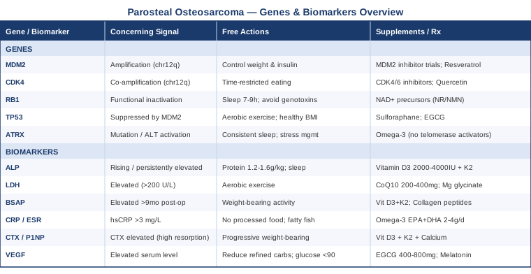
6 Biomarkers That Matter Most in Parosteal Osteosarcoma
Biomarkers are measurable signals — in blood, tissue, or imaging — that reflect biological activity at a specific moment. In parosteal osteosarcoma, tracking the right markers can provide ongoing visibility into disease status before and after surgery, flag early signals of recurrence, and assess systemic factors that influence healing and immune function. The six markers below are selected for relevance to this specific tumor biology, practical measurability, and actionability.
1. Alkaline Phosphatase (ALP)
Why it matters. Alkaline phosphatase is an enzyme produced primarily in bone, liver, kidneys, and bile ducts. In osteosarcoma, elevated ALP reflects increased osteoblastic activity — the bone-forming process that drives tumor-related new bone production. ALP elevation at diagnosis is one of the most consistently studied prognostic markers across osteosarcoma types, and persistently elevated levels after surgery may signal residual disease or recurrence. Multiple cohort studies have found elevated preoperative ALP correlates with worse event-free survival in osteosarcoma patients.
What it may reveal. In parosteal osteosarcoma specifically, elevations tend to be milder than in conventional high-grade disease — which reflects the lower-grade behavior of this tumor. But the trend over time matters more than any single value. A rising ALP after a period of post-surgical stability warrants investigation even when values remain below the laboratory upper limit of normal, particularly if accompanied by imaging changes.
How to measure it
ALP is included in a standard comprehensive metabolic panel (CMP), available at any laboratory. Cost typically ranges from $20–$60 for the full panel without insurance. Total ALP is the most common form reported, but requesting bone-specific ALP (BSAP, covered below) adds diagnostic precision. Frequency of monitoring is typically every 3–6 months post-surgery in standard follow-up protocols, though your oncologist may adjust this based on individual risk.
If the score is elevated: the plan without supplements
When ALP remains persistently elevated after treatment and hepatic causes have been ruled out (by checking liver-specific enzymes such as GGT and ALT), the priority is restaging imaging — typically MRI of the surgical site and chest CT to exclude local recurrence or pulmonary metastasis. Weight-bearing and high-impact activity over the surgical region should be reviewed with your surgeon. Adequate protein intake (1.2–1.6 g/kg body weight per day) and consistent sleep support normal bone healing physiology and should be assessed honestly.
If the score is elevated: the plan with supplements or equipment
The goal with bone-related ALP elevation is to support normal bone remodeling quality, not suppress it — unless hepatic contributions are identified, which require a different approach. For bone-specific elevation during the post-surgical healing period: Vitamin D3 (2000–4000 IU/day) combined with K2 (MK-7, 100–200 mcg/day) supports calcium utilization and bone matrix quality. Vitamin D status should always be confirmed first by 25-OH vitamin D blood test; target serum level is 40–60 ng/mL. Frequency: continuous daily use with reassessment every 3 months. Side effects at standard doses are minimal when dietary calcium is not excessive. Important: supplement use during or after cancer treatment must be discussed with your oncologist, as interactions with specific therapies are possible.
2. Lactate Dehydrogenase (LDH)
Why it matters. LDH is an enzyme involved in cellular energy metabolism that is released when cells are damaged or undergo rapid turnover. In osteosarcoma, elevated serum LDH at diagnosis has been consistently associated with worse outcomes in clinical cohort studies, and it remains part of standard prognostic assessment at staging. In parosteal osteosarcoma, LDH is less frequently elevated than in high-grade variants — which is one biological reason this tumor carries a better prognosis — but tracking it remains worthwhile for what it might signal over time.
What it may reveal. LDH elevation can signal tumor necrosis, accelerated cell turnover, or — when elevation is disproportionate to the known disease extent — dedifferentiation toward a higher-grade component. Dedifferentiated parosteal osteosarcoma is a recognized and clinically important entity with substantially worse prognosis. A patient with confirmed low-grade parosteal disease who develops rising LDH without an obvious benign explanation (infection, injury) deserves prompt clinical attention and consideration of re-biopsy.
How to measure it
LDH is a standard blood test, typically included in a metabolic panel or ordered separately. Cost ranges from $15–$40. It should be established at diagnosis and monitored at regular follow-up intervals. Normal range varies by laboratory but typically falls below 200–240 U/L in adults; the trend over serial measurements is often more informative than any single value.
If the score is elevated: the plan without supplements
Persistently elevated LDH following surgical treatment should prompt clinical review for recurrence or dedifferentiation before attributing it to other causes. Coordination with your oncology team for restaging imaging is the appropriate first step. Separately, progressive aerobic exercise within the limits set by surgical recovery has modest evidence for supporting mitochondrial efficiency and reducing systemic lactate accumulation at rest — but this is a background supportive measure, not a substitute for medical evaluation.
If the score is elevated: the plan with supplements or equipment
Evidence for supplement-driven LDH normalization specifically in cancer is limited and mostly indirect. Coenzyme Q10 (200–400 mg/day) supports mitochondrial function and has been studied as a supportive agent in cancer patients, with some positive signals on oxidative stress markers — direct LDH impact data in osteosarcoma is not yet available. Magnesium glycinate (300–400 mg/day) supports cellular energy metabolism broadly and is commonly deficient in cancer patients on active treatment. Discuss any supplementation with your oncology team; some agents interact with chemotherapy or affect wound healing parameters.
3. Bone-Specific Alkaline Phosphatase (BSAP)
Why it matters. Total ALP can rise from liver, kidney, or intestinal sources, making it a nonspecific signal when trying to understand what is happening in bone specifically. Bone-specific ALP isolates the osteoblast-derived component and provides a cleaner, more interpretable picture of skeletal activity. This distinction is particularly valuable in parosteal osteosarcoma follow-up, where distinguishing between normal post-surgical bone remodeling and pathological tumor-driven osteoblastic activity requires specificity.
What it may reveal. BSAP tracks osteoblast activity in isolation. Some elevation is expected and appropriate in the months following limb-salvage surgery as bone heals and remodels around implants or graft material. A BSAP that remains elevated or rises again well after the expected healing window is a more specific and meaningful signal than total ALP alone, and represents a justified indication for clinical review.
How to measure it
BSAP is measured by immunoassay and requires a specific laboratory request — it is not automatically included in standard panels. Cost typically ranges from $40–$100. It is available through major reference labs (Quest Diagnostics, LabCorp). It is particularly useful when serial monitoring of bone turnover is part of the follow-up strategy, and worth specifically requesting from your physician at follow-up appointments. Establishing a baseline value approximately 6 months post-surgery — when healing is expected to have stabilized — gives future values a meaningful reference point.
If the score is elevated: the plan without supplements
Temporal interpretation is key: elevation in the first 3–6 months post-surgery is expected and generally reassuring that healing is occurring. Elevation persisting or appearing beyond 6–9 months post-surgery, or a rising value after a period of stability, warrants imaging review. Avoiding excessive immobilization and engaging in graduated, supervised physical activity supports normal bone remodeling patterns.
If the score is elevated: the plan with supplements or equipment
Vitamin D3 + K2 as outlined above remain the foundational pairing. Adequate dietary calcium (targeting 1000–1200 mg/day from food where possible, supplemental calcium as a backup) and protein intake (1.2–1.6 g/kg/day) support healthy bone matrix formation. Collagen peptides (10–15 g/day of a hydrolyzed bovine or marine source) have emerging evidence for supporting bone repair quality in non-cancer orthopedic settings; direct osteosarcoma-specific evidence is absent, but the mechanism — providing substrates for type I collagen synthesis — is sound. Low-load weight-bearing activity, as tolerated and explicitly approved by your surgical team, supports bone density and quality over time.
4. C-Reactive Protein (CRP) and Erythrocyte Sedimentation Rate (ESR)
Why it matters. Systemic inflammation, measured by CRP and ESR, plays a documented role in tumor biology, wound healing, treatment tolerance, and immune function. In osteosarcoma patients, elevated inflammatory markers at diagnosis have been associated with worse outcomes in several cohort analyses. More broadly, the inflammatory tumor microenvironment is increasingly recognized as a modifiable biological factor — one where lifestyle and nutritional interventions have meaningful, measurable effects.
What it may reveal. In the post-treatment context, CRP and ESR help distinguish wound complications and infection from chronic low-grade inflammatory states that may reflect immune activation, residual tumor activity, or treatment side effects. High-sensitivity CRP (hsCRP) — the form recommended by physicians like Peter Attia for metabolic and cardiovascular risk tracking — is more sensitive for detecting low-grade inflammation than standard CRP, and provides useful added signal for cancer follow-up when serially tracked. ESR adds complementary information and is less affected by acute-phase variability.
How to measure it
Standard CRP is included in many inflammation panels; hsCRP is a separate test with similar cost ($15–$40). ESR runs $10–$25. Both are widely available at standard labs. Targets for general health interpretation: hsCRP below 1 mg/L is ideal; 1–3 mg/L represents intermediate risk; above 3 mg/L warrants attention; above 10 mg/L indicates acute inflammation requiring investigation to identify the source. In cancer patients, these thresholds serve as rough orientations, not absolute rules.
If the score is elevated: the plan without supplements
Non-supplement approaches to reducing chronic inflammation have the strongest and most consistent evidence base: reducing ultra-processed food and added sugar intake reduces inflammatory cytokines measurably within weeks. Increasing fatty fish intake (2–3 portions per week of salmon, sardines, or mackerel) provides EPA and DHA at levels shown in human trials to reduce hsCRP. Improving sleep duration and consistency (7–9 hours, same bedtime window) reduces inflammatory marker levels in ways measurable at standard labs. Progressive physical activity — even walking — reduces CRP in cancer patients at a level comparable to some pharmacological interventions.
If the score is elevated: the plan with supplements or equipment
Omega-3 fatty acids (EPA+DHA combined, 2–4 g/day) have the strongest evidence of any supplement for reducing hsCRP and inflammatory cytokines, with extensive human randomized trial data across populations including cancer patients. The anti-inflammatory evidence is robust enough that this is considered a first-line supplement intervention by multiple evidence-based clinicians. Frequency: continuous daily. Side effects at this dose range include mild GI effects and a modest effect on platelet function — relevant to discuss with your surgeon around procedural dates. Curcumin (500–1000 mg/day in a bioavailable form such as BCM-95 or Longvida) has anti-inflammatory evidence in human studies; discuss with your oncologist before adding, as CYP450 interactions can affect drug metabolism during chemotherapy.
5. C-Terminal Telopeptide (CTX) and Procollagen Type 1 N-terminal Propeptide (P1NP)
Why it matters. CTX measures bone resorption (breakdown), while P1NP measures bone formation. Together, these markers give a complete picture of bone turnover balance — whether the skeletal environment is tilting toward breakdown or construction. In parosteal osteosarcoma, this matters for two distinct reasons: first, the tumor itself alters local bone biology in ways that disrupt normal turnover; second, chemotherapy agents used when systemic treatment is indicated — notably methotrexate — have documented effects on bone metabolism that merit ongoing monitoring.
What it may reveal. Disproportionately elevated CTX after treatment may suggest abnormal resorption in the surgical region, secondary osteoporosis from treatment-related hormonal or metabolic changes, or in recurrence scenarios, new osteolytic activity. These markers are widely used in osteoporosis monitoring and are being adopted progressively in cancer-related bone disease follow-up, particularly in patients treated with agents that affect bone health.
How to measure it
Both CTX and P1NP are blood tests available through major reference laboratories. CTX should be drawn fasting in the morning — it rises significantly after eating and with circadian rhythmicity, making standardized collection critical for serial comparisons. Cost ranges from $50–$120 for each marker. These are not universally included in standard parosteal osteosarcoma follow-up protocols, but they add meaningful signal when bone health optimization is a clinical priority, particularly in patients who received systemic chemotherapy.
If the score is elevated (CTX high, resorption dominant): the plan without supplements
High CTX indicating accelerated bone breakdown can reflect immobilization-related bone loss during recovery — a real and measurable consequence of prolonged reduced weight-bearing after limb-salvage surgery. Progressive, supervised weight-bearing activity is the single most effective non-pharmaceutical intervention for normalizing CTX and improving bone mineral density. Impact and resistance-based mechanical loading stimulates osteoblastic activity and suppresses osteoclast-driven resorption through the RANK-RANKL pathway. The progression timeline must be directed by your rehabilitation and surgical team based on fixation status.
If the score is elevated (CTX high): the plan with supplements or equipment
The vitamin D3 + K2 + calcium triad forms the foundation of pharmaceutical-grade bone support without prescription drugs. K2 as MK-7 (100–200 mcg/day) specifically activates matrix Gla-protein and osteocalcin, directing calcium into bone rather than soft tissue. If CTX remains elevated despite optimized nutrition and activity, bisphosphonate therapy (zoledronic acid, alendronate) is an evidence-based prescription option your oncologist or endocrinologist can evaluate. Strontium ranelate has dual anti-resorptive and pro-formative evidence but has a more complex risk-benefit profile and is not available in all countries; discuss with a bone specialist if advanced support is warranted.
6. Vascular Endothelial Growth Factor (VEGF)
Why it matters. VEGF is a signaling protein that drives the formation of new blood vessels — angiogenesis — a process tumors depend on for sustained growth and eventual metastasis. In osteosarcoma, elevated VEGF expression in tumor tissue has been associated with increased metastatic risk and worse outcomes in multiple study cohorts. While VEGF is most commonly analyzed from tumor tissue samples, serum VEGF can also be measured and provides ongoing signal about systemic angiogenic activity that may not be captured by imaging alone.
What it may reveal. Elevated serum VEGF may reflect active tumor vascularity, systemic inflammatory states, or tissue ischemia in the post-surgical wound bed. In the follow-up context, trending serum VEGF values — particularly in patients whose imaging is equivocal — may offer supplementary signal about residual or recurrent disease activity. Most direct evidence comes from conventional high-grade osteosarcoma studies; parosteal-specific data is limited, but the biological mechanism is shared.
How to measure it
Serum VEGF is measured by ELISA assay and requires specific ordering through reference labs. Cost ranges from $80–$150. Specimen handling matters significantly: blood must be collected in a serum separator tube (SST) and processed promptly, as platelet degranulation during clotting artificially elevates VEGF if processing is delayed beyond 30 minutes. Tissue VEGF expression from biopsy specimens is assessed by immunohistochemistry and is typically reviewed as part of the pathology workup.
If the score is elevated: the plan without supplements
Hyperglycemia and hyperinsulinemia are among the most potent known stimulants of VEGF expression in human tissues — operating through HIF-1α and mTOR pathways. Reducing refined carbohydrate intake, prioritizing fiber, protein, and whole food sources, and managing fasting blood glucose (targeting below 90 mg/dL) reduces the primary metabolic driver of excess VEGF signaling. Chronic intermittent hypoxia from untreated sleep apnea also drives HIF-1α/VEGF upregulation; screening for sleep-disordered breathing in cancer patients is underutilized but clinically relevant.
If the score is elevated: the plan with supplements or equipment
Green tea extract (EGCG) at 400–800 mg/day with food inhibits VEGF receptor signaling in preclinical models and has supportive but preliminary evidence in human cancer studies. Melatonin (3–10 mg at night) has shown anti-VEGF properties in several cancer cell line studies and small human trials, though direct osteosarcoma evidence is lacking. Critically: any compound with anti-angiogenic activity should be discussed with your surgeon and oncologist before perioperative use, as wound healing requires intact vascular function. Cycling for botanical agents: 4–8 week periods with breaks; reassess with repeat serum VEGF measurement.
Tracking these biomarkers over time builds a picture that single-point measurements cannot. With a running record of ALP, LDH, BSAP, CRP, CTX/P1NP, and VEGF, conversations with your care team become more grounded and responsive to what is actually changing. The genetic picture below explains why these signals behave the way they do.
The Genetic Blueprint of Parosteal Osteosarcoma: 5 Key Genes
Parosteal osteosarcoma has one of the most distinctive and consistent genetic profiles in bone oncology. Its core alterations are highly reproducible across patients, which makes this tumor unusual among solid tumors — and makes the genetic information below more directly applicable than genetic findings in more heterogeneous cancers. For each gene, the focus is on what it means in practical and biological terms, and what the evidence currently supports for response.
Gene 1: MDM2 (Mouse Double Minute 2 Homolog)
What it does. MDM2 is the primary regulator of p53 — the "guardian of the genome." It acts as a brake on p53's tumor-suppressing function by binding to it and marking it for proteasomal degradation. When MDM2 is amplified (extra gene copies produce excess protein), p53 is effectively silenced despite remaining structurally intact. Cells can accumulate DNA damage and receive abnormal growth signals without triggering the repair and death responses that p53 would otherwise activate.
Why it defines this tumor. MDM2 amplification, located on chromosome 12q13-15, is found in approximately 70–95% of parosteal osteosarcomas — making it the most reliable molecular diagnostic marker for this tumor type. Ring chromosomes containing amplified 12q sequences are the chromosomal hallmark of parosteal disease, and MDM2 FISH (fluorescence in situ hybridization) testing is now standard in the diagnostic workup of any surface bone lesion where parosteal osteosarcoma is in the differential. MDM2 amplification also distinguishes parosteal osteosarcoma from periosteal osteosarcoma and from atypical cartilaginous tumors that can look similar radiologically.
If the gene is altered: the plan without supplements
The primary clinical response to MDM2 amplification in parosteal osteosarcoma is surgical — wide resection with clear margins remains the standard of care, and the prognostic impact of MDM2 status is primarily diagnostic rather than immediately therapeutic at the low-grade stage. For patients with recurrent or dedifferentiated disease, consultation with a sarcoma specialist about MDM2 inhibitor clinical trials (nutlin-class compounds, RG7112, AMG-232) is worthwhile — these agents work by blocking the MDM2-p53 interaction, restoring p53 function without requiring gene correction. Maintaining normal body weight and managing dietary sugar reduces insulin/IGF-1 signaling, which indirectly promotes MDM2 expression through AKT phosphorylation.
If the gene is altered: the plan with supplements or equipment
Direct MDM2 suppression through available supplements is not established in human clinical evidence. Several natural compounds have been studied in preclinical models. Resveratrol (trans-resveratrol, 500–1000 mg/day with food) has demonstrated MDM2 inhibitory properties in cancer cell line studies, including osteosarcoma lines, through SIRT1 and p53 pathway modulation. Berberine (500 mg twice daily with meals) has shown multi-pathway anti-tumor activity including AKT suppression, which indirectly reduces MDM2-mediated p53 degradation. Both agents are under active preclinical investigation but lack phase II human trial data in osteosarcoma specifically. Cycling: 4–8 weeks on, 2–4 weeks off; side effects include GI discomfort with both. These should not be used near surgical dates or concurrent with active chemotherapy without oncology clearance.
Gene 2: CDK4 (Cyclin-Dependent Kinase 4)
What it does. CDK4 is a key kinase in cell cycle regulation. In complex with cyclin D, it phosphorylates and inactivates the RB1 protein — releasing transcription factors that allow cells to pass the G1/S checkpoint and commit to division. When CDK4 is amplified, this brake on cell division is chronically released, driving accelerated proliferation regardless of external growth signals.
Why it matters here. CDK4 amplification co-occurs with MDM2 amplification in 40–70% of parosteal osteosarcomas, sitting on the same 12q13-15 chromosomal segment. This co-amplification pattern is pathognomonic and has direct therapeutic implications: CDK4/6 inhibitors — palbociclib (Ibrance), ribociclib (Kisqali), and abemaciclib (Verzenio) — are FDA-approved for breast cancer and are actively being studied in sarcoma subtypes, with parosteal and dedifferentiated liposarcoma (which shares the same 12q amplicon) being the most biologically rational targets.
If the gene is altered: the plan without supplements
For patients with CDK4-amplified disease, particularly at recurrence or in dedifferentiated disease, inquiry with a sarcoma oncology specialist about CDK4/6 inhibitor eligibility and available clinical trials is a concrete and well-justified next step. In the metabolic domain, caloric restriction and time-restricted eating reduce circulating insulin and IGF-1, both of which drive cyclin D1 production — the binding partner CDK4 requires to become active. This is a biologically sound indirect approach, though direct human osteosarcoma evidence for this mechanism is not yet available from clinical trials.
If the gene is altered: the plan with supplements or equipment
Quercetin (500–1000 mg/day, bioavailability enhanced when combined with bromelain) has demonstrated CDK4 inhibitory properties in multiple cancer cell line studies including osteosarcoma lines. It operates in part by preventing cyclin D1 accumulation and by downregulating transcription factors that drive CDK4 expression. Human evidence in cancer is preliminary and confined to smaller studies. Cycling: 4–8 weeks on with 2–4 week breaks; side effects are generally mild GI. Note that quercetin inhibits certain CYP450 drug-metabolizing enzymes — discuss with your oncologist if receiving systemic therapy, as drug exposure levels could be affected.
Gene 3: RB1 (Retinoblastoma Protein Gene)
What it does. RB1 encodes the retinoblastoma protein, a fundamental tumor suppressor that operates as the gate to S-phase entry. When phosphorylated by CDK4/6 complexes, it releases E2F transcription factors needed to initiate DNA replication. When RB1 is deleted or mutated, this gate is permanently open — cells divide without restraint regardless of CDK4 activity.
Why it matters in this context. Parosteal osteosarcoma typically achieves functional RB1 inactivation not by deleting the gene but by overwhelming it with CDK4 amplification — a distinctions with real clinical significance. Because the RB1 gene itself remains structurally intact, blocking CDK4 with CDK4/6 inhibitors can restore RB1 function and reinstate the cell cycle checkpoint. This is the same molecular rationale underlying the use of palbociclib in ER+/RB1-intact breast cancer — and it applies here. Outright RB1 deletion is more common in dedifferentiated parosteal osteosarcoma or recurrent disease, where the biology has shifted.
If the gene is altered: the plan without supplements
Supporting the CDK4-RB1 axis through the same dietary and sleep strategies outlined for CDK4 applies directly here. Avoiding additional genotoxic exposures — tobacco products, excessive alcohol, unnecessary radiation — reduces the mutational pressure on an already stressed cell cycle regulation system. Adequate sleep (7–9 hours with consistent timing) supports systemic DNA repair activity during slow-wave sleep phases and has been shown to maintain telomere length and genomic stability over time.
If the gene is altered: the plan with supplements or equipment
Supplements supporting DNA repair pathway activity: NAD+ precursors — nicotinamide riboside (NR) or nicotinamide mononucleotide (NMN), 250–500 mg/day — support PARP-dependent DNA repair mechanisms. NAD+ depletion is well-documented in cancer contexts and during aging, and restoration of NAD+ levels supports the enzymatic machinery that maintains genomic stability. Direct evidence in RB1-pathway dysfunction in osteosarcoma is not established, but the mechanistic rationale is sound. Frequency: continuous daily use. Side effects are minimal at standard doses; mild flushing occasionally with niacin-adjacent forms. Discuss with your oncologist given potential interactions with chemotherapy in timing.
Gene 4: TP53
What it does. TP53 encodes p53 — a transcription factor and tumor suppressor that activates DNA damage response pathways, halts cell cycle progression at checkpoints when damage is detected, and triggers apoptosis when damage is too severe to repair. It is involved in an extraordinary range of cellular stress responses and is the most commonly mutated gene across all human cancers.
Why it matters in parosteal osteosarcoma. Parosteal osteosarcoma is notable for having relatively low rates of direct TP53 mutation compared to conventional high-grade osteosarcoma — which is consistent with its lower-grade behavior and more indolent course. However, MDM2 amplification achieves functional p53 suppression without directly mutating the gene — meaning p53 is silenced but structurally intact. When parosteal osteosarcoma dedifferentiates into high-grade disease, TP53 mutations become progressively more common, suggesting TP53 loss is a driver of progression. This trajectory makes preserving what p53 function exists a meaningful supportive goal.
If the gene is altered: the plan without supplements
Lifestyle factors that support p53 activity are not hypothetical — they are measurable. Regular aerobic exercise has been shown in human studies to upregulate p53 pathway activity in peripheral blood mononuclear cells and in colonic epithelium, independently of weight change. Chronic obesity and metabolic syndrome are associated with blunted p53 responses, likely through insulin/IGF-1 pathway suppression of p53 transcriptional activity. Maintaining a healthy weight and managing insulin levels are therefore directly relevant to p53 function, not merely general cancer risk reduction.
If the gene is altered: the plan with supplements or equipment
Sulforaphane — from broccoli sprout extract standardized to 10–30 mg sulforaphane/day — has shown p53 stabilization effects in human clinical studies in colorectal cancer via NRF2 pathway activation and HSP90 chaperone modulation. Evidence specific to osteosarcoma is very early; the mechanism is biologically plausible. Frequency: continuous daily use is well-tolerated; side effects include occasional GI effects at higher doses. EGCG from green tea extract (400 mg/day standardized extract) also modulates p53 stability via HSP90 interference and shows overlapping NRF2 activity. As with all supplements, discuss timing around chemotherapy administration.
Gene 5: ATRX (Alpha-Thalassemia/Mental Retardation X-Linked)
What it does. ATRX is a chromatin remodeling protein with two interconnected functions: it maintains telomere integrity during replication, and it helps regulate gene expression through heterochromatin organization. When ATRX is lost or mutated, cells frequently activate an alternative telomere lengthening mechanism (ALT pathway) that allows unlimited replication without telomerase — and the associated chromatin dysregulation alters expression of hundreds of genes.
Why it matters in parosteal osteosarcoma. ATRX mutations are identified in a meaningful subset of osteosarcomas, occurring more commonly in recurrent or treatment-resistant disease. In osteosarcoma, ATRX loss and ALT activation are associated with genomic instability and may contribute to the dedifferentiation events that worsen prognosis in parosteal disease over time. ATRX status is not yet part of routine clinical reporting for parosteal osteosarcoma in most centers, but research is converging on it as a meaningful prognostic variable, particularly in recurrent settings.
If the gene is altered: the plan without supplements
ATRX loss-driven ALT activation does not currently have a direct pharmaceutical countermeasure in standard clinical use. The practical focus is on strategies that minimize overall genomic stress. Adequate, consistent sleep is directly relevant here: human studies consistently demonstrate shorter telomere length in individuals with chronic sleep deprivation or poor sleep quality, independent of age. Since ATRX-deficient cells rely on ALT-dependent telomere maintenance, reducing additional telomere erosion from sleep-related stress makes mechanistic sense. Psychological stress management through evidence-based approaches also reduces telomere erosion — this is one of the stronger arguments for including MBSR in the care plan of this patient population.
If the gene is altered: the plan with supplements or equipment
Astragalus-derived compounds, notably cycloastragenol (commercially as TA-65), have been studied for telomere support through modest telomerase activation — with some human evidence in immune cells. However, in an active cancer context, activating telomere elongation is inherently double-edged: while normal tissue telomere maintenance is beneficial, cancer cells that have activated ALT may also benefit from increased telomere stability. This is not a supplement to use without explicit oncology guidance in a cancer context. For general cellular health support: omega-3 fatty acids (as above for CRP) have been associated with longer telomere length in several large human observational studies, making them a low-risk, multi-benefit option worth maintaining.
The Cancer Code: 10 Things That May Change How You Think About This Disease
The Cancer Code by Dr. Jason Fung (Harper Wave, 2020) is among the most evidence-grounded books available for patients who want to understand the metabolic and evolutionary biology behind their cancer — without oversimplification or overclaiming. Fung, a nephrologist and researcher known for his work on fasting and insulin, applies those frameworks to cancer biology in ways that directly connect to the genetic and biomarker picture described in this article. The book challenges the view that cancer is purely a gene mutation disease and presents it instead as a product of disrupted cellular ecology. Below are the ten ideas with the most practical relevance for parosteal osteosarcoma patients.
1. Cancer Is an Ancient Survival Program Reactivated
Fung argues that cancer cells do not develop random new behaviors — they reactivate ancient, conserved survival mechanisms that normal multicellular cooperation suppresses. Understanding this changes the framing: cancer is not the body going haywire, it is one program overriding another. Treatment, and everything around it, can address both levels.
2. The Warburg Effect Is Not a Side Effect — It Is a Central Vulnerability
Cancer cells preferentially use glucose through anaerobic glycolysis even when oxygen is abundant — the Warburg effect. This glucose dependency creates a metabolic vulnerability that dietary modification and fasting can partially exploit. Fung presents this as one of the most underutilized insights in clinical oncology.
3. Insulin Is One of the Strongest Pro-Cancer Signals in the Human Body
Chronically elevated insulin — the result of high refined carbohydrate intake and metabolic syndrome — activates mTOR, IGF-1R, and AKT/PI3K pathways. These pathways directly promote MDM2 expression and CDK4 activity — both central drivers of parosteal osteosarcoma. Fung makes the case that insulin control is among the most overlooked aspects of oncology support.
4. Periodic Fasting Creates Conditions Hostile to Tumor Cell Survival
Fasting reduces insulin, depletes glucose, activates autophagy, and creates oxidative stress selectively in cells that are less metabolically flexible — which describes cancer cells well. Human trials in cancer patients show that short-term fasting around chemotherapy administration reduces toxicity and may sensitize tumor cells to treatment, without compromising patient nutrition overall.
5. The Tumor Microenvironment Matters as Much as the Tumor Itself
The inflammatory, hypoxic, and insulin-rich environment surrounding a tumor shapes its behavior as much as its intrinsic genetics. This directly validates the biomarker approach in this article — tracking CRP, VEGF, and insulin resistance markers is tracking that environment in real time, and modifying it through diet, exercise, and sleep is a legitimate biological intervention.
6. The Immune System Is the Body's Most Powerful Anti-Cancer Defense
Immunotherapy's rise has validated what Fung outlines mechanistically: immune surveillance is the difference between cancer contained and cancer progressing. Chronic inflammation suppresses that surveillance. Sleep, exercise, and stress reduction are not lifestyle recommendations — they are immune function interventions with measurable biological effects.
7. Standard Cancer Treatment and Metabolic Support Are Not in Conflict
Fung addresses directly the concern that dietary changes will interfere with surgery or chemotherapy. For the dietary modifications with the strongest evidence — carbohydrate reduction, protein adequacy, fish oil, fasting protocols — the evidence points toward complementary or neutral effects on treatment outcomes. The non-negotiable is coordination with your oncology team, not avoidance of support entirely.
8. mTOR Is the Central Node Where Multiple Anti-Cancer Strategies Converge
Fasting, exercise, metformin, rapamycin, and berberine all converge on mTOR inhibition. This protein complex regulates cell growth and is aberrantly activated in osteosarcoma. The fact that multiple independent strategies — dietary, pharmaceutical, and supplemental — hit the same node is meaningful evidence that the target is real and modifiable.
9. Obesity and Metabolic Syndrome Are Active Tumor Promoters, Not Passive Risk Factors
The epidemiological association between metabolic syndrome and cancer outcomes is now supported by biological mechanism — excess adipose tissue produces inflammatory cytokines, aromatizes hormones, and maintains the insulin/IGF-1 environment that feeds tumor proliferation. For bone cancer patients, this makes managing weight, blood sugar, and triglycerides a direct contribution to favorable biology.
10. You Have More Biological Agency Than Most Patients Are Told
This is perhaps the book's most clinically important message: the daily choices a patient makes — food quality, sleep timing, movement type, fasting pattern — genuinely alter the tumor microenvironment in measurable ways. Not as a replacement for surgery, radiation, or chemotherapy, but as legitimate biological levers that most standard oncology protocols leave entirely to chance.
Complementary Approaches with Clinical Evidence Relevant to Bone Cancer Patients
The following modalities have been selected based on availability of human clinical evidence applicable to osteosarcoma patients or bone cancer populations more broadly. They are presented as supportive adjuncts to standard medical care, with realistic assessments of evidence quality.
Mindfulness-Based Stress Reduction (MBSR)
MBSR is an 8-week structured program developed by Jon Kabat-Zinn that combines formal meditation, body scanning, and mindful movement into a standardized protocol. Its relevance for parosteal osteosarcoma patients lies at the intersection of two domains: managing the considerable psychological burden of a bone cancer diagnosis and treatment, and the documented biological downstream effects of stress reduction on inflammatory cytokines, cortisol, and immune function — all of which intersect directly with the biomarkers tracked in this article.
A widely cited randomized trial by Carlson and colleagues in cancer patients demonstrated significant reductions in salivary cortisol, improvements in natural killer cell activity, and reductions in anxiety and depression at 12-month follow-up following MBSR participation. Multiple subsequent meta-analyses of MBSR in mixed oncology populations confirm benefits for anxiety, depression, fatigue, and quality of life, with effect sizes categorized as moderate to large. The biological effects on inflammatory markers — including CRP — are increasingly supported by trial data.
For practical implementation: structured MBSR programs are available through many comprehensive cancer centers, often at reduced or no cost as part of survivorship programs. App-based formats (Insight Timer, Waking Up, UCLA Mindful) provide accessible entry points for those without local program access. Starting with 10–15 minutes of breath-focused practice daily and progressing over 4–8 weeks toward longer body scan sessions is a feasible protocol. Post-surgical recovery periods, when physical activity is restricted, offer a natural window to establish this practice.
Breathing-Based Therapies
Controlled breathing practices — from diaphragmatic breathing and box breathing to cyclic sighing and coherent breathing — act directly on the autonomic nervous system, shifting the balance from sympathetic activation toward parasympathetic tone. This produces measurable downstream effects on heart rate variability, cortisol levels, inflammatory cytokines, and subjective pain tolerance — all of which are relevant during and after osteosarcoma treatment.
A randomized study by Balban and colleagues, published in Cell Reports Medicine (2023), demonstrated that cyclic sighing — two nasal inhales followed by a full extended exhale, performed for five minutes — produced the largest improvements in mood and the greatest reductions in physiological arousal among several breathing protocols tested, including mindfulness meditation. For cancer patients whose inflammatory markers and stress biology are already activated by diagnosis and treatment, a five-minute daily intervention with documented autonomic effects represents an extremely accessible and low-burden tool.
Practical application: five minutes of cyclic sighing or diaphragmatic breathing twice daily — once upon waking and once before sleep — is a practical protocol with no equipment requirements. Box breathing (4 counts inhale, 4 hold, 4 exhale, 4 hold) is a strong alternative for procedural stress management around chemotherapy infusions or clinical appointments. The only common side effect is mild light-headedness from breath pattern changes; this resolves with ensuring full, relaxed exhales rather than forced ones.
Low-Level Laser Therapy / Photobiomodulation
Photobiomodulation (PBM) uses low-power red and near-infrared light in the 630–1100 nm range to stimulate mitochondrial cytochrome c oxidase, increasing cellular ATP production and reducing oxidative stress. Its applications most relevant to parosteal osteosarcoma patients are post-surgical wound healing, reduction of chemotherapy-induced oral mucositis, and management of musculoskeletal pain during recovery.
A systematic review and meta-analysis of PBM for chemotherapy-induced oral mucositis confirmed significant reductions in mucositis severity, pain, and duration — directly applicable to patients receiving MAP protocol chemotherapy (methotrexate, adriamycin, cisplatin). Multiple randomized trials in orthopedic contexts have shown PBM accelerates bone healing markers and reduces post-surgical pain at operative sites. The legitimate concern about PBM stimulating tumor cell proliferation in vitro does not appear to translate to clinical harm at standard therapeutic doses in available human data, but the reasonable precaution of avoiding direct irradiation over active tumor sites or unresected disease remains appropriate.
Practical implementation: PBM for mucositis and surgical wound care is best delivered by trained oncology rehabilitation specialists or physical therapists using medical-grade devices. Home devices in the 660–850 nm range are commercially available but should not be applied over or adjacent to any active disease site without oncology clearance. Standard protocols involve 10–20 minute sessions, 3–5 times per week. Discuss with your surgical and oncology team before initiating, particularly regarding device parameters and treatment location.
Music Therapy
Music therapy delivered by board-certified therapists encompasses live music, guided listening, improvisation, and songwriting. Its evidence base in oncology settings is substantial, with documented effects on pain perception, procedural anxiety, cancer-related fatigue, and quality of life across multiple tumor types and treatment settings. The mechanisms include endogenous opioid release, cortisol suppression, and autonomic regulation.
A Cochrane Systematic Review by Bradt and colleagues analyzing over 30 randomized trials in cancer patients found significant reductions in anxiety, pain, heart rate, respiratory rate, and blood pressure compared to control conditions — with moderate to large effect sizes for anxiety and pain specifically. Effects on pain during procedural interventions (biopsies, infusions, dressing changes) were particularly notable — directly applicable to the intensive procedural burden of osteosarcoma treatment.
Practical application: formal music therapy through a certified music therapist is available at many comprehensive cancer centers, typically as part of integrative oncology or palliative care programs. Self-directed intentional music listening — using personally meaningful, emotionally familiar music at 60–80 bpm for relaxation — produces measurable autonomic effects in studies and can be implemented immediately. Dedicated listening of 20–30 minutes around procedurally stressful treatment days, with intentional focus on the music rather than passive background use, captures most of the documented benefit without requiring clinical delivery.
Conclusion
Parosteal osteosarcoma is a rare diagnosis, but it is not an opaque one. Its genetic drivers — MDM2 and CDK4 amplification, the functional suppression of RB1 and p53, and the emerging story around ATRX — are among the most consistently characterized in all of bone oncology. The biomarkers that matter most for follow-up can be measured, tracked serially, and influenced through evidence-based interventions. That combination of specificity and actionability is worth using.
The most productive next step is a concrete one: bring a list of specific biomarkers — bone-specific ALP, hsCRP, LDH, and CTX at minimum — to your next follow-up appointment and ask which are already in your monitoring plan and which could be added. Review the genetic markers from your biopsy or surgical pathology report with your oncologist and ask about MDM2 and CDK4 amplification status if it has not been explicitly addressed. Begin one of the complementary practices in this article tonight — five minutes of cyclic sighing costs nothing and starts working immediately. This condition rewards steady, informed attention more than heroic interventions. You now have a specific map.
Cancer & Oncology Endocrine & Metabolic
Musculoskeletal: Bone Conditions
Autoimmune: Inflammatory Conditions
Cancer & Oncology: Bone Cancer