This article contains AI generated content.
· UpdatedPasteurella Multocida Septic Arthritis — 5 Genes And 7 Biomarkers To Track
Introduction
A cat bite rarely registers as a serious medical event in the moment it happens. It stings, you clean it, and you move on. But for a subset of people — particularly those with immune vulnerabilities, pre-existing joint conditions, or certain genetic profiles — that small wound becomes the entry point for Pasteurella multocida, a gram-negative bacterium found in the oral flora of most cats and a significant proportion of dogs. When this organism enters a joint space, either through a direct wound or via bloodstream spread, the result is septic arthritis: a rapidly progressing, destructive infection that can cause permanent joint damage within days if not treated aggressively. The timeline is unforgiving, and the stakes are higher than most patients initially understand.
What makes this infection especially difficult to navigate is the variability in how people respond — both to the infection itself and to treatment. Standard care involves prompt joint aspiration, blood cultures, and intravenous antibiotics, typically beta-lactams such as ampicillin-sulbactam, to which P. multocida is usually sensitive. But whether someone develops the infection at all after exposure, how severe the joint destruction becomes, how quickly inflammatory markers normalize, and how much residual damage remains — these outcomes differ dramatically from person to person. Standard protocols do not explain that variability. Biomarkers and genetics do.
This article focuses on what laboratory data and genetic information can reveal about individual risk, disease severity, and recovery trajectory in P. multocida septic arthritis. This is not about replacing standard antibiotic treatment, which is the non-negotiable clinical priority. It is about using available tools more completely — tracking the right numbers, understanding what they mean at each stage, and knowing which genetic variants may be shaping your experience of this infection far more than the average clinical presentation would suggest.
With that grounding in place, this article walks through seven biomarkers that can be measured from initial diagnosis through the full recovery period, each one offering a different window into what is happening in the joint and systemically. It then examines five genes whose variants are associated with differences in innate immune response, inflammatory intensity, and joint damage risk — along with practical steps for each. Beyond that, a section draws on the most impactful immune science insights from the Huberman Lab podcast, and three complementary modalities with genuine clinical backing round out the recovery framework. Better information does not guarantee a perfect outcome — but it consistently leads to better decisions.
Summary
This article examines Pasteurella multocida septic arthritis through two evidence-based lenses rarely addressed in depth by standard clinical protocols. The primary lens covers 7 actionable biomarkers — from CRP and procalcitonin to synovial fluid lactate and IL-6 — that track bacterial burden, inflammatory severity, treatment response, and joint damage risk at every stage. For each biomarker, you will find what the number reveals, how to measure it with cost ranges, and specific protocols for improving it both with and without supplements. The second lens covers 5 key genetic variants — including TLR4, TNF-alpha, MMP-3, and IL-10 — that help explain why some people experience severe, prolonged infections while others recover quickly, along with practical compensation strategies for each. The article also distills the most impactful insights on immune function and recovery from the Huberman Lab podcast, and identifies three complementary approaches — photobiomodulation, mindfulness-based stress reduction, and microbiome-directed therapy — with meaningful clinical backing for the recovery phase. Each section is practical, specific, and designed to be useful whether you are navigating active treatment or long-term joint recovery.
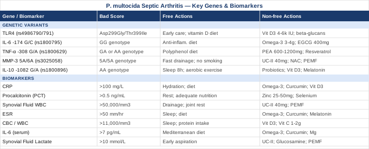
7 Biomarkers to Track From Diagnosis Through Recovery
The clinical decision-making in P. multocida septic arthritis is largely driven by laboratory data, yet the full value of these tests is rarely communicated to patients in a usable form. Understanding what each marker measures, how it moves over time, and what drives it in the wrong direction provides a far more useful map of recovery than waiting for symptoms alone to guide every decision. These seven markers cover the full arc — from confirming the diagnosis to verifying complete resolution and detecting complications early.
Biomarker 1: C-Reactive Protein (CRP)
Why it matters: CRP is produced by the liver in response to interleukin-6 released during infection and tissue damage. It is the most commonly used acute-phase reactant in clinical practice and one of the most responsive markers for tracking the trajectory of bacterial infections. In P. multocida septic arthritis, CRP rises dramatically within 24–48 hours of onset, often exceeding 100 mg/L in active disease. More importantly, it falls predictably with successful antibiotic treatment — making it one of the most useful markers for confirming that therapy is working. Elevated CRP persisting beyond 48–72 hours of appropriate antibiotic therapy signals something is not resolving as expected: inadequate drainage, a treatment-resistant organism, or a secondary complication emerging.
How to measure it: CRP is a standard blood test ordered as part of an inflammatory panel or standalone. High-sensitivity CRP (hs-CRP) is a more precise version used in cardiovascular risk contexts, but standard CRP is used in acute infectious settings and is universally available. Cost ranges from approximately $15 to $50 USD depending on the setting, with results typically available within 2–4 hours in a hospital. Reference range: less than 1 mg/L (some labs use less than 5 mg/L as the upper normal limit).
If the score is elevated, the plan without supplements: In the acute phase, elevated CRP should drive clinical action — confirming antibiotic coverage, ensuring adequate joint drainage (repeat aspiration or surgical washout if needed), maintaining joint rest, and monitoring at 48-hour intervals. Dietary priorities that support resolution without supplements include adequate hydration (minimum 2–2.5 liters of fluid daily to support renal clearance of inflammatory metabolites), reduction of refined sugars and ultra-processed foods (strong drivers of CRP elevation), and high protein intake (at least 1.5g/kg body weight per day) to support immune cell production and tissue repair. Mediterranean-pattern eating with emphasis on olive oil, fatty fish, vegetables, and legumes has well-documented CRP-lowering effects suitable for the recovery phase.
If the score is elevated, the plan with supplements or equipment: During the acute infectious phase, no supplement replaces antibiotics or drainage. Once the acute phase is controlled — typically after 72 hours of appropriate therapy — several supplements have evidence for supporting inflammatory resolution. Omega-3 fatty acids (EPA and DHA) at 2–4 grams per day have been shown in multiple randomized trials to reduce CRP. A typical protocol is 3 grams per day for 8–12 weeks, cycling to maintenance dose (1–2g) thereafter. Mild anticoagulant effect at higher doses; avoid very high doses if on warfarin. Curcumin with piperine (500–1000mg curcumin with 10–20mg piperine, twice daily for 8–12 weeks) has demonstrated CRP reduction in inflammatory conditions; cycle off for 4 weeks after each 12-week course. GI sensitivity can occur in some individuals. Vitamin D3 (maintain serum 25-OH-D at 40–60 ng/mL, typically requiring 2000–5000 IU/day with K2 at 100–200mcg) supports immune regulation and is consistently associated with lower CRP in observational data. Test serum levels before dosing.
Biomarker 2: Procalcitonin (PCT)
Why it matters: Procalcitonin is a precursor to calcitonin released in response to bacterial infections by virtually all nucleated cells in the body. Unlike CRP, which rises with any significant inflammation — viral, autoimmune, or sterile — PCT is considerably more specific to bacterial infection. This specificity is particularly valuable in distinguishing P. multocida septic arthritis from reactive arthritis or crystal-induced arthritis, which can present with overlapping clinical features. PCT also rises and falls faster than CRP, making it a useful early signal and a responsive tracking marker during treatment. In septic arthritis, persistently elevated PCT after 48–72 hours of antibiotic treatment suggests inadequate bacterial clearance and warrants clinical re-evaluation.
How to measure it: PCT is measured via blood test and available in most hospital settings, with turnaround times of 1–3 hours. It is less routinely ordered than CRP in community settings. Cost: approximately $30 to $100 USD. Reference values: less than 0.1 ng/mL (no infection or very localized), 0.1–0.5 ng/mL (possible local infection), 0.5–2.0 ng/mL (suggests systemic bacterial infection), greater than 2.0 ng/mL (consistent with severe bacterial infection or early sepsis).
If the score is elevated, the plan without supplements: Elevated PCT in the context of P. multocida septic arthritis primarily guides the intensity of antibiotic management. High values (above 2 ng/mL) should prompt consideration of bacteremia and joint compartment culture review. The decline curve is clinically informative: PCT typically halves every 24–48 hours with effective treatment. A slow decline should trigger review of antibiotic spectrum coverage and consideration of repeat aspiration. Non-supplement measures that support faster bacterial clearance include allowing low-grade fever when clinically tolerated (fever impairs bacterial replication and enhances neutrophil motility), maintaining adequate rest, and ensuring nutritional adequacy — particularly protein and micronutrients that support phagocyte function.
If the score is elevated, the plan with supplements or equipment: Zinc (25–50mg elemental zinc per day during the active infection phase, cycling off after 4 weeks to avoid copper displacement) supports neutrophil function and has data for reducing duration and severity of bacterial infections. Take with food to reduce GI upset; pair with 2mg copper per day if using longer than 4 weeks. Selenium (100–200mcg per day as selenomethionine) is a cofactor for glutathione peroxidase, the enzyme critical for neutrophil oxidative burst — the primary mechanism by which neutrophils kill gram-negative bacteria like P. multocida. Risk of selenosis above 400mcg/day; do not supplement long-term without measuring serum selenium. Doses return to dietary levels once PCT normalizes.
Biomarker 3: Synovial Fluid White Blood Cell Count and Differential
Why it matters: This is the gold standard biomarker for diagnosing septic arthritis and is indispensable for confirming P. multocida joint infection specifically. While all other markers discussed here are systemic, synovial fluid WBC measures what is happening directly inside the infected joint. The key thresholds: a synovial fluid WBC above 50,000 cells/mm³ is highly suggestive of bacterial infection; above 100,000 cells/mm³ is virtually diagnostic in the appropriate clinical context. The differential is equally important — a polymorphonuclear cell (PMN/neutrophil) percentage above 90% strongly supports bacterial etiology over crystal or autoimmune causes. According to a landmark diagnostic accuracy study published in JAMA, the synovial fluid WBC is the single most diagnostically useful test for septic arthritis, outperforming serum markers in specificity for joint infection (Margaretten et al., JAMA 2007).
How to measure it: Synovial fluid analysis is performed as part of joint aspiration (arthrocentesis), a procedure done under local anesthesia at the bedside or under ultrasound guidance. The aspirated fluid is sent for cell count, differential, Gram stain, and culture. The procedure itself typically costs $100–500 USD in an outpatient or emergency setting; the laboratory analysis adds another $50–150. Cell count results are typically available within 1–4 hours; culture results take 24–72 hours.
If the score is elevated, the plan without supplements: High synovial WBC drives urgent clinical decisions: confirm antibiotic coverage against P. multocida specifically (beta-lactamase-producing strains require amoxicillin-clavulanate), repeat aspiration daily until WBC falls to below 50,000 cells/mm³, and consider surgical irrigation and debridement if daily aspiration does not produce progressive improvement within 3–4 days. Joint rest is critical — immobilization in a comfortable position with elevation where possible. Range-of-motion exercises are contraindicated in the acute phase and are reintroduced carefully by physical therapy once the WBC is substantially reduced.
If the score is elevated, the plan with supplements or equipment: During the acute phase, joint-specific interventions beyond standard treatment are limited by necessity. Post-acute, once synovial WBC has normalized, undenatured type II collagen (UC-II) at 40mg per day has randomized trial data supporting cartilage protection in inflammatory joint conditions through oral tolerance mechanisms. Pulsed electromagnetic field (PEMF) therapy home devices have evidence for synovial fluid metabolism support and cartilage protection post-inflammation. Protocol: 20–30 minutes daily applied to the affected joint area; use only after confirmed infection resolution. Home PEMF devices cost $150–500 USD.
Biomarker 4: Erythrocyte Sedimentation Rate (ESR)
Why it matters: ESR measures the rate at which red blood cells settle in a vertical tube over one hour — a process accelerated when high concentrations of inflammatory proteins (fibrinogen, immunoglobulins, acute-phase reactants) are present in the plasma. In P. multocida septic arthritis, ESR is almost universally elevated, often exceeding 50–100 mm/hr. Unlike CRP, which peaks at 24–48 hours and falls rapidly, ESR rises more slowly (peak at 3–5 days) and normalizes more slowly — sometimes remaining elevated for weeks after infection resolution. This makes it less useful for acute tracking but valuable for monitoring prolonged recovery and detecting persistent low-grade infection that a normalizing CRP might mask. The combination of both markers — elevated ESR with normalizing CRP — may suggest incomplete recovery rather than recurrence, guiding different clinical decisions.
How to measure it: ESR is one of the most affordable laboratory tests available, typically costing $10 to $30 USD. It is ordered standalone or as part of an inflammatory marker panel. Normal values: less than 15–20 mm/hr for men under 50; less than 20–30 mm/hr for women under 50. Values increase slightly with normal aging and should be interpreted accordingly.
If the score is elevated, the plan without supplements: Given that ESR normalizes slowly, an isolated elevated ESR during recovery (weeks after infection) should be interpreted in context with other markers. If CRP has normalized and the patient is clinically well, a modestly elevated ESR (40–60 mm/hr) may simply reflect ongoing resolution. Persistent ESR above 80–100 mm/hr at 4–6 weeks post-treatment warrants re-evaluation for treatment failure, occult re-infection, or superimposed reactive arthritis. Non-supplement strategies: continue anti-inflammatory dietary approach, maintain adequate sleep (7–9 hours per night, as immune regulation is highly sleep-dependent), and avoid high-intensity exercise during the recovery phase as it temporarily elevates inflammatory markers.
If the score is elevated, the plan with supplements or equipment: The anti-inflammatory supplement protocol that applies to CRP applies equally here: omega-3 fatty acids (2–4g EPA+DHA daily for 8–12 weeks, cycling thereafter), curcumin with piperine (500–1000mg twice daily for 8–12 weeks, cycling off for 4 weeks between courses). Melatonin at physiological doses (0.5–1mg, 30 minutes before sleep) has modest anti-inflammatory properties and directly supports the restorative sleep that is essential for inflammatory resolution. Avoid high melatonin doses (5–10mg), which exceed physiological ranges and carry grogginess risk. Use for 4–8 weeks during active recovery, then reassess.
Biomarker 5: Complete Blood Count with Differential (CBC)
Why it matters: A CBC provides the systemic immune response view that other markers do not. In active P. multocida septic arthritis, the most relevant findings are: elevated total WBC (typically 12,000–20,000+ cells/mm³ with neutrophil predominance), increased band cells (immature neutrophils indicating a "left shift" as the bone marrow is pushed beyond capacity), and sometimes reactive thrombocytosis (elevated platelets as an acute-phase response). Lymphopenia may indicate significant physiological stress or overwhelming bacterial load. Serial CBC monitoring every 2–3 days during acute treatment documents the immune response trajectory. A normalizing WBC with resolving neutrophilia is a reassuring sign of effective treatment; persistent leukocytosis or a worsening left shift demands urgent clinical review.
How to measure it: CBC with differential is ordered routinely in virtually all clinical settings and is among the most affordable panels available. Cost: approximately $10 to $30 USD. Results are typically available within 1–2 hours. Normal WBC: 4,500–11,000 cells/mm³. Neutrophils: 50–70% of total WBC. Band cells (immature neutrophils): less than 5% in normal individuals.
If the score is elevated, the plan without supplements: Persistent leukocytosis or a worsening left shift during treatment demands clinical review of antibiotic adequacy, drainage status, and the possibility of a secondary infection site or bacteremia. Non-supplement immune support: prioritize sleep (during deep NREM sleep, cytokines coordinating bacterial clearance are preferentially released), ensure protein intake of at least 1.5g/kg body weight per day to support leukocyte production and turnover, and manage psychological stress (which drives sustained cortisol release that suppresses neutrophil phagocytic capacity via glucocorticoid receptor pathways).
If the score is elevated, the plan with supplements or equipment: Vitamin D3 (maintaining serum 25-OH-D at 40–60 ng/mL, typically requiring 2000–5000 IU/day with K2 at 100–200mcg as cofactor) is among the most evidence-supported micronutrients for immune cell function — vitamin D receptors are expressed on virtually every immune cell type, including neutrophils, monocytes, and natural killer cells. Vitamin C (1–2g per day in divided doses, post-acute phase) supports neutrophil motility and phagocytic capacity. Very high oral vitamin C doses (above 2g/day) can cause GI distress and, rarely, oxalate kidney stone formation in susceptible individuals. Cycling: maintain vitamin D year-round with quarterly serum monitoring; use therapeutic vitamin C (1–2g) for 6–8 weeks post-acute, then return to dietary amounts.
Biomarker 6: Interleukin-6 (IL-6)
Why it matters: IL-6 sits upstream of CRP in the inflammatory cascade — it is the signal that drives the liver to produce CRP, and IL-6 itself is released from macrophages, synoviocytes, and other immune cells within hours of P. multocida infection. Elevated IL-6 rises faster than CRP (within 6–12 hours of infection onset), making it potentially useful as an earlier diagnostic marker in ambiguous presentations. More importantly for clinical management, persistently elevated IL-6 after antibiotic treatment can help differentiate post-infectious reactive arthritis — where IL-6 may remain elevated through immune-mediated mechanisms — from ongoing bacterial infection requiring further antibiotic treatment. IL-6 is also the direct therapeutic target of drugs like tocilizumab (used in autoimmune arthritis), making familiarity with this marker increasingly relevant as precision medicine evolves.
How to measure it: IL-6 is not routinely ordered in standard septic arthritis management but is available in most hospital laboratory settings and as a reference lab send-out test. Cost: approximately $50 to $200 USD depending on the setting. Normal serum IL-6: less than 7 pg/mL. In active septic arthritis, levels typically exceed 50–100 pg/mL and can reach several hundred pg/mL in severe cases.
If the score is elevated, the plan without supplements: Elevated IL-6 in the post-acute phase warrants assessment to determine whether the driver is residual infection or immune-mediated inflammation. Clinical evaluation, repeat synovial fluid analysis, and MRI (the most sensitive imaging for detecting residual joint infection versus reactive changes) are the appropriate next steps. Diet plays a meaningful direct role in IL-6 regulation: high-sugar, high-refined-carbohydrate intake promotes chronic IL-6 elevation, while Mediterranean-pattern eating demonstrably reduces it. Extra-virgin olive oil contains oleocanthal with documented IL-6-modulating properties. Regular moderate exercise — walking, swimming, cycling — reduces baseline IL-6 over time through myokine pathways once the patient is cleared for activity.
If the score is elevated, the plan with supplements or equipment: Omega-3 fatty acids (EPA/DHA) are the highest-priority supplement for elevated post-acute IL-6: 3–4g combined EPA+DHA daily for 8–12 weeks, reducing to 2g maintenance, with the same cycling approach described above. Curcumin directly inhibits NF-κB, the transcription factor that drives IL-6 gene expression — 500mg curcumin complex with piperine, twice daily, cycling every 12 weeks with a 4-week break. Magnesium glycinate (200–400mg at night) has modest evidence for reducing IL-6 in inflammatory states and also supports sleep quality — itself a major biological regulator of IL-6 via HPA axis modulation. Magnesium is commonly deficient and generally well-tolerated for continuous use.
Biomarker 7: Synovial Fluid Lactate
Why it matters: Synovial fluid lactate is an emerging marker gaining recognition for its ability to differentiate bacterial septic arthritis from crystal-induced arthritis (gout, pseudogout) and other inflammatory arthritis forms — a distinction that is clinically critical when making real-time treatment decisions before culture results are available. In bacterial septic arthritis including P. multocida infection, the metabolic activity of bacteria and activated immune cells within the joint creates an anaerobic environment, driving lactate production well above normal joint fluid levels. Synovial fluid lactate above 10 mmol/L is strongly associated with bacterial infection, while levels below 5 mmol/L suggest crystal or autoimmune etiology. This differentiation affects antibiotic initiation, drainage decisions, and patient counseling in a clinically meaningful time window that other markers cannot fill.
How to measure it: Synovial fluid lactate is measured from the same joint aspiration sample as the cell count. It must be specifically ordered — not all laboratories include it automatically. Cost: approximately $20 to $50 USD as an add-on. Not universally available; tertiary care centers typically offer it. Normal synovial fluid lactate: less than 2.5 mmol/L. In bacterial septic arthritis: typically 10–25 mmol/L or higher.
If the score is elevated, the plan without supplements: Elevated synovial lactate is principally a diagnostic signal that should accelerate antibiotic initiation and optimization of drainage. The lactate level itself does not respond to supplement or dietary interventions during active bacterial infection — it tracks bacterial metabolic activity, not host inflammation. Once infection is resolved, monitoring shifts to serum CRP, ESR, and CBC. Non-supplement recovery measures that support joint metabolic normalization include graduated range-of-motion exercises introduced by a physiotherapist, avoidance of prolonged immobility (which reduces synovial fluid circulation and impairs cartilage nutrient delivery), and adequate sleep with the affected joint elevated in a supported position.
If the score is elevated, the plan with supplements or equipment: The post-resolution phase is where joint metabolic support becomes relevant. Undenatured type II collagen (UC-II) at 40mg per day provides the mechanism for cartilage matrix reconstruction after proteolytic destruction. Glucosamine sulfate and chondroitin sulfate (1500mg and 1200mg respectively, daily for 3–6 months post-acute) have evidence for cartilage support in post-inflammatory joint conditions; minimal side effects with occasional mild GI sensitivity. PEMF devices used post-resolution provide a non-invasive physical intervention with evidence for synovial fluid metabolism improvement and chondrocyte protection; 20–30 minutes daily over the affected joint.
With the biomarker picture now clearly established, the natural next question is why some people experience more severe infections, slower recovery, or greater joint damage than others — and that is where genetics offers genuinely useful answers.
What Recent Genetics Research Reveals About Individual Susceptibility
The immune response to Pasteurella multocida is not identical across individuals. Subtle differences in the genes that encode pattern recognition receptors, inflammatory cytokines, and joint-protective enzymes can meaningfully shift the trajectory of the same infection in two different people. These variants do not predetermine outcomes — they shift probabilities. Understanding where those shifts occur creates a foundation for targeted, personalized compensation strategies. The five genes below are among the most directly relevant to bacterial joint infection biology.
Gene 1: TLR4 — The First Sensor for Gram-Negative Bacteria
What it affects: Toll-like receptor 4 (TLR4) is the primary pattern recognition receptor for lipopolysaccharide (LPS), the endotoxin embedded in the outer membrane of gram-negative bacteria — including P. multocida. When TLR4 binds LPS, it activates the NF-κB signaling cascade, producing pro-inflammatory cytokines (TNF-alpha, IL-6, IL-1β) that recruit neutrophils to the infection site. Two well-studied single nucleotide polymorphisms — Asp299Gly (rs4986790) and Thr399Ile (rs4986791) — are associated with reduced LPS sensitivity. Carriers may mount a slower or blunted initial innate immune response to P. multocida, potentially allowing the infection to establish more deeply in the joint before clinical symptoms prompt medical attention. Research across gram-negative bacterial infection models has consistently associated these variants with altered disease susceptibility and severity in human populations. Evidence is strongest in gram-negative sepsis and recurrent bacterial infection datasets.
If the gene variant is present, the plan without supplements: Because TLR4 variants may blunt early warning signals — pain, swelling, and fever may be less dramatic in the initial 12–24 hours — individuals with these variants should be especially vigilant after any animal bite. A lower threshold for seeking early medical evaluation (within 4–6 hours of a cat bite over a joint) is clinically warranted. If septic arthritis is suspected, advocating for early joint aspiration rather than a watchful-wait approach is appropriate. Discussion with a clinician about antibiotic prophylaxis after high-risk animal bites is more relevant for TLR4 variant carriers than for the general population. Dietary priorities: consistent vitamin D sufficiency (which supports TLR4 downstream antimicrobial pathways), adequate zinc (a necessary cofactor for innate immune receptor function), and avoidance of excessive saturated fat intake (which can trigger non-infectious TLR4 activation and desensitize the receptor pathway over time).
If the gene variant is present, the plan with supplements or equipment: Vitamin D3 is the highest-priority supplement for TLR4 variant carriers. Vitamin D directly upregulates antimicrobial peptide production — cathelicidins and defensins — that provide innate immune defense independent of TLR4 receptor function. Dose: maintain serum 25-OH-D at 50–70 ng/mL (typically requiring 4000–6000 IU/day depending on baseline; test quarterly). Take with vitamin K2 (200mcg MK-7 form) and magnesium glycinate (200–400mg/day) as cofactors for optimal utilization. Beta-glucans (250–500mg/day from Saccharomyces cerevisiae or oat sources) stimulate innate immunity through Dectin-1 and complement receptors — pathways that are entirely independent of TLR4, potentially compensating for blunted TLR4 signaling. Human trials of beta-glucans in recurrent infections show meaningful reductions in infection severity. Cycling: use continuously during periods of animal exposure risk; cycle off (2 weeks off for every 3 months on) to prevent potential receptor adaptation.
Gene 2: IL-6 Gene Variant — Calibrating the Inflammatory Volume
What it affects: The IL-6 -174 G/C polymorphism (rs1800795) affects the promoter region of the IL-6 gene, regulating how much IL-6 is produced in response to infection. The GG genotype is associated with higher IL-6 transcriptional activity, meaning GG carriers produce more IL-6 in response to P. multocida infection. In the short term, this may actually accelerate clinical recognition: more IL-6 means faster CRP rise, more dramatic systemic symptoms, and more obvious joint inflammation — signals that prompt earlier medical care. But in the context of recovery, higher IL-6 also means more prolonged synovial inflammation, higher risk of chondrocyte apoptosis (cartilage cell death driven by IL-6 directly), and more significant post-infectious joint damage. The CC genotype is associated with a blunted IL-6 response — potentially slower initial recognition but lower post-infectious inflammatory burden. Evidence for this variant in joint infection outcomes comes primarily from autoimmune arthritis literature, with mechanistic extrapolation to infectious arthritis.
If the gene variant is present (GG genotype), the plan without supplements: GG carriers should prioritize an anti-inflammatory dietary approach not only during active infection but as an ongoing baseline practice. The key dietary levers for IL-6 regulation: eliminate ultra-processed food and refined sugar (among the strongest dietary drivers of IL-6 production), increase polyphenol-rich foods (berries, dark chocolate, green tea, extra-virgin olive oil), and maintain healthy body weight (adipose tissue — particularly visceral fat — is itself a significant source of circulating IL-6). Post-acute, a structured return to moderate exercise (walking, swimming, low-impact cycling — not high-intensity training until clinically cleared) drives long-term IL-6 downregulation through skeletal muscle myokine pathways.
If the gene variant is present, the plan with supplements or equipment: Omega-3 fatty acids (EPA/DHA) are the highest-priority supplement for GG carriers: 3–4g combined EPA+DHA per day for 12 weeks post-acute, reducing to 2g maintenance thereafter. Multiple randomized controlled trials confirm dose-dependent IL-6 reduction with omega-3 supplementation. Anticoagulant effect at high doses — relevant if on warfarin or other anticoagulants; GI discomfort is manageable by taking with meals. Cycling: reduce to maintenance dose for 4 weeks every 3 months. Green tea extract (EGCG) at 400–600mg per day has dose-dependent IL-6-inhibiting effects via NF-κB modulation. Avoid on empty stomach (nausea risk); do not use with existing liver conditions. Cycling: 8 weeks on, 4 weeks off.
Gene 3: TNF-Alpha — The Volume Knob for Systemic Inflammation
What it affects: Tumor necrosis factor-alpha (TNF-α) is one of the most potent pro-inflammatory cytokines in the body and a primary driver of systemic inflammatory response in bacterial infections. The -308 G/A polymorphism (rs1800629) in the TNF-alpha promoter region is among the most extensively studied inflammatory gene variants in human medicine. The A allele (GA or AA genotype) is associated with significantly higher TNF-alpha transcriptional activity — with some studies reporting 2–3 times greater gene expression compared to the GG genotype. In P. multocida septic arthritis, high TNF-alpha production means more severe systemic symptoms (higher fever, more pronounced fatigue, greater risk of bacteremia), more aggressive joint inflammation, and potentially slower inflammatory resolution even with effective antibiotics. This variant has been associated with more severe outcomes in gram-negative bacterial infections across multiple human population studies. Early evidence from septic arthritis cohorts shows that high TNF-alpha producers have longer hospitalization and more residual joint damage.
If the gene variant is present (AA or GA genotype), the plan without supplements: High TNF-alpha producers benefit from more aggressive early management and closer monitoring during the acute phase. This means more frequent inflammatory marker checks (CRP, WBC, PCT at 48-hour rather than 72-hour intervals), a lower threshold for escalating antibiotic therapy if markers fail to decline as expected, and early physical therapy consultation for the post-acute phase to prevent joint stiffness and muscle wasting that high TNF-alpha accelerates through its direct catabolic effects. A strictly anti-inflammatory diet during and after infection is more important for AA/GA carriers: eliminate trans fats and industrial seed oils, emphasize polyphenol-rich whole foods, and support antioxidant status (rapidly depleted by TNF-alpha-mediated oxidative stress) through colorful vegetables, berries, and nuts.
If the gene variant is present, the plan with supplements or equipment: Palmitoylethanolamide (PEA) at 600–1200mg per day (post-acute phase only) has anti-inflammatory properties relevant to TNF-alpha modulation and is gaining evidence in chronic inflammatory conditions. Side effects are minimal; cycle for 8–12 weeks with 4-week break. Resveratrol (150–500mg per day of trans-resveratrol, taken with a fatty meal for bioavailability) inhibits NF-κB, directly reducing TNF-alpha gene expression. Cycling: 8 weeks on, 4 weeks off. Critical timing note: avoid high-dose resveratrol during the acute infection phase — its NF-κB-inhibiting effects could theoretically interfere with bacterial clearance. The post-infection recovery window (after antibiotics are complete and infection is confirmed resolved) is the appropriate period for both PEA and resveratrol.
Gene 4: MMP-3 — The Joint Destruction Risk Factor
What it affects: Matrix metalloproteinase-3 (MMP-3, also called stromelysin-1) is an enzyme that degrades extracellular matrix components including collagen, proteoglycans, fibronectin, and laminin — the structural building blocks of joint cartilage and connective tissue. MMP-3 is upregulated during joint infection as part of the inflammatory response, and its excessive activity is a primary mechanism of permanent cartilage damage in septic arthritis. The 5A/6A promoter polymorphism (rs3025058) has a clinically significant effect on expression levels: the 5A allele is associated with higher MMP-3 transcriptional activity. Individuals with the 5A/5A genotype (homozygous high expressers) are at substantially greater risk for cartilage degradation during infectious and inflammatory joint events. This is particularly important in P. multocida septic arthritis, where the intensity and speed of joint destruction can be extreme and the therapeutic window for preventing permanent damage is narrow. MMP-3 5A/5A genotype has been associated with more severe joint damage in rheumatoid arthritis and infectious arthritis cohorts in multiple human studies.
If the gene variant is present (5A/5A or 5A/6A), the plan without supplements: For MMP-3 high expressers, the clinical priority during the acute infection is speed: earlier antibiotic initiation, more aggressive early joint drainage (lower threshold for surgical washout rather than aspiration-only management), and closer follow-up during the early recovery phase with imaging (MRI is preferable to plain radiograph for detecting early cartilage damage). After the acute phase, structured physical therapy becomes particularly important — emphasizing low-impact, high-repetition movement rather than loading, to stimulate cartilage nutrition via synovial fluid circulation without excessive mechanical stress. A collagen-rich dietary approach (bone broth, skin-on fish, gelatin-containing foods) provides substrate for connective tissue repair. Avoid smoking (a potent direct MMP-3 upregulator) and excess alcohol (disrupts collagen synthesis pathways).
If the gene variant is present, the plan with supplements or equipment: Undenatured type II collagen (UC-II) at 40mg per day has the strongest evidence for joint cartilage support specifically via oral tolerance mechanisms — it trains regulatory T cells to reduce immune-mediated cartilage attack while providing substrate for repair. Multiple randomized trials support its use in inflammatory joint conditions; use continuously for 3–6 months post-infection. Side effects are minimal. N-acetyl cysteine (NAC) at 600mg twice daily provides glutathione precursor support and has modest MMP inhibitory effects at relevant doses via Nrf2 pathway activation. Cycling: 8 weeks on, 4 weeks off. PEMF therapy (home device, 20–30 minutes daily) has evidence for stimulating type II collagen gene expression in chondrocytes — directly relevant for cartilage recovery after MMP-3-mediated degradation during infection. Use exclusively post-resolution of active infection.
Gene 5: IL-10 — The Inflammatory Brake
What it affects: Interleukin-10 (IL-10) is a potent anti-inflammatory cytokine produced by regulatory T cells, macrophages, and other immune cells. Its primary role is to limit the inflammatory response — suppressing pro-inflammatory cytokine production and providing a resolution signal once an infection is being cleared. The -1082 G/A polymorphism (rs1800896) is one of the most studied IL-10 gene variants. The A allele is associated with lower IL-10 production. Individuals with the AA genotype (low IL-10 producers) may mount adequate bacterial killing responses but fail to adequately resolve the inflammatory cascade afterward — resulting in prolonged joint inflammation, more extensive tissue damage, and higher risk of post-infectious arthritis even after bacterial clearance is confirmed. In clinical terms, AA carriers are at greater risk for the frustrating "resolved infection, still inflamed joint" pattern. This variant has been associated with more severe inflammatory outcomes in infectious and autoimmune joint disease cohorts in human population studies. Evidence is currently strongest from rheumatoid arthritis and inflammatory bowel disease literature, with mechanistic extrapolation to post-infectious arthritis that is biologically well-grounded.
If the gene variant is present (AA or AG genotype), the plan without supplements: Low IL-10 producers particularly benefit from an anti-inflammatory lifestyle during the entire recovery period. Sleep quality is among the most powerful natural IL-10 inducers — deep NREM sleep drives regulatory T cell activity and IL-10 release. Prioritizing 7–9 hours per night with consistent timing is especially important for AA carriers post-infection. Moderate aerobic exercise (once clinically cleared) drives skeletal muscle IL-10 production through a well-documented myokine pathway: working muscle releases IL-6, which subsequently triggers anti-inflammatory IL-10 secretion in a tightly regulated cascade — a counter-intuitive but robust finding from exercise immunology research. Stress management is biologically relevant because chronic psychological stress suppresses regulatory T cell function and IL-10 production through cortisol-mediated mechanisms, extending the post-infectious inflammatory window.
If the gene variant is present, the plan with supplements or equipment: Probiotics — specifically Lactobacillus rhamnosus GG and Bifidobacterium longum strains — have documented ability to stimulate IL-10 production via gut-immune axis signaling through dendritic cell IL-10 induction. This is especially relevant because antibiotic treatment for P. multocida (typically 2–3 weeks of beta-lactam therapy) profoundly disrupts gut microbiome diversity, compounding the IL-10 deficit in genetically low-expressing individuals. Protocol: 50–100 billion CFU multi-strain probiotic daily during antibiotic treatment and for 6–8 weeks afterward. Vitamin D3 upregulates IL-10 production through vitamin D receptor signaling on dendritic cells and macrophages — particularly relevant for AA carriers (maintain serum 25-OH-D at 50–70 ng/mL). Melatonin at 0.5–1mg before sleep has direct IL-10-stimulating properties and supports deep NREM sleep that drives regulatory T cell IL-10 production. Use for 4–8 weeks during recovery.
The genetic picture adds meaningful texture to why outcomes vary so dramatically in this infection. But genetics is only one layer of the biological story. Understanding how to optimize the physiological conditions that allow the immune system to function at its best during recovery is equally practical — and few communicators have translated that research more clearly than Andrew Huberman.
Ten Insights on Immune Function and Infection Recovery Worth Knowing
Andrew Huberman, a neuroscientist at Stanford University, has dedicated multiple episodes of the Huberman Lab podcast to the science of immune function, infection response, and recovery optimization. Drawing across dozens of primary research studies — from sleep immunology to breathing physiology to temperature biology — his discussions offer an accessible but rigorously grounded translation of immune science that directly applies to recovering from serious bacterial infections like P. multocida septic arthritis. What follows is a synthesis of the most impactful and clinically relevant points from those discussions.
1. Sleep Is the Most Powerful Immune Tool Available
Deep NREM sleep is when the brain and immune system coordinate most intensively. During slow-wave sleep, glymphatic clearance removes inflammatory metabolites, regulatory T cells proliferate, and IL-10 is preferentially produced. Huberman consistently cites research showing that even partial sleep deprivation — less than 6 hours per night for a week — substantially impairs natural killer cell activity, neutrophil phagocytic capacity, and antibody response. For recovering from P. multocida septic arthritis, this means treating sleep not as passive rest but as the primary biological recovery intervention: consistent sleep-wake timing, a dark and cool environment (65–68°F), no screens in the 60 minutes before bed, and deliberate morning light exposure to anchor the circadian rhythm.
2. Fever Is a Feature, Not a Bug — With Important Nuance
Huberman discusses the adaptive function of fever in bacterial infections: elevated body temperature directly impairs bacterial replication (including gram-negative organisms like P. multocida), enhances neutrophil chemotaxis, and accelerates cytokine signaling cascades. Aggressively suppressing mild-to-moderate fevers (below 39°C/102°F) with antipyretics may slow immune resolution by removing an adaptive mechanism. He references research suggesting that allowing the body to maintain low-grade fever, combined with physical cooling measures (cool compresses, hydration) rather than pharmaceutical suppression where tolerated, can be more conducive to recovery. This should always be coordinated with the treating clinician, particularly given the severity of P. multocida infections.
3. Nasal Breathing Activates Innate Immune Defenses Systemically
Nasal passages produce nitric oxide, which has direct antimicrobial properties and modulates innate immune tone in airways and mucosa. Huberman explains that nasal breathing increases nasal nitric oxide by approximately 15-fold compared to mouth breathing, supporting innate immune surveillance beyond the respiratory system through vagal tone and parasympathetic modulation. The improved parasympathetic regulation achieved through consistent nasal breathing directly reduces the chronic cortisol elevation that suppresses neutrophil and lymphocyte function — a meaningful but simple intervention during any recovery from systemic bacterial infection.
4. Morning Sunlight Exposure Primes the Immune Circadian Clock
The immune system has its own circadian architecture — immune cell trafficking, cytokine production, and inflammatory gene expression are all time-of-day regulated by internal clocks synchronized to light. Huberman emphasizes that 10–20 minutes of outdoor morning light exposure (ideally within 30–60 minutes of waking) anchors the body's master circadian pacemaker, which in turn synchronizes immune circadian rhythms across all tissues. Research in circadian immunology demonstrates that bacterial infections encountered at certain times of day are cleared with different efficiency, and that repair and regeneration processes are also circadian. Morning light exposure is a zero-cost daily habit with meaningful immune synchrony benefits during recovery.
5. Stress Hormones Are the Immune System's Thermostat
Epinephrine mobilizes natural killer cells rapidly, causing their trafficking to lymph nodes — a short-term immune enhancement. But chronic cortisol elevation from sustained psychological stress suppresses virtually all immune functions: lymphocyte proliferation, neutrophil phagocytosis, and regulatory T cell IL-10 production. Huberman consistently draws the critical distinction between acute stress (adaptive, can briefly enhance immune readiness) and chronic stress (systematically immune-suppressive). For patients recovering from P. multocida septic arthritis, managing the psychological burden of a serious and painful infection is not a secondary concern — it is a direct immunological intervention with measurable effects on inflammatory marker trajectories.
6. Hydration Is Non-Negotiable for Immune Cell Function
Huberman references research showing that even 2% dehydration significantly impairs lymphocyte function, mucosal IgA secretion, and phagocytic capacity. Synovial fluid — the medium within which immune cells are battling P. multocida — is critically dependent on systemic hydration for its volume and composition. Adequate hydration (minimum 2–3 liters per day for an average adult, more with fever or in warm environments) supports joint synovial fluid volume, facilitates inflammatory metabolite clearance from the joint space, and maintains the osmotic environment necessary for optimal neutrophil function. This is a frequently overlooked variable during hospitalization when fluid management is organized around antibiotic infusion schedules.
7. Cold Exposure Builds Immune Resilience — But Timing Is Critical
Regular deliberate cold exposure (cold showers or cold water immersion at 55–60°F, 3–5 minutes, 3–5 times per week) has been shown in human studies to reduce incidence of upper respiratory infections and enhance epinephrine-mediated natural killer cell mobilization. Huberman advocates this as a long-term immune resilience protocol. However, cold exposure is contraindicated during the acute infection phase and for several weeks after — it temporarily suppresses adaptive immune function in the immediate post-exposure window and diverts physiological resources toward thermoregulation at the expense of active immune resolution. Reintroduce cold exposure only 4–6 weeks after full clinical resolution, confirmed by normalized inflammatory markers.
8. Heat Exposure Drives Heat Shock Protein Production Relevant to Recovery
Sauna exposure (15–20 minutes at 80–100°C, 3–4 times per week) induces heat shock proteins (HSPs) that protect cells from proteolytic damage, modulate inflammatory signaling, and support clearance of misfolded proteins generated during bacterial infection and tissue damage. Huberman discusses research showing that regular sauna use is associated with measurably lower inflammatory biomarkers including CRP and IL-6 in prospective cohort studies. For P. multocida recovery, sauna may be appropriate in the convalescent phase — after fever is fully resolved, antibiotics are complete, and the joint is no longer actively inflamed. It should not be used during active infection. Always confirm with the treating clinician before reintroducing sauna after a serious joint infection.
9. Nutrition Timing and Immune Circadian Synchrony Are Interconnected
Huberman references research on time-restricted eating showing that eating within a consistent 8–10 hour window, aligned with daylight hours, enhances immune circadian synchrony and reduces baseline inflammatory tone. For P. multocida recovery, this means avoiding late-night eating (which disrupts circadian-immune rhythms), maintaining protein adequacy within the eating window (1.5–2g/kg body weight for tissue repair), and prioritizing whole food sources of the micronutrients most critical for immune function: zinc (red meat, shellfish, pumpkin seeds), vitamin D (fatty fish, fortified foods, sunlight exposure), and vitamin C (bell peppers, citrus, kiwi). The quality and timing of nutrition meaningfully influences the immunological environment in which recovery is happening.
10. Social Connection Has Documented Effects on Immune Gene Expression
Huberman has discussed research — particularly the work of the late John Cacioppo — showing that social isolation produces measurable effects on immune gene expression: upregulating pro-inflammatory pathways while downregulating adaptive immune response genes. The effect is not trivial. For a patient recovering from P. multocida septic arthritis — which often involves hospitalization, restricted mobility, disruption of normal activity, and significant pain — maintaining meaningful social connection is immunologically relevant, not merely psychologically supportive. Practical steps: regular in-person or voice contact with supportive individuals throughout the recovery period, and reintegration into light social activities as soon as mobility allows.
These behavioral and biological inputs create the physiological foundation in which recovery happens. The next section identifies three complementary modalities with specific clinical evidence for the post-infectious joint recovery phase.
Complementary Approaches With Meaningful Clinical Evidence
The following three modalities are selected for their direct relevance to P. multocida septic arthritis recovery — not as substitutes for antibiotic and surgical management, but as post-acute adjuncts with human clinical data supporting their use in relevant biological contexts.
Low-Level Laser Therapy (Photobiomodulation)
Low-level laser therapy (LLLT), also called photobiomodulation, uses red and near-infrared light at non-thermal doses (typically 600–1000nm wavelength) to stimulate mitochondrial cytochrome c oxidase, driving ATP production, reducing reactive oxygen species, and modulating inflammatory signaling in target tissue. It is directly relevant to post-infectious joint recovery because of its demonstrated effects on reducing synovial inflammation, promoting cartilage healing, and reducing joint pain in conditions involving significant joint inflammation. After P. multocida septic arthritis, where residual synovitis and cartilage damage can persist for weeks to months after bacterial eradication, LLLT provides a non-invasive mechanism for supporting joint tissue healing with an excellent safety profile.
A Cochrane systematic review on LLLT in musculoskeletal joint conditions found consistent pain-reducing and functional improvement effects. While specific evidence in post-infectious arthritis is limited (most studies focus on osteoarthritis and autoimmune joint disease), the biological mechanisms — mitochondrial stimulation, NF-κB modulation, and anti-inflammatory prostaglandin shifts — are directly applicable to post-infectious synovial tissue.
Practical protocol: use only after active infection is clinically resolved and antibiotic treatment is complete. Home devices with 810–980nm wavelength, minimum 100mW output, applied to the affected joint for 15–20 minutes per session, 3–5 times per week. Treatment course: 4–8 weeks minimum. Contraindications: do not use over active infection sites, malignancy areas, or directly over the eyes. Cost: home devices range from $150 to $600 USD; clinical LLLT sessions cost $50–150 USD each.
Mindfulness-Based Stress Reduction (MBSR)
MBSR is an 8-week structured program developed by Jon Kabat-Zinn that combines meditation, body scan practice, and gentle yoga in a format extensively validated in randomized clinical trials for chronic pain management, immune function, and inflammatory regulation. Its relevance to P. multocida septic arthritis recovery is twofold: direct pain management during the prolonged recovery period, and modulation of the cortisol-immune axis — directly relevant to IL-10 production and regulatory T cell function, as discussed in the IL-10 genetics section above. The mechanism is well-characterized: reduced HPA axis activation lowers chronic cortisol, which restores lymphocyte and natural killer cell function suppressed by chronic stress.
A meta-analysis published in Psychoneuroendocrinology found that MBSR consistently reduced inflammatory biomarkers including CRP and IL-6 across multiple randomized trials in diverse clinical populations. Separate analyses have documented improvements in natural killer cell activity and regulatory T cell function. For recovering patients whose immune systems are dealing with the aftermath of serious joint infection, these are not marginal benefits.
Practical application for P. multocida recovery: MBSR programs are available through hospitals, integrative medicine centers, and validated online platforms. The standard 8-week protocol includes 2-hour weekly group sessions plus 45 minutes of daily home practice. During the early recovery phase, 20–30 minutes of daily mindfulness practice — starting with breath awareness and body scan adapted for limited mobility — is achievable even with significant joint restriction. Formal MBSR training is more effective than self-guided apps (Insight Timer, Calm), though apps offer accessible entry points for those unable to access a program immediately.
Microbiome-Directed Therapy
Microbiome-directed therapy in the context of P. multocida septic arthritis is primarily about restoring gut microbiome diversity following antibiotic-induced disruption. Beta-lactam antibiotics — the primary treatment for P. multocida — have significant disruptive effects on gut bacterial diversity, reducing the commensal bacterial populations that support IL-10 production, regulatory T cell function, and systemic immune regulation. This disruption can persist for weeks to months after antibiotic completion and may directly contribute to the prolonged inflammatory state some patients experience. This overlap with the IL-10 genetics section is intentional — low IL-10 producers face a combined genetic and antibiotic-induced deficit in gut-immune axis signaling.
A Cochrane review on probiotic use during antibiotic therapy found significant reductions in Clostridioides difficile-associated complications (a real risk with prolonged beta-lactam courses), and multiple trials have documented accelerated restoration of gut microbial diversity with multi-strain probiotic use during and after antibiotic courses. The evidence for systemic immune effects from post-antibiotic probiotic therapy is supported by research on gut-associated lymphoid tissue signaling.
Practical protocol: begin a high-quality multi-strain probiotic during antibiotic treatment, separated by 2–3 hours from the antibiotic dose to prevent direct inactivation. Dose: 50–100 billion CFU per day containing both Lactobacillus and Bifidobacterium strains, plus Saccharomyces boulardii (a yeast-based probiotic resistant to antibacterial antibiotics). Continue for 6–8 weeks after antibiotic completion. Pair with prebiotic fiber (15–25g daily from whole food sources: oats, legumes, garlic, leeks, and asparagus) to support probiotic colonization and diversity restoration. For IL-10 AA variant carriers specifically, this protocol carries heightened importance given the combined genetic and antibiotic-induced deficit.
Conclusion
Pasteurella multocida septic arthritis is a rapidly progressing infection that demands urgent medical treatment — and that remains the non-negotiable starting point. But the story does not end with antibiotics and joint drainage. Understanding which biomarkers to track at each stage, which genetic variants may be shaping the inflammatory trajectory, and which behavioral and supplemental interventions carry genuine evidence behind them gives patients and caregivers a far more complete and useful picture of what recovery actually requires.
The seven biomarkers covered here — from synovial fluid WBC to IL-6 to procalcitonin — are actionable numbers that, tracked serially, reveal whether treatment is working, whether inflammation is resolving on schedule, and whether the joint is at risk for longer-term damage. The five genetic variants are not destiny, but they are meaningful context: knowing whether you carry the MMP-3 5A allele or the IL-10 AA genotype changes the recovery approach in practical, specific ways that standard clinical follow-up rarely addresses.
The next concrete step depends on where you are in this process. If you are in active treatment, request serial CRP and procalcitonin monitoring to track the trajectory of your infection. If you are in recovery, discuss with your clinician when physical therapy and post-acute anti-inflammatory support — dietary, supplemental, and behavioral — are appropriate to introduce. If you have repeated exposure to animals professionally and have had a previous infection, genetic panel testing for the variants discussed here and a prophylactic antibiotic discussion with your physician are both worthwhile conversations to initiate. Better information, applied methodically and in collaboration with qualified clinicians, is the foundation of better outcomes.
Musculoskeletal: Joint Conditions
Autoimmune: Inflammatory Conditions
Infectious: Bacterial Infections