This article contains AI generated content.
· UpdatedPatella Alta: 5 Genes and 7 Biomarkers to Track
Introduction
Patella alta — a condition where the kneecap rides abnormally high in the femoral groove — tends to arrive in people's lives as a measurement on a radiology report, followed by a list of generic exercises and a vague instruction to strengthen the quadriceps. If you live with patellar instability, chronic anterior knee pain, or a kneecap that feels like it belongs somewhere else, you likely already know that the standard advice only takes you so far. What most clinical protocols don't address is why your knee behaves the way it does biologically, not just mechanically.
Two people can share identical patellar height measurements on imaging and have entirely different outcomes: one remains pain-free and active for decades, while the other develops progressive chondromalacia, recurrent subluxations, and pain that limits daily function. That gap is not purely about exercise compliance or surgical candidacy — it reflects differences in connective tissue biology, inflammatory load, cartilage repair capacity, and hormonal environment. These are measurable and, in many cases, modifiable.
This is where biomarker science and basic genetics offer something that a radiology report cannot: a window into the biological conditions that determine how your knee responds to load, how efficiently it repairs, and how quickly it degrades. Tracking the right biomarkers can reveal whether your joint is operating in a proinflammatory, tissue-degrading environment — and give you specific, actionable targets. Exploring key gene variants can clarify whether your connective tissue laxity, cartilage vulnerability, or enzymatic matrix degradation has a heritable component that shapes which interventions will serve you best.
This article covers both. The primary section examines seven biomarkers worth tracking for patella alta, each with a practical protocol for measurement and improvement. The second section explores five gene variants associated with connective tissue integrity, cartilage homeostasis, and joint stability — with concrete plans for compensating for unfavorable variants. Better information doesn't fix anatomy, but it consistently leads to better decisions.
7 Biomarkers That Help Decode Your Patella Alta Biology
Biomarkers don't diagnose patella alta — imaging does that. What they reveal is the biological terrain in which your knee is operating: whether the environment favors repair or degradation, whether foundational inputs are adequate, and whether cartilage stress is already generating measurable signals. Each of the following seven markers captures something distinct about that terrain.
Biomarker 1: High-Sensitivity CRP (hs-CRP)
Why it matters
Chronic low-grade systemic inflammation accelerates virtually every tissue-degrading process in musculoskeletal biology. In patella alta, the malaligned kneecap generates repetitive microtrauma across the patellofemoral cartilage surface — and if systemic inflammatory tone is already elevated, the body's repair response is consistently outpaced by degradation. High-sensitivity CRP (hs-CRP) is the most accessible and clinically validated marker of systemic inflammation available. Peter Attia has identified it as one of the core panels in any serious health optimization protocol, and for joint health specifically, it serves as a proxy for the inflammatory milieu that either protects or erodes cartilage over years.
How to measure it
A standard hs-CRP blood draw is available at any clinical laboratory. Out-of-pocket cost typically runs $10–$30 and is frequently covered by insurance. The conventional clinical cutoff of 3 mg/L misses a large population running at subclinical inflammatory elevation. Attia's functional target is below 0.5 mg/L. Values between 1 and 3 mg/L — where many adults quietly reside — represent a meaningful inflammatory background that erodes joint tissue on a years-long timescale. Test annually at minimum, or quarterly if actively addressing inflammation.
If the score is bad, the plan without supplements
Sleep quality is the most impactful free lever: targeting 7–9 hours of consistent, high-quality sleep reduces hs-CRP through multiple mechanisms, including cortisol normalization and improved glymphatic clearance. Eliminating ultra-processed foods, refined seed oils, and high-fructose products reduces the dietary inflammatory substrate. Zone 2 aerobic exercise — a pace where conversation remains possible — for 150–200 minutes per week has robust evidence for reducing hs-CRP over 8–12 weeks without the inflammatory spike that very high intensity exercise produces. Extended exhale breathing (4 counts in, 6–8 counts out) for 5–10 minutes daily activates the vagal anti-inflammatory pathway. Body fat reduction, particularly visceral adiposity, is cumulative and self-reinforcing. Retest every 8–12 weeks.
If the score is bad, the plan with supplements or equipment
If lifestyle changes alone do not bring hs-CRP below 1 mg/L within 12 weeks, targeted supplementation adds measurable effect. Curcumin with piperine (500–1000 mg curcumin, 5–10 mg piperine, twice daily with food) has multiple randomized trials supporting its anti-inflammatory action. Fish oil (2–4g EPA+DHA daily) is synergistic and reduces prostaglandin-driven inflammation. For equipment, a continuous glucose monitor (CGM) worn for 2–4 weeks identifies hidden glucose excursions that chronically elevate CRP — often revealing food sensitivities that standard dietary advice misses entirely. Curcumin can be taken continuously; fish oil is generally continuous unless digestive tolerance requires enteric-coated capsules. Both are safe at these doses; fish oil modestly thins blood at higher doses and warrants a physician conversation if anticoagulants are in use.
Biomarker 2: 25-OH Vitamin D
Why it matters
Vitamin D receptor (VDR) expression has been confirmed in skeletal muscle, chondrocytes, and tendon cells — making adequate vitamin D status directly relevant to the three tissue types most compromised in patella alta. Quadriceps strength, and particularly vastus medialis oblique (VMO) activation, is critical for maintaining proper patellar tracking and opposing the lateral tracking bias intrinsic to patella alta. Multiple studies have linked vitamin D deficiency with reduced muscle force production, impaired patellar stability, and poor rehabilitation outcomes after knee injury. Despite being easily correctable, vitamin D deficiency remains extraordinarily common in people with musculoskeletal pain — particularly in northern latitudes and among those with limited outdoor exposure.
How to measure it
A 25-OH vitamin D blood test costs $25–$60 out of pocket and is widely available. The clinical deficiency threshold of 20 ng/mL is far too conservative as a target. Attia's functional range — and the range associated with optimal musculoskeletal outcomes in the literature — is 50–80 ng/mL. Test at least annually; ideally in late winter when levels are at their seasonal low. Many people who test in summer within "normal" range will be deficient in winter without realizing it.
If the score is bad, the plan without supplements
Midday sun exposure of 15–30 minutes on bare arms and legs (without sunscreen) on clear days can generate 2,000–10,000 IU of vitamin D depending on latitude, season, and skin tone. Darker skin types require meaningfully longer exposure for the same synthesis. Dietary contributions from fatty fish (wild salmon, mackerel, sardines), egg yolks, and liver are real but modest. For most people outside tropical latitudes, dietary and sun exposure sources alone cannot reliably maintain levels above 50 ng/mL year-round. If your baseline is below 40 ng/mL, supplementation is functionally necessary, not optional.
If the score is bad, the plan with supplements or equipment
Vitamin D3 at 5,000 IU daily paired with Vitamin K2 MK-7 at 100–200 mcg is the standard protocol. K2 directs calcium metabolism appropriately and reduces the theoretical risk of soft-tissue calcification at higher D3 doses. Crucially, magnesium is a required cofactor for vitamin D activation — without adequate RBC magnesium (see below), supplemented D3 may not fully convert to its active form. Retest after 90 days to calibrate dose. For levels below 20 ng/mL, some clinicians use 10,000 IU/day for 8–12 weeks before transitioning to maintenance dosing. Toxicity risk is limited to sustained doses above 40,000 IU/day without K2 cofactoring — at 5,000 IU with K2, risk is negligible. Retest every 90 days while optimizing.
Biomarker 3: CTX-II (C-Terminal Telopeptide of Type II Collagen)
Why it matters
CTX-II is one of the most specific markers of articular cartilage collagen breakdown available without invasive testing. When type II collagen — the primary structural collagen of joint cartilage — is degraded, its telopeptide fragments are released into urine and serum. In patella alta, the abnormal contact pattern distributes pressure unevenly across the patellofemoral cartilage, particularly at the proximal and lateral facets. CTX-II measures whether that mechanical stress is generating actual cartilage degradation at a biochemical level — often before changes become visible on MRI. In precision medicine circles, this marker has been flagged as systematically underutilized in routine clinical knee assessment.
How to measure it
Urinary CTX-II (first morning void, standardized to urinary creatinine) is the preferred measurement format. This is a specialty test not included in standard panels; it requires a laboratory offering musculoskeletal biomarker assays. Cost: $50–$150 depending on the lab. Lower values within age- and sex-matched reference ranges are always preferable. Critically, because intense exercise transiently raises CTX-II, testing should follow a rest day to capture the true resting baseline rather than exercise-induced cartilage turnover.
If the score is bad, the plan without supplements
Reducing cumulative high-impact joint loading is the most direct intervention. For patella alta patients, this means substituting running, stair descent at speed, and plyometric activity during elevated CTX-II periods with lower-impact alternatives: cycling (saddle height adjusted to minimize patellofemoral compression in extreme flexion), swimming, and water walking. Time-under-tension (TUT) resistance training — slow eccentrics of 3–4 seconds, lighter loads, 3 sets of 6–8 repetitions at 50–65% of maximum — provides the mechanical stimulus that supports chondrocyte health without the degradative peak forces of ballistic movement. Three to four sessions per week of lower-limb TUT training is a sustainable protocol. Retest CTX-II every 90 days while adjusting load.
If the score is bad, the plan with supplements or equipment
Undenatured type II collagen (40 mg daily, taken on an empty stomach) has randomized trial data supporting reduced cartilage degradation markers. Its mechanism is oral tolerance — an immunological pathway distinct from hydrolyzed collagen — and it should not be confused with collagen protein powder. Crystalline glucosamine sulfate (1500 mg daily) has the strongest evidence base among joint supplements for reducing urinary CTX-II, particularly in populations with patellofemoral stress. Boswellia serrata standardized to 30% AKBA (200 mg twice daily) reduces MMP-3 activity that drives type II collagen degradation. These three can be cycled in pairs rather than stacked simultaneously: undenatured collagen continuously, with glucosamine or boswellia in alternating 8–12 week cycles. For equipment, a patellar tracking brace during high-activity periods measurably reduces patellofemoral contact stress and may secondarily lower CTX-II in monitoring. Side effects at these doses are mild; glucosamine may modestly affect blood glucose in diabetics.
Biomarker 4: COMP (Cartilage Oligomeric Matrix Protein)
Why it matters
COMP is a structural glycoprotein of the cartilage and tendon extracellular matrix. Serum COMP rises when these tissues are under abnormal mechanical stress or early-stage degeneration — it reflects matrix distress before frank collagen degradation generates CTX-II signal. This makes COMP and CTX-II functionally complementary: COMP captures upstream stress and early chondrocyte distress, while CTX-II captures downstream collagen breakdown. For patella alta, where both the patellar cartilage and patellar tendon endure abnormal loading, serum COMP provides an earlier and broader early-warning signal. Importantly, COMP rises transiently after exercise in healthy individuals — it is chronically elevated resting values that indicate pathological matrix stress.
How to measure it
Serum COMP via a specialty musculoskeletal biomarker panel. Cost: $80–$200. Best measured in the morning before physical activity to capture the resting baseline rather than the post-exercise transient rise. Some integrative medicine labs include COMP in their joint health panels. Compare values against sex- and age-matched reference norms — an absolute number is less informative than relative positioning within population reference ranges.
If the score is bad, the plan without supplements
Planned deloading periods — reducing total weekly training volume by 30–50% for two-week blocks every 8–12 weeks — allow COMP levels to normalize if chronic training volume has been outpacing matrix repair. Sleep quality is directly relevant: COMP-mediated matrix repair is concentrated during restorative sleep, and chronically poor sleep maintains elevated COMP. Adequate total caloric intake matters too — energy restriction suppresses matrix repair. Full hydration is non-negotiable: cartilage is approximately 70% water, and even mild systemic dehydration degrades COMP function within the matrix. These interventions are not passive; retest after 8 weeks of deliberate deloading.
If the score is bad, the plan with supplements or equipment
Hydrolyzed collagen peptides (10–15g daily) with 50–100 mg of vitamin C taken 30–60 minutes before exercise maximizes delivery to mechanically stimulated tissue — this is the protocol investigated by Shaw, Baar, and colleagues for tendon repair that has been extended to cartilage contexts. High-molecular-weight oral hyaluronic acid (80–200 mg daily) supports the synovial and cartilage matrix environment and modestly reduces synovial inflammation. For equipment, dry needling or percutaneous needle electrolysis of the patellar tendon by a qualified physiotherapist has evidence in tendinopathy contexts for normalizing structural abnormality and may reduce chronically elevated patellar tendon COMP contribution. Collagen peptides can be taken continuously; no established cycling protocol exists, but periodic 4-week breaks to assess baseline are reasonable.
Biomarker 5: Omega-3 Index
Why it matters
The Omega-3 Index measures the percentage of EPA and DHA in red blood cell membranes — a stable 2–3 month reflection of long-term omega-3 integration that is far more meaningful than serum levels, which fluctuate with recent intake. Peter Attia considers the Omega-3 Index one of his highest-priority panels because of its systemic anti-inflammatory relevance across virtually every chronic disease process. For patella alta specifically, adequate omega-3 status modulates the inflammatory response to cartilage microtrauma, affects chondrocyte and tenocyte cell membrane function, reduces prostaglandin-mediated joint pain amplification, and directly influences COMP and CTX-II trajectories when optimized. Most Western adults test at 4–6% — a range consistently associated with a proinflammatory baseline.
How to measure it
The gold standard is the OmegaQuant finger-prick home test kit, developed by omega-3 researcher William Harris — the same assay used in most peer-reviewed research. Cost: $50–$80 for the home kit with mailed results. Attia's functional target is 8–12%. Retest every 3–4 months when actively optimizing dosing.
If the score is bad, the plan without supplements
Two to three servings per week of fatty fish — wild-caught salmon, sardines, mackerel, anchovies, or herring — provides meaningful EPA and DHA. Reaching an Omega-3 Index above 8% through diet alone requires very consistent fatty fish consumption that most people do not sustain. Simultaneously reducing linoleic acid (LA) intake from refined seed oils (soybean, corn, sunflower, canola) improves omega-3 incorporation because LA and omega-3 compete for the same elongase and desaturase enzymes. A real-food diet with significant fatty fish and eliminated seed oils can meaningfully move the index over 3–4 months, though supplementation is usually required to reach the 8–12% target.
If the score is bad, the plan with supplements or equipment
Fish oil or algal oil providing 2–4g of combined EPA+DHA daily is the standard intervention. Triglyceride-form fish oil (labeled as rTG or TG) absorbs 70% better than ethyl ester form — this distinction matters at the label level. Krill oil provides phospholipid-form omega-3 with high bioavailability at lower doses and may suit those with fishy aftertaste issues. Take with the largest meal of the day for maximum absorption. Cycling: fish oil is taken continuously with no established cycling protocol — this is a steady-state nutritional intervention, not a periodic supplementation strategy. Side effects: mild fishy aftertaste mitigated by enteric-coated or refrigerated capsules; modest blood-thinning at 3g+ daily warrants a physician conversation for anticoagulant users. Use an OmegaQuant kit every 4 months while dose-optimizing to remove guesswork entirely.
Biomarker 6: RBC Magnesium
Why it matters
Standard serum magnesium — the test routinely ordered in clinical practice — is a poor indicator of actual magnesium status because the body tightly regulates serum levels by extracting from intracellular stores, masking deficiency until it is severe. RBC magnesium (erythrocyte magnesium) is the functionally meaningful measurement. Magnesium is a cofactor in over 300 enzymatic reactions, but its relevance to patella alta is specific: it governs muscle contraction quality and efficiency in the VMO — the quadriceps head most responsible for medial patellar pull and proper tracking. Low intracellular magnesium impairs VMO activation, reduces pain threshold by increasing neurological sensitization, and blocks the conversion of supplemented D3 to its active hormonal form.
How to measure it
Request specifically as "RBC magnesium" or "erythrocyte magnesium" — serum magnesium is not a valid substitute. Cost: $30–$80 depending on laboratory. Optimal range: 5.2–6.5 mg/dL (approximately 2.1–2.7 mmol/L). A serum magnesium appearing "normal" at 1.8–2.5 mg/dL does not rule out intracellular deficiency — clinicians who rely on serum alone systematically miss this.
If the score is bad, the plan without supplements
Dietary magnesium from whole foods: dark leafy greens (spinach, Swiss chard), pumpkin seeds, dark chocolate (85%+), black beans, almonds, and avocado provide the richest sources along with synergistic cofactors absent from isolates. Reducing alcohol intake dramatically reduces urinary magnesium wasting — even moderate alcohol consumption meaningfully depletes intracellular magnesium. Reducing ultra-processed carbohydrates and stress load (cortisol increases renal magnesium excretion) are additive. Many people with poor dietary patterns can bring RBC magnesium into the adequate range within 60–90 days of focused dietary change alone. Retest every 90 days.
If the score is bad, the plan with supplements or equipment
Magnesium glycinate or magnesium malate at 300–400 mg elemental magnesium daily has substantially better gastrointestinal tolerability than magnesium oxide, which causes loose stools at effective doses. Magnesium L-threonate crosses the blood-brain barrier more efficiently and may carry additional benefits for pain sensitivity and sleep architecture. Take in the evening — the mild relaxation effect supports sleep quality as a secondary benefit. Epsom salt baths (2 cups in warm water, 20-minute soak) offer a transdermal delivery route with plausible but limited absorption data; they are a useful complement to oral supplementation, not a replacement. Magnesium is safe for continuous long-term use; loose stools at higher doses signal a need to reduce dose or switch to glycinate form. No cycling required — this is a nutritional repletion protocol.
Biomarker 7: Estradiol and Relaxin
Why it matters
The relationship between sex hormones and connective tissue laxity is among the most important and least discussed dimensions of patellar instability — particularly in women. Relaxin, produced during the luteal phase and dramatically elevated during pregnancy, directly increases extensibility and reduces the stiffness of ligamentous tissue, including the medial patellofemoral ligament (MPFL) and the patellar retinacula. Estradiol has a similar but smaller effect. This explains the well-documented clinical observation that many women with patella alta experience markedly worse instability in the second half of their menstrual cycle, during and after pregnancy, and in perimenopause. In men, testosterone's role in supporting collagen synthesis and muscle mass means low testosterone can indirectly worsen the connective tissue support structure around the patella.
How to measure it
Estradiol (E2): standard blood draw, $20–$50. In women, day 3 of the cycle (early follicular phase) provides the most stable baseline reference. Testosterone (total and free): standard blood draw, $30–$80. Relaxin assays are primarily available in research and fertility laboratory contexts and are not routine clinically — but tracking instability symptoms across the menstrual cycle using a symptom log or app provides a free and practical proxy for the relaxin contribution. In women with clearly cyclical patellar instability, this tracking itself often generates the most actionable information.
If the score is bad, the plan without supplements
For women: cyclical training modification — reducing jump training, loaded squats at depth, and lateral cutting movements in the 5–7 days before and during menstruation (when ligamentous laxity is at its cyclical peak) — is a practical, evidence-informed harm-reduction strategy. Systematic resistance training maintains collagen synthesis and builds compensatory active stabilizer strength independent of hormonal fluctuation. Tracking symptom-load correlation over two to three full cycles reveals the personal pattern clearly enough to guide programming. For men with low testosterone: resistance training, adequate protein intake (1.6–2.2g per kg body weight), sleep quality, and stress management all support endogenous testosterone within normal physiological range.
If the score is bad, the plan with supplements or equipment
Vitamin C (500–1000 mg daily) supports collagen crosslinking in ligamentous tissue and provides modest biochemical buffering of connective tissue vulnerability during high-laxity phases. Taurine (1–2g daily) has emerging evidence for collagen fiber stability that may be particularly relevant during cyclical laxity periods. For women experiencing clear cyclical patellar instability tied to the menstrual cycle, a discussion with a sports medicine or integrative medicine physician about luteal phase hormonal evaluation — and in some cases, progesterone support — is medically appropriate. This is a specialist conversation, not a self-management decision. A proprioceptive training device (wobble board, perturbation board, or BOSU) used for 10–15 minutes daily builds the reactive neuromuscular compensation that partially mitigates the mechanical consequences of relaxin-mediated laxity — this is among the highest-return free tools available.
What Your Genes Reveal About Your Patella Alta
The genetic basis of patella alta is not mapped to a single gene — this condition arises from a complex interaction of developmental anatomy, soft tissue biology, and loading history. But specific gene variants influence the quality of the biological materials your knee depends on: the patellar tendon, the articular cartilage, the retinacula, and the synovial environment. Knowing which variants you carry doesn't change your anatomy, but it can clarify why your connective tissue behaves as it does and sharpen your choice of interventions.
Gene 1: COL5A1 (Collagen Type V Alpha 1)
COL5A1 encodes collagen type V, a quantitatively minor but structurally critical component of tendons and ligaments. Type V collagen acts as a nucleating template for the assembly of type I collagen fibrils, regulating fiber diameter and mechanical properties. The rs12722 C-to-T polymorphism in the 3' UTR of COL5A1 has been associated in multiple human studies with altered tendon stiffness and elevated risk of soft tissue injuries in the lower limb, including Achilles tendinopathy and ACL injury. Research from the Division of Exercise Science and Sports Medicine at the University of Cape Town has produced the most consistent COL5A1 genotyping data in musculoskeletal populations (see PubMed: COL5A1 rs12722 tendon injury). For patella alta, the implication is direct: risk allele carriers may have inherently more extensible patellar tendons and retinacula, contributing to a higher resting patellar position and greater susceptibility to maltracking under load.
If the gene is bad, the plan without supplements
Heavy slow resistance (HSR) tendon loading is the most evidence-supported approach for improving tendon mechanical properties and is especially important for COL5A1 risk carriers, who may have a wider gap between passive tissue quality and functional requirements. HSR protocol: slow controlled movement at 3 seconds concentric and 3 seconds eccentric, 70–80% of maximum effort, 3 sets of 6–8 repetitions, 3 times per week. For the patellar tendon specifically: decline board squats (15–25 degrees) for eccentric loading, single-leg press, and terminal knee extensions isolating the VMO. Twelve weeks of consistent HSR has been shown to increase functional tendon stiffness. Avoid prolonged deconditioning periods — tendon properties regress quickly with disuse.
If the gene is bad, the plan with supplements or equipment
The Shaw-Baar collagen loading protocol — 10–15g hydrolyzed collagen peptides with 50 mg vitamin C taken 30–60 minutes before tendon-loading exercise — has direct evidence for increased collagen synthesis in mechanically stimulated tissue, referenced in Shaw and Baar's work on tendon adaptation. Lysine supplementation (1–3g daily) supports the lysyl oxidase-mediated collagen crosslinking that gives tendon fibers their mechanical resistance — particularly relevant for COL5A1 variants that compromise fibril nucleation. A decline board ($30–$80) more effectively isolates patellar tendon eccentric load than flat-surface exercise and is a high-return low-cost equipment investment for this genotype. Cycling: collagen + vitamin C continuously pre-exercise; lysine in 8-week cycles with 2-week breaks.
Gene 2: GDF5 (Growth Differentiation Factor 5)
GDF5, also known as CDMP-1 (Cartilage-Derived Morphogenetic Protein 1), plays a fundamental role in joint formation, chondrocyte differentiation, and cartilage matrix homeostasis. The rs143384 A-to-G polymorphism in the 5' UTR of GDF5 reduces GDF5 expression in joint tissue by approximately 27%, impairing the cartilage's intrinsic repair capacity. This variant reached genome-wide significance in the landmark GOAL study (Miyamoto et al., Nature Genetics, 2007 — PMID 17952077) as a risk factor for knee and hip osteoarthritis in Japanese and European populations. For patella alta, this matters directly: if your patellofemoral cartilage is already under abnormal mechanical loading due to maltracking, a GDF5 risk allele narrows the margin between cumulative stress and the capacity for cellular repair. Early chondromalacia becomes more likely, not inevitable.
If the gene is bad, the plan without supplements
Cartilage-preserving load management is essential for GDF5 risk carriers: avoiding sustained deep knee flexion under heavy axial load, managing total weekly training volume over intensity, and prioritizing low-impact modalities (aquatic exercise, cycling, elliptical) that provide the intermittent compression signal favorable to chondrocyte health without destructive peak forces. Time-restricted eating (16:8 fasting protocol) has evidence for upregulating autophagy in chondrocytes — a cellular renewal mechanism that may partially compensate for reduced GDF5-driven repair. Consistent daily implementation amplifies the effect; weekend-only fasting produces negligible autophagy benefit.
If the gene is bad, the plan with supplements or equipment
Crystalline glucosamine sulfate (1500 mg daily) combined with chondroitin sulfate (800–1200 mg daily) represents the most evidence-supported supplement combination for cartilage matrix preservation and directly supports the proteoglycan environment that GDF5 signaling helps maintain. Curcumin phytosome formulation (500 mg twice daily) has randomized trial data for knee cartilage protection and reduces inflammatory cytokine interference with GDF5 signaling. Aquatic resistance training — pool running, water resistance exercises — is particularly valuable for GDF5 risk carriers who need to maintain lower-limb muscle mass and cardiovascular fitness while minimizing impact load on cartilage with compromised repair capacity. Cycling: glucosamine and chondroitin continuously; curcumin in 8–12 week on/off cycles.
Gene 3: MMP3 (Matrix Metalloproteinase 3)
MMP3 (stromelysin-1) is a matrix-degrading enzyme that cleaves collagens, proteoglycans, and fibronectin as part of normal connective tissue remodeling. Its overexpression becomes pathological — and the 5A/6A promoter polymorphism (rs3025058) drives precisely this: the 5A allele produces significantly higher MMP3 expression than the 6A allele under inflammatory conditions. Carriers of the 5A/5A homozygous genotype have consistently shown greater cartilage degradation under equivalent mechanical stress and more aggressive progression to osteoarthritis in longitudinal studies. In patella alta, where the abnormal tracking pattern chronically stresses the patellofemoral cartilage and medial patellofemoral ligament, high MMP3 activity accelerates surface cartilage loss and degrades the structural restraints that would otherwise limit patellar migration.
If the gene is bad, the plan without supplements
Anti-inflammatory dietary strategy targeting NF-κB suppression — the transcription factor upstream of MMP3 expression — forms the cornerstone. Mediterranean dietary pattern, polyphenol-rich foods (berries, dark chocolate, green tea, cruciferous vegetables), and elimination of trans fats and ultra-processed foods have documented NF-κB suppressing and MMP3-moderating effects. Sleep quality directly regulates MMP3: chronic sleep disruption increases matrix metalloproteinase activity across joint tissue through elevated cortisol and inflammatory cytokine activation. Moderate-intensity resistance training (not training to failure, adequate recovery between sessions) supports MMP3 regulation; high-volume, high-frequency overtraining amplifies the inflammatory signal that triggers MMP3 overexpression in 5A carriers.
If the gene is bad, the plan with supplements or equipment
Boswellia serrata standardized to 30% AKBA (200 mg twice daily) is one of the best-documented MMP3 inhibitors in the supplement category, with randomized trial evidence for significant reductions in MMP3 activity in joint tissue specifically. EGCG (green tea extract, 400–800 mg daily) inhibits NF-κB activation upstream of MMP3 transcription and is synergistic with Boswellia. Curcumin (500–1000 mg twice daily with piperine) adds overlapping MMP3-suppressive effects via a distinct pathway. Rotate these in pairs across 8–12 week cycles rather than stacking all three simultaneously — Boswellia continuously, EGCG or curcumin alternating. Side effects are mild; Boswellia can cause occasional GI discomfort; EGCG on an empty stomach may cause nausea at higher doses. Take all three with food.
Gene 4: TNXB (Tenascin-X)
Tenascin-X (encoded by TNXB) is a large extracellular matrix glycoprotein that maintains the structural integrity and hierarchical organization of collagen fibers throughout connective tissue. Haploinsufficiency — partial functional loss from a single-copy variant — produces a clinically recognizable hypermobility phenotype: excessive joint laxity, skin hyperextensibility, and chronic musculoskeletal pain. This phenotype overlaps substantially with hypermobile Ehlers-Danlos Syndrome (hEDS) and is more common in the general population than historically recognized. For patella alta, TNXB variants are specifically relevant because the patellar retinacula — the lateral and medial fibrous structures that hold the patella within its trochlear groove — depend on tenascin-X for their mechanical stiffness and resistance to deformation. Reduced expression means these structures allow more superior migration and lateral drift under loading.
If the gene is bad, the plan without supplements
Proprioceptive and neuromuscular training is the central compensatory strategy when passive restraints are mechanically compromised: the VMO, hip abductors, and hip external rotators must be systematically built to substitute for insufficient retinacular stiffness. Daily single-leg balance drills on progressively unstable surfaces (balance pad, BOSU, perturbation board) combined with VMO-isolated exercises (terminal knee extensions, adductor-loaded squats, single-leg press) replicate the proprioceptive signaling that lax connective tissue fails to provide. McConnell patellar taping technique during high-demand activities provides immediate passive compensation while neuromuscular capacity is developing. Frequency: daily proprioceptive training, resistance loading 3 times per week.
If the gene is bad, the plan with supplements or equipment
Type I and III hydrolyzed collagen peptides (10–15g daily) with vitamin C provide substrate for connective tissue remodeling — while TNXB-related laxity cannot be fully corrected by supplementation, supporting collagen turnover reduces the secondary degradation that follows from mechanical insufficiency. Blood flow restriction (BFR) training at 20–30% of 1RM with a tourniquet cuff allows significant VMO hypertrophy stimulus at loads that avoid excessive patellofemoral cartilage compression — critical for TNXB variants where cartilage tolerance is often reduced alongside ligamentous laxity. A rigid patellar tracking brace or unloader brace during sport and exercise provides functional substitution for mechanically insufficient retinacula — a legitimate long-term tool, not a crutch. Collagen peptides continuously; BFR training 2–3 times per week ongoing.
Gene 5: ACAN (Aggrecan)
Aggrecan (encoded by ACAN) is the primary load-bearing proteoglycan of articular cartilage. Its densely sulfated glycosaminoglycan chains attract water molecules, generating the osmotic swelling pressure that gives cartilage its compressive resistance. ACAN gene variants — particularly repeat number variations in the variable number tandem repeat (VNTR) region — have been associated with differences in stature, intervertebral disc degeneration, and articular cartilage composition. Variants that reduce aggrecan synthesis or alter glycosaminoglycan chain structure compromise the patellofemoral cartilage's capacity to absorb and redistribute the compressive forces that converge on the patella during knee flexion. In patella alta, where those forces are abnormally distributed, ACAN-related cartilage insufficiency is a meaningful amplifier of long-term risk.
If the gene is bad, the plan without supplements
Cartilage loading quality and hydration are the two free levers most directly relevant to ACAN-related insufficiency. Intermittent, moderate compression — as occurs in controlled resistance exercise and cycling — stimulates aggrecan synthesis by chondrocytes through mechanotransduction pathways. Prolonged static compression or high-impact repetitive loading suppresses it. Cycling at moderate resistance, swimming, and elliptical training provide the favorable intermittent loading signal without destructive peak forces. Sustained deep kneeling or loaded squatting at depth is the loading pattern most likely to accelerate aggrecan loss in compromised cartilage. Full systemic hydration daily is non-negotiable: aggrecan's biological function depends directly on its water-binding capacity, and even mild chronic dehydration degrades its performance.
If the gene is bad, the plan with supplements or equipment
Chondroitin sulfate (1200 mg daily) provides precursor substrate for the glycosaminoglycan side chains on aggrecan and is the supplement with the most direct biochemical rationale for ACAN-related cartilage insufficiency. High-molecular-weight hyaluronic acid (80–200 mg daily oral) supports the proteoglycan-rich matrix environment that ACAN products occupy. MSM (methylsulfonylmethane, 1–3g daily) provides bioavailable sulfur for glycosaminoglycan synthesis — a cofactor-level intervention for the same biochemical pathway as chondroitin. These three form a coherent cartilage matrix support stack for ACAN risk variants. All three are safe for continuous use at standard doses; a 4-week break annually to assess baseline is reasonable. No significant drug interactions at standard doses; chondroitin has mild blood-thinning potential that warrants awareness if anticoagulants are in use.
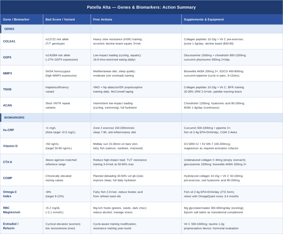
The table above consolidates the action framework for all five genes and seven biomarkers discussed in this article. For the genes section, the "bad score" column represents the risk allele or unfavorable variant; for biomarkers, it represents the out-of-range threshold warranting intervention.
The Book That Challenges Everything You Were Told About Knee Rehabilitation
"Knee Ability Zero" by Ben Patrick (the Knees Over Toes Guy) — published 2021 — is a protocol manual built around a principle that contradicts most conventional knee rehabilitation: that knees benefit from being systematically loaded through their full range of motion, including with the knee tracking well past the toes, under progressively increasing resistance. For patella alta patients who have been told to avoid deep knee flexion, limit quad loading, and rest their way to recovery, this framework is genuinely disorienting — and backed by a compelling evidence base that has attracted serious attention in sports science circles.
1. The Knees-Over-Toes Doctrine Is Safer Than Conventional Avoidance
The "knees should not go past the toes" rule — which dominated physical therapy for decades — has no strong biomechanical basis and actually increases compressive load on the knee by restricting natural forward lean. Ben Patrick's system teaches gradual, controlled progression of knee-over-toe movements that build tendon and cartilage tolerance rather than enforcing avoidance that produces deconditioning.
2. Tibialis Anterior Strength Is the Foundation of Knee Health
Patrick begins every program with tibialis anterior strengthening — the muscle on the front of the shin — via backward walking and toe raises. This muscle governs dorsiflexion and directly determines how far the knee can track forward over the foot safely. Weak tibialis anterior is the hidden upstream cause of impaired knee mechanics in a large proportion of people with anterior knee pain.
3. Full Range of Motion Strengthening Reverses Degeneration
When tendons and cartilage are loaded progressively through full range, they adapt and strengthen. Patrick documents numerous cases of structural cartilage improvement on imaging following his protocol — challenging the clinical assumption that cartilage degradation is a one-way process. This aligns with the mechanobiology literature on chondrocyte adaptation to controlled loading.
4. Sled Pushing Is the Safest Knee Exercise That Exists
Sled push — forward and backward — loads the quadriceps, VMO, and patellofemoral joint with minimal compressive force and near-zero eccentric braking stress. Patrick considers it the single most important exercise in his system for rebuilding knee tissue tolerance without exacerbating existing irritation. Even severely compromised knees can typically tolerate sled pushing from day one.
5. Nordic Hamstring Curls Protect the Entire Posterior Chain
Patrick places high importance on the Nordic hamstring curl for building eccentric hamstring strength — a critical stabilizer of the knee that counters anterior tibial translation. Weak hamstrings increase patellofemoral stress during dynamic activity. The Nordic curl specifically develops the long-length eccentric hamstring capacity that conventional leg curls completely miss.
6. ATG Squatting Done Progressively Is Rehabilitative
Ass-to-Grass (ATG) squatting — reaching full knee flexion — done incrementally with appropriate load progression, actually develops cartilage and tendon tolerance that restricted-range squatting cannot. Patrick's protocol uses bodyweight and elevated heel positions initially before progressively loading the full range. For patella alta, this is a long-term rather than early-phase tool and should not be rushed.
7. Step-Up Progressions Mirror Real-Life Knee Mechanics
Patrick's extensive step-up protocol — from low steps to progressively higher platforms, loading a single leg through the full hip-knee-ankle chain — builds the eccentric quad and VMO strength that has direct transfer to stair descent, walking, and daily-life activities. These patterns are among the most undertrained in conventional physiotherapy.
8. Patellar Tracking Improves Through Strength, Not Stretching Alone
The emphasis in patella alta management on stretching the IT band and hip flexors addresses symptoms at best; it does not change tracking mechanics. Patrick's system argues — and the biomechanics literature supports — that VMO strength and hip external rotator strength are the primary determinants of patellar trajectory under load. Building strength changes tracking; stretching provides temporary symptom relief.
9. Blood Flow Restriction Allows Loading Injured Knees That Cannot Handle Normal Resistance
BFR training at 20–30% of maximum with tourniquet cuff restriction produces hypertrophic muscle stimulus at loads that patellofemoral joints can tolerate during acute phases of irritation. Patrick integrates BFR throughout his programming for athletes returning from knee surgery, managing active chondromalacia, or dealing with flare periods. For patella alta patients, BFR quad work is among the most effective tools during periods when standard loading would exacerbate symptoms.
10. Reverse Nordics Rebuild the Patellar Tendon From the Inside Out
The reverse Nordic curl — a quad-dominant eccentric movement performed kneeling and slowly lowering the trunk back — builds the patellar tendon at its most vulnerable range. This exercise is absent from nearly all conventional knee programs and directly addresses the tendon quality deficit that contributes to abnormal patellar height and instability in patella alta. Patrick's protocol introduces it early and progresses it slowly, with documented improvements in patellar tendon structure over months.
Complementary Approaches With Clinical Evidence
Several evidence-supported modalities can complement biomarker-guided and genetics-informed interventions for patella alta. The three below have the most direct clinical relevance for this condition.
Biofeedback
Electromyographic (EMG) biofeedback is a technique in which surface electrodes placed over the VMO provide real-time visual or auditory feedback of muscle activation. For patella alta, the VMO is the single most critical active stabilizer of the patella — its selective activation relative to the vastus lateralis determines whether the kneecap tracks medially and inferiorly (correct) or drifts laterally and superiorly (pathological). Many patients with patellar instability have difficulty selectively activating the VMO without visual feedback; EMG biofeedback directly addresses this neuromuscular deficit.
A randomized controlled trial by Ng and Cheng (2002) published in Archives of Physical Medicine and Rehabilitation demonstrated that EMG biofeedback training produced significantly greater VMO-to-VL activation ratios and greater pain reduction compared to exercise alone in patellofemoral pain syndrome — a condition sharing considerable overlap with patella alta presentations. EMG biofeedback for VMO is included in clinical guidelines for patellofemoral pain management internationally.
Clinically, EMG biofeedback is applied in 15–20 minute sessions, 2–3 times per week, guided by a physiotherapist initially. Home biofeedback units are available for $150–$400 for patient-directed ongoing practice. The protocol involves progressing from isometric VMO activation to dynamic terminal knee extensions to partial squats, always with biofeedback confirmation of selective VMO activation. The benefit is cumulative over 6–12 weeks and then transitions to a maintenance exercise program without the device as the neuromuscular pattern is internalized.
Low-Level Laser Therapy / Photobiomodulation
Low-level laser therapy (LLLT), also called photobiomodulation (PBM), delivers specific wavelengths of red and near-infrared light to tissue at non-thermal intensities, stimulating cytochrome c oxidase in mitochondria and triggering anti-inflammatory and tissue-repair cascades. For patella alta, where patellar tendon and patellofemoral cartilage undergo chronic mechanical stress, LLLT's evidence for reducing tendon pain, decreasing local inflammatory mediators, and potentially supporting cartilage metabolism makes it a relevant adjunct.
A meta-analysis by Stausholm et al. (2019) published in BMJ Open Sport and Exercise Medicine — PMID 31673384 — reviewed evidence for photobiomodulation in knee conditions and found clinically significant pain reduction versus sham treatment with a low adverse event profile. Evidence is strongest for knee osteoarthritis and patellar tendinopathy — both mechanistically adjacent to the cartilage and tendon stress encountered in patella alta.
Practical application requires a class III or class IV laser device at a physiotherapy or sports medicine clinic, or a quality home device in the 100–500 mW range at 630–850 nm wavelength. Treatment is typically applied for 60–120 seconds per point over the patellar tendon and infrapatellar fat pad, 3 times per week for 6–8 weeks. Home devices at medical-grade power output cost $300–$1200; consumer devices below 50 mW are unlikely to deliver therapeutic tissue doses. Evidence is limited and mixed for patella alta specifically — results are more consistent for pain reduction than for structural change.
Massage Therapy
Therapeutic massage targeting the quadriceps, iliotibial band, and lateral retinaculum addresses the soft tissue tension that actively worsens patellar maltracking in patella alta. Lateral patellar tilt and excessive superior displacement are partly maintained by tight lateral retinacular and iliotibial structures that resist medial and inferior repositioning. Systematic soft tissue release of these structures reduces the passive lateral bias on the patella, directly complementing the active VMO strengthening approach.
A systematic review by van Middelkoop et al. published in British Journal of Sports Medicine identified soft tissue massage as a component of effective conservative multimodal management for patellofemoral pain, with evidence supporting its combination with exercise therapy over exercise alone (see PubMed: van Middelkoop patellofemoral systematic review). The evidence base for massage as a standalone treatment is more limited; its value is best established as part of a multimodal program.
Clinically, targeted deep tissue massage of the lateral retinaculum, IT band, and distal quadriceps by a sports physiotherapist or massage therapist, in sessions of 30–45 minutes, 1–2 times per week for 6–8 weeks, produces meaningful reductions in lateral patellar tension and pain during activity. Patients can supplement with self-massage using a foam roller on the IT band and lateral thigh daily (5–10 minutes). Important caveat: medial retinacular and MPFL structures should not be aggressively massaged in patients with documented medial instability, as release of these structures could worsen instability.
Conclusion
Patella alta is an anatomical reality, but the pain, instability, and cartilage vulnerability that may accompany it are not fixed outcomes. The biological environment in which your knee operates — its inflammatory load, nutritional status, cartilage repair capacity, and connective tissue quality — is measurable and, to a meaningful degree, modifiable. Seven biomarkers give you specific, actionable signals about that environment; five gene variants explain why some people's connective tissue and cartilage respond the way they do under comparable mechanical stress.
The most useful next step is not to try to implement everything at once. Start with the two most accessible and highest-yield interventions: getting an hs-CRP and a 25-OH vitamin D test, and addressing whatever is out of range. If patellar instability is cyclical or clearly linked to hormonal changes, add estradiol tracking. If conservative management has been inconsistent in its results, consider a COMP or CTX-II measurement to understand whether cartilage degradation is actively occurring. And if you have a history of hypermobility, easy sprains, or a family pattern of joint laxity, COL5A1 or TNXB genotyping through a consumer genomics platform is a low-cost way to clarify whether connective tissue biology is driving your presentation.
Discuss these findings with a sports medicine physician, physiotherapist, or integrative medicine practitioner who can interpret them in the context of your full clinical picture. Better data leads to better decisions — and for a condition as individually variable as patella alta, that specificity consistently makes the difference.
Musculoskeletal: Joint Conditions Tendon & Ligament Conditions Sports Injuries
Autoimmune: Inflammatory Conditions Connective Tissue Conditions