This article contains AI generated content.
Patellar Bone Contusion: 5 Genes and 7 Biomarkers to Track
Introduction
A bone bruise on the kneecap is a strange injury to live with. The X-ray comes back clean, the surgeon says there's nothing to operate on, and yet the knee still aches on stairs, still swells after a long walk, and still feels "not quite right" eight weeks later. Generic advice tends to stop at rest, ice, and time — true as far as it goes, but thin when you're the one waiting for the ache to disappear and wondering whether you're actually healing or just getting used to the pain.
The trouble with generic advice is that it treats every patella contusion as identical, when the underlying biology is not. Two people with the same MRI finding of bone marrow edema can heal at very different rates depending on their vitamin D status, their inflammatory baseline, how efficiently their body mineralizes new bone matrix, and even small differences in the genes that govern collagen and calcium handling. None of that shows up on a standard knee X-ray, and none of it gets addressed by "take it easy for six weeks."
This article takes a more specific approach. Instead of one-size-fits-all guidance, it walks through the blood markers that actually reflect how your bone is remodeling after a contusion, what the genetic research suggests about who is more prone to slow healing or recurring injury, and what a serious longevity-focused physician's playbook looks like when applied to joint resilience. Each section ends with a concrete plan — what you can do without spending money on tests or supplements, and what a more targeted approach with lab work and supplementation might look like.
None of this replaces a conversation with your physician or physical therapist, and none of it promises a faster recovery than your biology allows. But better information changes decisions — it tells you which questions to ask your doctor, which labs are worth requesting, and which daily habits are quietly working for or against your knee. That is a realistic kind of hope, and it's the one this article is built around.
Summary
A patellar bone contusion is not just "a bruise" — it's an active remodeling process happening inside the trabecular bone of your kneecap, and that process runs on measurable biology. Seven blood markers, from 25-hydroxyvitamin D to CTX-I, can tell you whether your body currently has the raw materials and the metabolic environment to mineralize new bone efficiently, or whether something is quietly working against you. Five genes — including VDR, COL1A1, and LRP5 — help explain why two people with an identical injury can heal at very different speeds, and what to do differently if you're carrying a less favorable variant. Beyond the biology, a longevity physician's framework for joint resilience and a short list of complementary approaches with real supporting evidence round out a plan that goes well past "rest and ice." The diagram below maps how these genes and biomarkers connect to the actual healing pathway inside the bone.
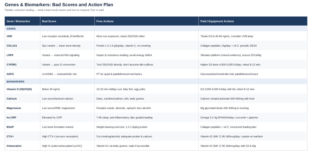
The 7 Biomarkers Worth Tracking After a Patellar Bone Contusion
A patellar contusion heals in overlapping phases: an initial inflammatory phase that clears damaged tissue, a proliferative phase where osteoblasts lay down new osteoid, and a mineralization/remodeling phase where that matrix hardens into mature bone. Each phase depends on specific nutrients, hormones, and a functioning inflammatory response — and each can be approximated with a blood test. Peter Attia and Thomas Dayspring have both argued publicly that standard reference ranges are built around population averages, not around what's optimal for someone actively trying to heal tissue — a theme that runs through every marker below.
1. 25-Hydroxyvitamin D
Vitamin D status is the single most-tested nutrient in bone biology, and for good reason. The active form of vitamin D drives intestinal calcium absorption and helps regulate the mineralization of the osteoid matrix that osteoblasts lay down at the contusion site. Low vitamin D has been associated with slower bone remodeling and delayed healing in stress-reaction and fracture research, which is directly relevant to a bone marrow edema injury like a patellar contusion. MedlinePlus's vitamin D overview is a useful starting point for understanding sources and deficiency risk.
How to measure it
A standard serum 25(OH)D blood draw, available through most primary care panels or direct-to-consumer labs, typically costs between $40 and $90 out of pocket, or $100+ through specialty functional-medicine labs that also report vitamin D–binding protein. Most conventional labs flag "sufficient" at 30 ng/mL; Attia and other longevity-focused physicians generally target 40-60 ng/mL for people actively repairing tissue.If the score is low: the plan without supplements
Aim for 15-20 minutes of midday sun exposure on bare arms and legs several times a week (adjusted for skin tone and latitude), and add vitamin D–rich foods such as fatty fish, egg yolks, and fortified dairy. Retest after 8-12 weeks, since sunlight-driven changes are slow and seasonal.If the score is low: the plan with supplements or equipment
Vitamin D3, typically 2,000-5,000 IU per day depending on how far below target you are, taken with a meal containing fat for absorption. No cycling is required — this is a daily, ongoing habit, not a short course. Retest at 8-12 weeks and adjust dose to land in range rather than guessing. In low-sunlight climates or winter months, a UVB lamp is a reasonable equipment-based alternative for people who prefer not to supplement, though it requires more consistent use than a capsule. The main side effect risk is hypercalcemia from over-supplementing without monitoring — levels above 100 ng/mL are not a "more is better" situation and should prompt a dose reduction.2. Serum or Ionized Calcium
Calcium is the literal building block of the hydroxyapatite crystal that gives bone its hardness. A contusion site actively laying down new mineral needs a steady calcium supply, and while the body regulates blood calcium tightly through parathyroid hormone, persistently low readings can flag malabsorption or an undiagnosed vitamin D problem before it becomes obvious elsewhere. MedlinePlus's calcium blood test page explains the difference between total and ionized calcium testing.
How to measure it
A total calcium test is part of almost every basic metabolic panel and costs $10-30; an ionized calcium test, which better reflects the biologically active fraction, runs closer to $30-50 and is more useful if total calcium is borderline.If the score is low: the plan without supplements
Increase dietary calcium through dairy, canned fish with edible bones (sardines, salmon), tofu set with calcium sulfate, and leafy greens like kale and bok choy. Correcting an underlying vitamin D deficiency often normalizes calcium on its own.If the score is low: the plan with supplements or equipment
Calcium citrate or calcium carbonate, dosed at 500-600 mg per single dose (absorption drops off above that), taken with food. Allan Sniderman and Peter Attia have both been cautious about high-dose calcium supplementation given unresolved questions around cardiovascular calcification risk, so the general guidance is to prioritize food sources first and use supplements only to close a documented gap, not as a default. Total intake from food and supplements combined should stay under roughly 2,000-2,500 mg/day.3. Magnesium
Roughly 60% of the body's magnesium is stored in bone, where it's involved in the structural crystal lattice and acts as a cofactor for the enzymes that activate vitamin D. A magnesium shortfall can therefore blunt the benefit of otherwise-adequate vitamin D and calcium status. MedlinePlus's magnesium blood test overview covers the basics of testing and interpretation.
How to measure it
Serum magnesium is inexpensive ($20-40) but only reflects about 1% of total body magnesium, so it can look normal even with a real tissue-level deficit. Red blood cell (RBC) magnesium, at $30-70, is a better proxy for overall status and worth requesting specifically if serum levels are borderline.If the score is low: the plan without supplements
Increase intake of pumpkin seeds, almonds, spinach, black beans, and whole grains. Reducing alcohol intake also helps, since alcohol increases urinary magnesium loss.If the score is low: the plan with supplements or equipment
Magnesium glycinate or magnesium citrate, 200-400 mg per day, taken in the evening (it also supports sleep quality, which matters for tissue repair). Avoid magnesium oxide, which is poorly absorbed and more likely to cause diarrhea. No cycling is needed for typical repletion doses; people with reduced kidney function should check with a physician before supplementing, since magnesium clearance depends on renal function.4. High-Sensitivity C-Reactive Protein (hs-CRP)
CRP is produced by the liver in response to inflammatory signaling, and while it's best known as a cardiovascular risk marker, it also gives a reasonable window into whether your body is still stuck in an active inflammatory phase longer than expected after a bone injury. A contusion that should be transitioning to the remodeling phase but shows persistently elevated CRP may be dealing with additional inflammatory load — from poor sleep, excess body fat, an unrelated infection, or simply doing too much too soon on the knee. MedlinePlus's CRP test page explains the distinction between standard CRP and hs-CRP.
How to measure it
An hs-CRP blood test costs $15-40 and is widely available through primary care or direct-to-consumer labs.If the score is high: the plan without supplements
Prioritize 7-9 hours of sleep, since sleep deprivation reliably raises inflammatory markers. Shift toward an anti-inflammatory dietary pattern emphasizing fatty fish, vegetables, and olive oil while cutting back on ultra-processed foods, and make sure knee loading during rehab is graded rather than aggressive — too much, too soon can keep local inflammation elevated.If the score is high: the plan with supplements or equipment
Omega-3 fish oil, 2-3 g combined EPA/DHA per day, and/or curcumin with piperine for absorption, taken daily with food. Recheck hs-CRP every 4-8 weeks rather than weekly, since it's a slow-moving marker. Side effects: fish oil can cause GI upset or mild blood-thinning at higher doses, so caution is warranted for anyone on anticoagulants or awaiting a procedure.5. Bone-Specific Alkaline Phosphatase (BSAP)
BSAP is produced by active osteoblasts and is one of the more direct markers of bone-formation activity. It's used more often in metabolic bone disease than in routine sports-medicine follow-up, but it's a reasonable way to confirm that the remodeling phase of a contusion is actually engaging, rather than assuming it based on time elapsed alone.
How to measure it
This is a specialty test, typically $50-100, ordered through a functional-medicine or endocrine lab rather than as part of a routine panel; it's not usually covered by insurance for an isolated bone bruise.If the score is low: the plan without supplements
Progressive, clinician-approved weight-bearing exercise is the strongest non-pharmacological driver of osteoblast activity — bone responds to mechanical load. Pair this with adequate dietary protein (roughly 1.2-1.6 g/kg of body weight per day) to supply the amino acids needed for new collagen matrix.If the score is low: the plan with supplements or equipment
Collagen peptides, around 15 g/day taken with a source of vitamin C (which is required for collagen crosslinking), alongside a structured loading progression — isometric quad sets progressing to leg press or step-up work as cleared by a physical therapist. No cycling needed; collagen peptides are well tolerated, with occasional mild GI upset as the main side effect.6. CTX-I (C-Terminal Telopeptide of Type I Collagen)
CTX-I is a breakdown product of type I collagen and reflects osteoclast-driven bone resorption. Comparing CTX-I to BSAP gives a rough sense of remodeling balance — if resorption markers are elevated well beyond formation markers, the bone may be breaking down faster than it's rebuilding, which is worth flagging to a physician.
How to measure it
CTX-I should be drawn in a fasted state in the morning, since it has a pronounced diurnal rhythm; the test costs $60-120 and is typically ordered through a specialty or endocrine lab.If the score is high: the plan without supplements
Reduce or eliminate smoking and excess alcohol, both of which accelerate bone resorption. Make sure protein and calcium intake are adequate, since resorption tends to increase when the body is scavenging minerals it isn't getting from the diet.If the score is high: the plan with supplements or equipment
Vitamin K2 (as MK-7), 90-180 mcg per day, helps direct calcium into bone matrix rather than soft tissue and has been studied for its role in bone turnover balance. Weight-bearing exercise remains central. Important caution: vitamin K2 interacts with warfarin and other vitamin K antagonists, so anyone on these medications should not supplement K2 without their prescribing physician's sign-off.7. Osteocalcin and Percent Undercarboxylated Osteocalcin
Osteocalcin is produced by osteoblasts and needs vitamin K–dependent carboxylation to properly bind calcium into the bone matrix. A high percentage of undercarboxylated osteocalcin (ucOC) signals that vitamin K status may be limiting how well newly formed bone actually mineralizes, even if calcium and vitamin D look adequate on their own.
How to measure it
This is a less standardized test, typically $80-150, available through specialty or functional-medicine labs rather than routine panels.If the score is high (poor carboxylation): the plan without supplements
Increase intake of vitamin K1 through leafy greens (kale, spinach, Swiss chard) and, where culturally accessible, fermented foods like natto, which is unusually rich in K2 (MK-7).If the score is high (poor carboxylation): the plan with supplements or equipment
Vitamin K2 (MK-7), 90-180 mcg daily, ideally alongside vitamin D and magnesium since the three work together in mineral metabolism. As above, this is contraindicated without medical supervision for anyone on warfarin. No cycling is required; this is a daily maintenance nutrient, not a short-term intervention.Taken together, these seven markers cover the three practical questions worth asking after a patellar contusion: do you have the raw materials to rebuild bone, is your inflammatory environment cooperating, and is the remodeling process actually balanced in favor of formation over breakdown. A reasonable starting panel is vitamin D, calcium, magnesium, and hs-CRP — all inexpensive and widely available — with BSAP, CTX-I, and osteocalcin added if healing seems slower than expected at the 6-8 week mark.
What Your Genes May Add to the Picture
Biomarkers tell you where things stand today; genetics can help explain why two people with the same injury and similar lab values still heal at different speeds. Ali Torkamani's research on genomic predictors of common conditions and Gary Brecka's work popularizing actionable genetic testing have both pushed a useful idea into the mainstream: a "bad" gene variant isn't a life sentence, it's a signal about where to compensate more deliberately. The evidence below ranges from well-replicated to early-stage, and it's labeled accordingly.
VDR — Vitamin D Receptor
The VDR gene encodes the receptor that lets cells actually respond to active vitamin D, regardless of how much is circulating in the blood. Certain common variants (often referred to by their restriction-enzyme names, such as FokI or BsmI) are associated in observational studies with differences in calcium absorption efficiency and bone mineral density, though the evidence across populations is moderate and not fully consistent — this is a "worth knowing," not a "definitely fated" gene. The NCBI Gene entry for VDR has the full technical summary.
If the gene is unfavorable: the plan without supplements
Be more diligent than average about sun exposure and dietary vitamin D sources, and consider testing serum 25(OH)D more frequently than someone without this variant would need to, since reduced receptor sensitivity means blood levels alone may understate what's needed.If the gene is unfavorable: the plan with supplements or equipment
The practical compensation is the same lever as in the biomarker section — titrate vitamin D3 dosing to reach the higher end of the optimal range (40-60 ng/mL) rather than settling for the low end, and retest every 8-12 weeks. A UVB lamp is a reasonable equipment option in low-sunlight seasons. Watch for hypercalcemia if pushing dosing upward without monitoring.COL1A1 — Collagen Type I Alpha 1
COL1A1 encodes a core component of type I collagen, the dominant structural protein in bone matrix. A well-studied promoter-region variant (the "Sp1" polymorphism) has been linked across multiple meta-analyses to lower bone mineral density and higher fracture risk, making this one of the more consistently replicated bone-genetics findings in humans. Details are available at the NCBI Gene entry for COL1A1.
If the gene is unfavorable: the plan without supplements
Make sure protein intake supports collagen synthesis (1.2-1.6 g/kg/day), include vitamin C–rich foods since vitamin C is a required cofactor for collagen crosslinking, avoid smoking (which directly impairs collagen crosslinking), and prioritize consistent weight-bearing activity once cleared for loading.If the gene is unfavorable: the plan with supplements or equipment
Collagen peptide supplementation (15 g/day) paired with vitamin C, combined with a progressive resistance-training program using basic equipment (resistance bands progressing to leg press or step-up loading). Periodic DEXA bone density scans are a reasonable way to track whether this variant is translating into measurably lower bone mass over time.LRP5 — LDL Receptor-Related Protein 5
LRP5 is a co-receptor in the Wnt signaling pathway, one of the primary pathways bone cells use to sense mechanical load and decide whether to build or resorb bone. This is genuinely strong human evidence — rare loss-of-function mutations cause osteoporosis-pseudoglioma syndrome, while gain-of-function mutations cause high bone mass phenotypes, both well documented in families carrying these variants. Common variants with smaller effects on general bone density are less dramatic but still relevant.
If the gene is unfavorable: the plan without supplements
Since this pathway is activated by mechanical loading, consistent impact and resistance exercise is the most direct lever — this is a gene where movement matters more than diet. Avoid prolonged energy deficits (under-eating relative to training load), which are known to suppress Wnt signaling and bone formation.If the gene is unfavorable: the plan with supplements or equipment
A vibration training platform is sometimes used as an adjunct to impact loading, though the equipment evidence here is mixed rather than definitive. Ensuring adequate vitamin D, calcium, and magnesium (see biomarker section above) supports the same downstream mineralization process this pathway feeds into.CYP2R1 — Vitamin D 25-Hydroxylase
CYP2R1 encodes the liver enzyme responsible for converting vitamin D into 25(OH)D, the form measured on standard blood tests. Genome-wide association studies have consistently linked variants in this gene to lower circulating vitamin D levels independent of sun exposure or dietary intake — this is one of the more robustly replicated findings in vitamin D genetics.
If the gene is unfavorable: the plan without supplements
Don't assume that "normal" sun exposure and diet will produce normal blood levels — test 25(OH)D rather than guessing, since the bottleneck here is enzymatic conversion, not intake.If the gene is unfavorable: the plan with supplements or equipment
This variant often requires a higher-than-typical D3 dose (commonly 4,000-5,000 IU/day) to reach the same blood level someone without the variant would hit on a standard dose. Retest every 8-12 weeks and adjust rather than assuming the standard dose is working. A UVB lamp can help but won't fully overcome a conversion bottleneck on its own.GDF5 — Growth Differentiation Factor 5
GDF5 plays a role in cartilage, joint, and ligament development. A specific promoter variant (rs143383) is one of the most consistently replicated genetic associations with osteoarthritis risk across populations, and joint tissue quality around the patella plausibly affects how the whole joint tolerates and recovers from a bone bruise — though direct evidence connecting GDF5 specifically to patellar contusion healing, rather than joint degeneration broadly, is still early and should be read as suggestive rather than established.
If the gene is unfavorable: the plan without supplements
Work with a physical therapist on quadriceps and patellofemoral mechanics to reduce abnormal loading on the joint during and after healing, since this variant is more about joint tissue quality than bone mineral content specifically.If the gene is unfavorable: the plan with supplements or equipment
Glucosamine and chondroitin are sometimes used here, though the supporting evidence is mixed and effect sizes are modest — treat this as a low-cost trial rather than a guaranteed fix. A patellofemoral brace during the rehab loading phase is a reasonable equipment-based option to reduce joint stress while tissue quality issues are addressed through movement work. Glucosamine's main side effect is mild GI upset, and it should be avoided or used cautiously by anyone with a shellfish allergy.Genetics here should be read as a modifier, not a verdict — even a fully unfavorable set of variants is compensated well by the same fundamentals of vitamin D sufficiency, adequate protein, progressive loading, and controlled inflammation covered in the biomarker section above. The two strategies are meant to work together, not separately.
The Longevity Book Reshaping How Serious Athletes Think About Joint Injuries
Peter Attia's Outlive: The Science and Art of Longevity isn't written about bone contusions specifically, but its central argument — that most of medicine is reactive when it should be proactive — maps directly onto how people typically handle a bone bruise: wait, rest, hope it resolves, and only investigate further if it doesn't. Attia's framework, built on decades of clinical practice and referencing a wide body of exercise-physiology and geriatric research, argues for the opposite approach, and several of its ideas are directly applicable to someone rehabbing a patellar injury.
1. Exercise Is the Most Potent Longevity Intervention Available
Attia argues that no drug or supplement comes close to matching the effect size of regular structured exercise on healthspan — and bone tissue specifically requires mechanical loading to remodel properly, which means appropriately dosed movement during rehab isn't just "not harmful," it's the primary driver of recovery.2. The Centenarian Decathlon
Attia recommends working backward from the physical tasks you want to still be able to do in your 80s and 90s — carrying groceries, getting off the floor, climbing stairs — and training toward those specific capacities now. Applied to a knee injury, this reframes rehab goals around real-world function (kneeling, stair descent, single-leg balance) rather than just "pain-free."3. Stability Is the Overlooked Foundation
Before strength or endurance, Attia emphasizes joint stability as a prerequisite — a joint that isn't stable will compensate in ways that create new problems. This is directly relevant to the patella, where quadriceps and hip stability largely determine how much stress the kneecap absorbs during daily movement.4. Zone 2 Training Supports the Metabolic Environment for Repair
Low-intensity aerobic work, sustained for extended durations, improves mitochondrial efficiency and metabolic flexibility — a supportive backdrop for tissue repair that doesn't load the injured joint the way higher-intensity work would.5. Muscle Mass Is a Buffer Against Joint Stress
Attia treats strength training as protective infrastructure — stronger quadriceps and hip muscles absorb force that would otherwise transfer directly to the patella, which is one reason rehab protocols emphasize progressive resistance work rather than passive rest alone.6. Rate of Force Development and Fall Prevention Matter More Than People Assume
Many patellar contusions originate from falls or sudden impacts. Attia's emphasis on power and reactive strength training as we age is framed around fall prevention — a lever that reduces the odds of a repeat injury to the same knee.7. Sleep Is a Non-Negotiable Repair Window
Attia repeatedly returns to sleep as an underrated recovery lever, citing its role in tissue repair and inflammatory regulation — directly relevant to the hs-CRP marker discussed earlier, since poor sleep is one of the more common reasons inflammation stays elevated longer than expected.8. Nutrition Should Serve Function
Rather than framing nutrition around weight, Attia frames it around whether it supports the muscle mass and metabolic health needed to perform physically — which lines up with the protein-sufficiency point made earlier for collagen and bone matrix synthesis.9. Think in Decades, Not Injury Cycles
Attia's "marginal decade" concept argues for addressing risk factors well before they become acute problems. For joints specifically, that means not waiting for a second contusion before addressing quadriceps weakness, vitamin D status, or gait mechanics that contributed to the first one.10. Medicine 3.0 Means Measuring What Matters
Attia's broader argument for what he calls "Medicine 3.0" is that proactive biomarker tracking — rather than waiting for a problem to become symptomatic — is how serious prevention actually works. That philosophy is exactly why the biomarker panel earlier in this article is worth taking seriously rather than treating as optional extra testing.Complementary Approaches Worth Considering
None of the following replace medical evaluation or structured physical therapy, and the evidence base for each is more modest than for the biomarker and loading fundamentals above. But each has some genuine human support for musculoskeletal recovery and pain management, which makes them reasonable additions rather than distractions.
Photobiomodulation / Low-Level Laser Therapy
Photobiomodulation uses low-level laser or LED light to stimulate cellular activity in injured tissue, and it has been studied specifically in the context of bone healing and fracture repair, making it one of the more directly relevant complementary options for a bone contusion rather than a general wellness add-on. The proposed mechanism involves stimulating mitochondrial activity in osteoblasts, which could theoretically support the remodeling phase discussed throughout this article.
Clinical and preclinical research on photobiomodulation for bone repair has shown promising results in some fracture and oral bone-healing studies, though the evidence in humans specifically for bone bruises (as opposed to fractures or dental bone grafts) is more limited, and protocols vary considerably in wavelength, dose, and duration across studies — a meaningful caveat given how much this affects outcomes.
In practice, this means treating photobiomodulation as a reasonable adjunct rather than a primary strategy: a session with a physical therapist or sports medicine clinic that already has the equipment and protocol experience is more sensible than purchasing a home device based on marketing claims, and it should be layered on top of — not instead of — the loading and nutrient fundamentals covered earlier.
Massage Therapy
Massage therapy is commonly used in orthopedic rehabilitation to manage compensatory muscle tension that develops around an injured joint — in this case, the quadriceps, IT band, and calf often tighten as gait changes to protect a sore kneecap, and that tension can itself become a secondary source of pain.
Human trials of massage therapy for musculoskeletal pain generally show modest but real short-term improvements in pain and perceived function, particularly when combined with active rehabilitation rather than used alone; it is best thought of as a comfort-and-mobility tool rather than something that accelerates bone remodeling directly.
The important caution for a patellar contusion specifically is timing and location: direct massage over the acute bruise itself should be avoided, especially in the first few weeks, and any work should focus on the surrounding musculature rather than the contusion site until a clinician confirms it's appropriate.
Mindfulness Meditation / MBSR
Mindfulness-based stress reduction (MBSR) is well studied for chronic and subacute musculoskeletal pain, and a slow-healing bone contusion — with its unpredictable flare-ups and uncertain timeline — is exactly the kind of frustrating, low-grade pain experience where pain-coping strategies tend to help most.
Randomized trials of MBSR for chronic musculoskeletal pain conditions have generally shown improvements in pain-related distress and coping, even when the underlying tissue pathology itself is unchanged — which is a meaningful distinction: MBSR is unlikely to speed bone remodeling, but it can measurably change how disruptive the recovery period feels day to day.
A realistic application is a short daily practice (10-15 minutes) using any reputable guided-meditation program during the weeks when pain is present but activity is still restricted — a low-cost, low-risk addition that pairs naturally with the more passive phases of rehab.
Guided Imagery / Motor Imagery
Motor imagery — mentally rehearsing movement without physically performing it — has been studied in orthopedic rehabilitation as a way to preserve neuromuscular patterns and reduce strength loss during periods of immobilization or restricted weight-bearing, which is directly relevant to the weeks when a patellar contusion limits normal knee loading.
Research in post-surgical and injury rehabilitation settings suggests motor imagery, when combined with standard physical therapy, can help preserve muscle activation patterns and may modestly support strength retention compared to rest alone, though it works best as a supplement to active rehab rather than a replacement for it.
Practically, this means spending a few minutes daily visualizing normal, pain-free knee movement — walking, stairs, squatting — during the period when actual loading is restricted, ideally guided by cues from a physical therapist about the movement patterns worth reinforcing.
Conclusion
A patellar bone contusion heals through a specific, trackable biological process — inflammation resolving, osteoblasts laying down matrix, minerals hardening that matrix into bone — and each stage leaves a fingerprint in blood work that generic "rest and ice" advice never looks at. Vitamin D, calcium, magnesium, and hs-CRP are a reasonable and affordable starting panel for anyone whose knee isn't progressing the way they expected; bone-specific alkaline phosphatase, CTX-I, and osteocalcin are worth adding if healing still seems slow past the six-to-eight-week mark. Genetics won't change what you do day to day as much as biomarkers will, but knowing you carry a variant in VDR, COL1A1, LRP5, CYP2R1, or GDF5 can explain why you need to be more deliberate than someone else about the same fundamentals.
None of this is a substitute for a proper clinical evaluation, and nothing here promises to compress a biological timeline that ultimately depends on your own physiology. What it does offer is a clearer set of questions to bring to your next appointment — which labs to ask for, which supplements are actually justified by a documented gap rather than a marketing claim, and which daily habits are quietly helping or hurting. The next reasonable step is a simple one: get the first four biomarkers checked, track your symptoms against them over the coming weeks, and talk through the results with your physician or physical therapist before deciding what, if anything, to change.
Musculoskeletal: Bone Conditions Joint Conditions