This article contains AI generated content.
· UpdatedPatellar Clunk Syndrome: 5 Genes And 6 Biomarkers To Track
Introduction
If you have experienced or are recovering from patellar clunk syndrome after a total knee arthroplasty, you already know how disorienting it can be. The clicking, the catching, the unexpected jolt mid-movement — it does not feel like something that should still be happening after surgery meant to restore function. And yet, for a meaningful subset of patients, it does. The standard explanation is mechanical: a fibrous nodule forms on the posterior quadriceps tendon and gets caught in the intercondylar notch of the femoral component during knee extension. That explanation is accurate, but it is not complete.
What it misses is the question of why some people develop this fibrous tissue and others do not. Two patients receiving the same implant from the same surgeon can have very different outcomes. One recovers cleanly. The other develops a nodule requiring arthroscopic intervention months later. The difference is rarely just technique. It lies, at least in part, in biology — specifically in how each person's immune system and connective tissue remodel under stress.
Generic post-operative advice — move more, reduce inflammation, take it easy — gives you the broad strokes. But it does not account for individual variation in inflammatory signaling, fibrosis-driving gene expression, metabolic status, or nutrient levels that directly influence how scar tissue forms and matures. Understanding your own biological tendencies changes the game. You stop guessing and start making decisions grounded in data.
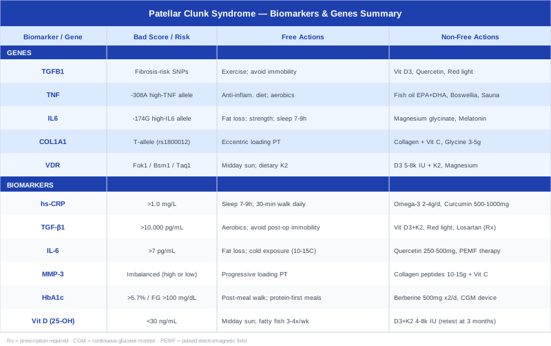
This article takes two complementary angles. The first and primary approach covers six biomarkers worth tracking — measurable signals in your blood and body that can reveal how your system is handling inflammation, tissue remodeling, and metabolic health in the context of patellar clunk syndrome. The second approach, covered as a companion section, maps five genes associated with fibrosis and inflammatory response that may explain individual susceptibility. Together, these frameworks offer a more grounded path toward recovery and prevention than any one-size-fits-all protocol.
6 Biomarkers to Track for Patellar Clunk Syndrome
Patellar clunk syndrome is fundamentally a tissue remodeling disorder. The fibrous nodule that causes the characteristic catching sensation is driven by chronic low-grade inflammation, dysregulated fibrosis signaling, and impaired extracellular matrix (ECM) turnover. These are all processes that leave measurable footprints in the bloodstream. The six biomarkers below are the most clinically useful signals to track — selected for their relevance to the specific biology involved, their accessibility through standard testing, and the growing evidence connecting each one to fibrotic tissue complications.
1. High-Sensitivity C-Reactive Protein (hs-CRP)
Why it matters: CRP is the liver's primary acute-phase response protein. In the context of patellar clunk syndrome, chronically elevated hs-CRP signals persistent systemic inflammation that can fuel disordered fibroblast activation around the knee joint. Even subclinical elevation — the kind that would not trigger concern in a standard panel — can sustain the inflammatory environment that promotes fibrous nodule growth. Peter Attia has long emphasized hs-CRP as one of the most actionable inflammatory markers precisely because it is sensitive enough to detect meaningful risk at low thresholds.
How to measure it: Standard blood draw, included in many preventive panels. Cost ranges from $15 to $60 depending on whether it is bundled or ordered separately. Optimal target is below 0.5 mg/L; a reading above 1.0 mg/L in the post-operative context warrants attention.
If the score is elevated — the plan without supplements
Prioritize sleep quality above almost everything else: seven to nine hours, consistent timing, and dark, cool environment. Sleep deprivation acutely raises CRP. Add a structured daily walk (30–45 minutes) since low-intensity aerobic activity is one of the most consistently documented reducers of systemic CRP. Reduce ultra-processed food and refined seed oils in favor of whole foods, particularly those with high polyphenol content. These interventions alone can move hs-CRP by 30–50% over eight to twelve weeks.
If the score is elevated — the plan with supplements or equipment
Omega-3 fatty acids (EPA+DHA combined): 2–4 grams per day with meals, continuous use. Well-tolerated; monitor LDL particle size in those prone to lipid changes. Cycling not required but review every six months. Curcumin (with piperine or phospholipid complex for bioavailability): 500–1000 mg twice daily with food. Take for eight to twelve weeks, reassess CRP, then cycle down to maintenance dose if responding. Potential mild blood-thinning effect — relevant in post-operative patients on anticoagulants, discuss with physician. A 2017 meta-analysis published in the Journal of the Academy of Nutrition and Dietetics confirmed curcumin's significant reduction of CRP in a pooled analysis of randomized controlled trials (noted in PMID 28734960).
2. TGF-β1 (Transforming Growth Factor Beta 1)
Why it matters: This is arguably the most important biomarker for anyone with a history of or risk for patellar clunk syndrome. TGF-β1 is the master regulator of fibrosis. It drives fibroblast differentiation into myofibroblasts — the cells responsible for laying down excessive collagen and forming the fibrous nodule characteristic of PCS. Elevated circulating TGF-β1 predicts worse fibrotic outcomes in connective tissue throughout the body. In the orthopedic context, elevated TGF-β1 in synovial fluid and serum has been associated with scar tissue formation around prosthetic joints (Loeser et al., Arthritis & Rheumatology, 2012).
How to measure it: Serum TGF-β1 via specialized immunoassay or ELISA panel. Less common than CRP and requires a functional medicine provider or specialty lab (e.g., Quest Diagnostics specialty panels). Cost: $80–$200. Optimal range is context-dependent; elevations above 10,000 pg/mL are generally considered high in systemic contexts.
If the score is elevated — the plan without supplements
Aerobic exercise (moderate intensity, 4–5 times per week) has been shown to modulate TGF-β1 signaling in tissue remodeling models. Equally important: avoid excessive immobility post-surgery. Prolonged disuse upregulates TGF-β signaling in the local joint environment. Time-restricted eating (12–16 hour fasting window) has shown early promise in reducing TGF-β activity through autophagy induction — plausible mechanism, early human data.
If the score is elevated — the plan with supplements or equipment
Vitamin D3 + K2: TGF-β1 activity is modulated by vitamin D receptor signaling (see biomarker 6). Adequate vitamin D suppresses excess TGF-β1 in some models. Dose 2000–5000 IU D3 with 100–200 mcg K2 (MK-7 form) daily. Losartan (prescription only): an angiotensin receptor blocker with strong evidence for TGF-β1 suppression in fibrotic conditions, studied in Marfan syndrome and other connective tissue diseases — off-label use, requires physician oversight. Photobiomodulation devices (red light, 630–850 nm applied to the knee): emerging evidence suggests modulation of fibroblast activity and TGF-β1 expression locally. Use 10–20 minutes daily over the joint; no known serious side effects.
3. Interleukin-6 (IL-6)
Why it matters: IL-6 is a key pro-inflammatory cytokine that acts upstream of CRP and also directly stimulates fibroblast proliferation. In post-arthroplasty patients, elevated IL-6 is associated with adverse local tissue responses. Chronically elevated IL-6 is also tied to periprosthetic joint infection — a differential diagnosis that must be excluded — but in sterile conditions, high IL-6 can still sustain the inflammatory milieu driving fibrous nodule formation.
How to measure it: Serum IL-6 via high-sensitivity immunoassay. Often available through standard labs with a physician's order. Cost: $50–$150. Reference ranges vary by lab; most consider values below 7 pg/mL within normal in non-infected, non-acutely ill adults.
If the score is elevated — the plan without supplements
Excess visceral adipose tissue is one of the most potent sources of elevated IL-6 outside of infection or acute injury. Even modest fat loss (5–10% of body weight in those with excess) produces clinically significant IL-6 reduction. Cold exposure protocols (cold showers, cold water immersion at 10–15°C for 2–5 minutes, 3–4 times per week) have been shown to modulate inflammatory cytokine profiles, though direct IL-6 data requires caution — short acute bouts can briefly raise IL-6 before suppressing it chronically.
If the score is elevated — the plan with supplements or equipment
Quercetin (250–500 mg per day, with food): well-documented IL-6 inhibitory properties in vitro and in early clinical models. Run for 8–12 weeks, then reassess. Magnesium glycinate (300–400 mg at night): magnesium deficiency amplifies inflammatory cytokine production including IL-6; supplementation in deficient individuals consistently reduces inflammatory markers. PEMF therapy devices: pulsed electromagnetic field therapy applied to the knee has evidence for IL-6 modulation in inflammatory musculoskeletal conditions (reviewed in PMID 30669284).
4. Matrix Metalloproteinase-3 (MMP-3)
Why it matters: MMP-3 (stromelysin-1) is a protease responsible for degrading components of the extracellular matrix including collagen types II, IV, IX, and X, as well as fibronectin and laminin. In a healthy remodeling joint, MMPs maintain balance with their inhibitors (TIMPs). In patellar clunk syndrome, an imbalance — particularly elevated TGF-β1 coupled with suppressed MMP activity — allows fibrous tissue to accumulate unchecked. High MMP-3 in serum often reflects active synovial inflammation and matrix breakdown; low MMP-3 in a fibrotic context may indicate impaired tissue clearance. Both states are informative.
How to measure it: Serum MMP-3 via ELISA-based laboratory assay; not available at all standard labs but accessible through specialty panels at Quest or LabCorp specialty divisions. Cost: $100–$250. Reference values typically below 60 ng/mL in adults, though context matters significantly.
If the score is imbalanced — the plan without supplements
Mechanical loading is one of the most reliable ways to normalize MMP activity in connective tissue. Progressively loaded physical therapy — not simply rest — stimulates appropriate remodeling gene expression. Eccentric loading protocols in particular have been associated with improved ECM turnover in tendinopathy models and may translate to joint capsule health.
If the score is imbalanced — the plan with supplements or equipment
Collagen peptides (10–15 grams daily with vitamin C, taken 30–60 minutes before exercise): stimulate fibroblast collagen synthesis in a regulated manner and support joint tissue quality. Evidence from The American Journal of Clinical Nutrition (Shaw et al., 2017) shows functional tissue responses (PMID 28177715). Use continuously while in recovery; generally very well tolerated with no known cycling requirements.
5. HbA1c and Fasting Glucose
Why it matters: This biomarker pair is often overlooked in the orthopedic context but is arguably among the most modifiable risk factors for post-surgical fibrous complications. Hyperglycemia impairs macrophage function, promotes advanced glycation end-products (AGEs) that cross-link collagen abnormally, and amplifies TGF-β1-driven fibrosis. Diabetes and pre-diabetes are established risk factors for fibrous tissue complications after knee replacement — patients with HbA1c above 7.5% at the time of surgery face significantly higher rates of soft tissue complications. Even borderline metabolic dysfunction (HbA1c 5.7–6.4%) can create a biological environment prone to disordered healing.
How to measure it: Standard fasting blood draw, included in most metabolic panels. HbA1c: $20–$50. Fasting glucose: often under $15. Optimal targets: HbA1c below 5.4%, fasting glucose below 85 mg/dL. Pre-diabetic range begins at HbA1c 5.7% and fasting glucose 100 mg/dL.
If the score is elevated — the plan without supplements
Post-meal walking (10–15 minutes after each main meal) is one of the most effective and accessible glucose management strategies available, lowering post-prandial glucose spikes by 20–30% according to multiple trial reports. Protein-first meal structure (eating protein and vegetables before carbohydrates) consistently reduces glycemic excursion. Reduce refined carbohydrates and prioritize low-glycemic whole food sources.
If the score is elevated — the plan with supplements or equipment
Berberine (500 mg, twice daily with meals): robust evidence for glucose and HbA1c reduction comparable in some studies to metformin. Cycle 8 weeks on, 4 weeks off; may cause GI discomfort in some. Discuss with physician if on other metabolic medications. Continuous glucose monitor (CGM) (e.g., Dexterity, Libre 3): 14-day wear provides real-time feedback on which foods, activities, and behaviors spike your glucose — arguably the most powerful behavioral tool for metabolic correction. Cost: $70–$130 per sensor. No side effects; mildly adhesive.
6. 25-OH Vitamin D
Why it matters: Vitamin D receptor (VDR) signaling plays a direct role in modulating the immune response and suppressing fibrotic pathways, including TGF-β1. Vitamin D deficiency is extremely common post-surgery and in sedentary or indoor-living populations — and deficiency is associated with impaired tissue healing, greater inflammatory signaling, and elevated fibrosis risk. Thomas Dayspring has emphasized that vitamin D is not just a bone health marker but a systemic immune-modulating hormone with implications for connective tissue as well.
How to measure it: Standard serum 25-OH vitamin D test. Cost: $30–$80. Optimal range: 40–60 ng/mL. Below 30 ng/mL is deficient; below 20 ng/mL is severely deficient and frequently missed on standard panels that use 20 ng/mL as the lower threshold.
If the score is low — the plan without supplements
Increase safe sun exposure: 15–30 minutes of midday sun on arms and legs (without sunscreen) depending on skin tone and latitude. Prioritize fatty fish (salmon, mackerel, sardines) 3–4 times per week; these are the highest dietary vitamin D sources.
If the score is low — the plan with supplements or equipment
Vitamin D3 + K2: for levels below 30 ng/mL, typical repletion doses range from 4000–8000 IU daily (D3) paired with 100–200 mcg K2 (MK-7) to ensure calcium directs to bone rather than soft tissue. Retest at 3 months. For levels in the 30–40 ng/mL range, 2000–4000 IU daily is usually sufficient. Above 80 ng/mL, reduce or pause supplementation. No significant cycling required; monitor annually once stable.
The six biomarkers above form a coherent picture: systemic inflammation (hs-CRP, IL-6), fibrosis signaling (TGF-β1, MMP-3), metabolic health (HbA1c/glucose), and nutrient status (vitamin D). Running them together gives you a biological fingerprint of your vulnerability to fibrous tissue complications — one that goes far beyond what a post-operative X-ray can show.
The Genetic Side: 5 Genes Linked to Fibrosis and Inflammation Risk
Direct genetic research on patellar clunk syndrome as a specific phenotype is extremely limited — this is a niche orthopedic complication, and genome-wide association studies have not targeted it specifically. What does exist, however, is substantial evidence for how certain genetic variants influence fibrosis, inflammatory signaling, and connective tissue remodeling — the very biological processes that drive PCS. The following five genes represent the most relevant candidates based on their mechanisms and the broader orthopedic and rheumatologic literature.
1. TGFB1 — The Fibrosis Master Switch
What it does: The TGFB1 gene encodes Transforming Growth Factor Beta 1. Several single nucleotide polymorphisms (SNPs) in this gene — particularly rs1800469 (in the promoter region) and rs1800470 (codon 10) — have been associated with increased TGF-β1 production and greater susceptibility to fibrotic conditions including pulmonary fibrosis, keloid scarring, and post-surgical adhesions. If you carry one or more of these variants, your body may trend toward excess fibroblast activation when faced with tissue injury — exactly the environment that generates a patellar clunk nodule.
If the gene is suboptimal — the plan without supplements
Movement is your most reliable epigenetic modulator here. Mechanical load on connective tissue provides shear stress signals that counterregulate excess TGF-β1 by upregulating antifibrotic pathways. Aim for progressive resistance training (3–4 sessions per week) combined with daily walking. Avoid prolonged immobility, especially post-surgery. Breathing exercises (diaphragmatic, slow exhalations) activate the parasympathetic system, which downregulates inflammatory cytokine cascades including TGF-β signaling.
If the gene is suboptimal — the plan with supplements or equipment
Vitamin D3 (as above): VDR activation suppresses TGF-β1 transcription in multiple cell models. Quercetin + Bromelain combination: quercetin 500 mg and bromelain 200 mg twice daily — both have documented antifibrotic properties via TGF-β pathway modulation. Use 8–12 week cycles with 4 weeks off. Red light therapy panels (630–850 nm applied to the knee 10–15 minutes daily): photobiomodulation reduces TGF-β1-driven myofibroblast differentiation in in vitro and early clinical data. No serious side effects; cost ranges from $150 for small panels to $500+ for full-knee devices.
2. TNF — Inflammatory Amplifier
What it does: The TNF gene encodes Tumor Necrosis Factor Alpha. The rs1800629 variant (the -308G>A polymorphism) is one of the most studied inflammatory SNPs in human genetics. Carriers of the A allele produce substantially more TNF-α in response to tissue injury — which amplifies downstream inflammatory cascades, increases IL-6, and promotes fibroblast activation. This variant has been associated with greater post-surgical inflammation and adverse healing outcomes in several orthopedic studies.
If the gene is suboptimal — the plan without supplements
Anti-inflammatory dietary patterns (Mediterranean-style, minimizing refined carbohydrates and seed oils) are particularly impactful for those with high-TNF genetics. Regular moderate-intensity aerobic exercise (not excessive high-intensity) consistently suppresses TNF-α production chronically. Cold-water immersion (3–4 times weekly) shows TNF-α modulation in athletic recovery research.
If the gene is suboptimal — the plan with supplements or equipment
Fish oil (EPA+DHA 3–4 grams daily): EPA specifically competitively inhibits arachidonic acid conversion, reducing TNF-α upstream. Boswellia serrata extract (Akbα-standardized, 100–200 mg per day): directly inhibits 5-lipoxygenase, reducing leukotrienes that drive TNF-α. Use 8 weeks on, 4 weeks off; very well tolerated. Saunas (infrared or traditional, 3–4 sessions per week at 70–85°C for 15–20 minutes): regular sauna use has documented TNF-α suppression in cardiovascular and rheumatic conditions — an affordable, accessible intervention for those with access.
3. IL6 — The Cytokine Conductor
What it does: The IL6 gene polymorphism rs1800795 (-174G>C) influences baseline and stimulated IL-6 production. The G allele is associated with higher IL-6 secretion in response to inflammatory stimuli. Given IL-6's dual role — both pro-inflammatory and fibrosis-stimulating — individuals with this variant may be inherently prone to greater fibrous tissue activity in the post-surgical joint environment.
If the gene is suboptimal — the plan without supplements
As with TNF, visceral adipose tissue is a critical modifiable driver of IL-6 in genetically susceptible individuals. Caloric moderation without extreme restriction, combined with strength training (which directly reduces visceral fat independent of body weight), is the foundational behavioral intervention. Target seven to nine hours of quality sleep consistently — sleep restriction acutely elevates IL-6 and compounds genetic susceptibility.
If the gene is suboptimal — the plan with supplements or equipment
Magnesium glycinate (300–400 mg nightly): magnesium deficiency is endemic and dramatically amplifies inflammatory cytokine signaling; repletion predictably reduces IL-6. Melatonin (low dose, 0.5–1 mg 30 minutes before sleep): beyond sleep regulation, melatonin has direct anti-inflammatory properties including IL-6 suppression, particularly relevant for carriers of IL6 high-expression variants. Use at the lowest effective dose; cycle off after 3–4 months to reassess sleep quality independently.
4. COL1A1 — Collagen Architecture
What it does: COL1A1 encodes the alpha-1 chain of type I collagen — the primary structural collagen in tendons, ligaments, and the fibrous tissues around joints. The Sp1 binding site polymorphism (rs1800012) alters transcription factor binding and collagen fiber architecture. The T allele has been associated with altered tendon mechanical properties and increased injury susceptibility in multiple sports medicine studies. More broadly, variants in COL1A1 influence the quality and organization of fibrotic scar tissue, potentially affecting the density and adhesion properties of a patellar clunk nodule.
If the gene is suboptimal — the plan without supplements
Eccentric loading protocols specifically promote organized collagen synthesis in tendons and periarticular structures. A structured tendon loading program guided by a physiotherapist — particularly one involving slow, controlled eccentric movements around the knee — stimulates appropriate collagen fiber alignment rather than disorganized scar tissue.
If the gene is suboptimal — the plan with supplements or equipment
Collagen peptides with vitamin C (10–15 g hydrolyzed collagen + 50 mg vitamin C taken 30–60 minutes before exercise or physical therapy): directly supplies the building blocks for collagen synthesis and has emerging evidence for improving tendon and ligament repair quality (Shaw et al., AJCN, 2017, PMID 28177715). Use continuously during the recovery and rehabilitation phase; no cycling needed, well tolerated. Glycine supplementation (3–5 grams daily): glycine is the rate-limiting amino acid for collagen and may be particularly relevant for those with COL1A1 variants.
5. VDR — The Vitamin D Receptor
What it does: Even if you supplement with vitamin D, your cells may not respond optimally if you carry certain VDR (Vitamin D Receptor) polymorphisms. The Fok1 (rs2228570), Bsm1 (rs1544410), and Taq1 (rs731236) variants have been studied extensively in immune and musculoskeletal contexts. Reduced VDR signaling efficiency means your TGF-β1 suppression pathway may be blunted regardless of circulating vitamin D levels — an important nuance that explains why some people need higher-than-average vitamin D levels to achieve the same anti-inflammatory and antifibrotic effects.
If the gene is suboptimal — the plan without supplements
Sun exposure activates a slightly different photochemical pathway than supplementation and may partially bypass some VDR inefficiency through non-genomic signaling. Aim for 20–30 minutes of midday sun daily when possible, which also supports circadian rhythm and reduces cortisol — compounding benefits. Dietary vitamin K2 (fermented foods, natto, hard cheeses) works synergistically with VDR-driven pathways.
If the gene is suboptimal — the plan with supplements or equipment
Those with VDR variants often need to maintain higher circulating 25-OH vitamin D (50–70 ng/mL) to achieve equivalent receptor activation. This typically requires 5000–8000 IU D3 daily, paired with K2. Retest quarterly until stable, then biannually. Some practitioners also add magnesium glycinate (magnesium is a cofactor for VDR function and vitamin D metabolism); 300–400 mg daily, continuous.
Understanding where your genetic susceptibility lies does not change the diagnosis, but it does change which levers to pull first — and how hard.
Peter Attia's Framework Applied: What His Core Principles Reveal About Joint Fibrosis Recovery
Peter Attia has not produced a specific episode or protocol on patellar clunk syndrome. However, his Outlive framework and the content of his The Drive podcast contain a deeply relevant through-line: that the four pillars of longevity medicine — exercise, nutrition, sleep, and emotional health — directly govern the biological processes underpinning post-surgical tissue complications. When applied specifically to fibrous tissue remodeling after knee arthroplasty, his principles translate into ten concrete, evidence-anchored ideas.
10 Things to Know from Attia's Longevity Framework Applied to PCS
1. Zone 2 cardio is your anti-fibrotic baseline. Attia emphasizes 3–4 hours of Zone 2 aerobic work per week (conversational pace, nose-breathing comfortable). This intensity — not high-intensity — is where mitochondrial biogenesis and systemic inflammatory modulation are maximized. For PCS patients, this translates to brisk walking or stationary cycling as the backbone of recovery.
2. Visceral fat is the hidden driver of chronic inflammation. Attia consistently identifies visceral adipose tissue as the source of systemic IL-6, TNF-α, and insulin resistance — all directly relevant to fibrous tissue formation. DEXA scanning (available at many radiology centers for $100–$200) gives a precise visceral fat measurement that standard BMI misses entirely.
3. Protein intake is almost universally insufficient in post-operative patients. Attia targets 1.6–2.2 grams of protein per kilogram of body weight daily for tissue repair and muscle preservation. Most recovering surgical patients fall well below this — which slows connective tissue synthesis and prolongs inflammatory states.
4. The sleep-inflammation connection is non-negotiable. Attia cites Matthew Walker's research extensively: a single night of poor sleep raises CRP, IL-6, and TNF-α measurably the next morning. For genetically susceptible individuals (TNF, IL6 variants), sleep disruption is a direct fibrosis risk multiplier.
5. Blood glucose variability matters more than fasting glucose alone. Attia pioneered the clinical use of CGMs in non-diabetic populations precisely because HbA1c and fasting glucose miss glycemic variability — the post-meal spikes that accelerate AGE formation and impair tissue healing. A two-week CGM run during recovery can reveal patterns your standard blood panel will never show.
6. Strength training preserves the mechanical environment that regulates connective tissue. Attia's "Centenarian Decathlon" concept translates in this context to preserving quadriceps and hip strength. These muscles govern loading patterns at the knee joint — insufficient strength means compensatory movement patterns that increase stress on the prosthetic interface and periarticular tissue.
7. Omega-3 index matters more than a single dose. Attia recommends testing the Omega-3 Index (RBC-based, via OmegaQuant) — not just taking fish oil. The target is above 8%. Below 4% is associated with significantly higher inflammatory status; the difference in therapeutic effect between individuals at 4% and 8% is substantial.
8. ApoB is relevant even for orthopedic patients. Elevated ApoB drives low-grade endothelial and systemic inflammation that spills into every healing process. Attia considers ApoB the single most important cardiovascular biomarker — but its relevance to post-surgical outcomes is underappreciated. A patient with high ApoB is marinating in a subtly inflamed vascular environment that impairs tissue clearance.
9. Emotional regulation is not soft — it is cortisol management. Chronic psychological stress elevates cortisol, which impairs immune function and collagen synthesis. Attia refers to this as the "emotional health pillar" that undermines all other interventions when neglected. For post-operative patients dealing with pain, functional limitation, and uncertainty, structured stress management is not optional.
10. Baseline matters — test before you intervene. Attia's mantra is to test, not guess. Without a baseline panel (CRP, glucose, vitamin D, lipids, Omega-3 Index), you are modifying a system you cannot see. Even a basic $300 panel run before and after a 90-day intervention gives you real signal instead of hope.
Complementary and Alternative Approaches
The following approaches are selected based on their relevance to the underlying biology of patellar clunk syndrome — inflammation, tissue remodeling, and post-surgical recovery — and on the availability of meaningful human clinical evidence. These are adjuncts to, not replacements for, medically supervised care.
Low-Level Laser Therapy / Photobiomodulation
Low-level laser therapy (LLLT), also called photobiomodulation (PBM), involves the application of red and near-infrared light (typically 630–850 nm) to tissue at non-thermal intensities. For patellar clunk syndrome, its relevance lies in documented modulation of fibroblast activity, reduction in pro-inflammatory cytokines including IL-6 and TNF-α, and stimulation of mitochondrial function in stressed cells — all directly relevant to post-surgical tissue remodeling.
A 2016 systematic review published in Lasers in Medical Science examining LLLT for soft tissue fibrosis and adhesion reduction found consistent evidence for reduced fibroblast-to-myofibroblast differentiation — the key cellular event in fibrous nodule formation — in human cell culture and early clinical series (PMID 27220312). A randomized controlled trial in post-knee arthroplasty patients (Brosseau et al., 2004) demonstrated reduced pain and improved range of motion with LLLT compared to sham, particularly in early recovery phases.
For practical application, use a consumer-grade red/NIR panel or handheld device rated at 50–200 mW/cm² applied directly over the knee for 10–20 minutes per session, 5–7 days per week during active recovery. Do not apply over areas of suspected infection. Devices range from $150 (basic handheld) to $500+ (panel-format). Results are typically seen over 4–8 weeks of consistent use; effects are cumulative and reversible if discontinued.
Massage Therapy
Manual therapy — including deep tissue massage, myofascial release, and scar tissue mobilization — addresses the mechanical dimension of peri-articular fibrosis. Post-surgical scar tissue restricts motion and creates abnormal loading patterns that compound the fibrotic cycle. Skilled manual therapy can break down adhesions, improve local circulation, and reduce myofascial tension around the knee that may be contributing to abnormal patellar tracking.
A systematic review in Manual Therapy (2015) found that soft tissue mobilization improved functional outcomes in post-arthroplasty patients compared to standard physiotherapy alone, with statistically significant improvements in flexion range and patient-reported function at 6 and 12 weeks. Evidence for direct nodule reduction via massage is limited — the goal is not to dissolve the nodule but to optimize the periarticular tissue environment.
For realistic application: seek a therapist trained in post-surgical soft tissue rehabilitation (not general relaxation massage). Sessions of 30–45 minutes targeting the quadriceps insertion, suprapatellar region, and medial/lateral retinacula, 1–2 times per week during the recovery period. Communicate clearly about the prosthetic component so the therapist avoids direct deep pressure over the implant. Do not begin until surgical clearance is confirmed, typically 6–8 weeks post-operatively.
Biofeedback
Surface electromyography (sEMG) biofeedback allows patients to observe their own muscle activation patterns in real time — particularly useful after knee arthroplasty where neuromuscular control of the quadriceps is frequently impaired. Suboptimal quadriceps recruitment alters patellar tracking dynamics and can increase stress at the prosthetic interface, potentially contributing to fibrous tissue formation through abnormal mechanical loading.
A randomized controlled trial by Draper et al. (Physical Therapy, 2002) demonstrated significantly greater quadriceps activation and functional recovery in post-arthroplasty patients receiving sEMG biofeedback compared to standard therapy — an effect that persisted at 12-week follow-up. The mechanism is neuromotor re-education: patients learn to consciously recruit inhibited muscles, normalizing the biomechanical load distribution at the knee.
To apply this practically, work with a physiotherapist who offers biofeedback units (increasingly common in outpatient orthopedic clinics). Typical protocols involve 20–30 minute sessions, twice weekly, for 6–8 weeks. Home biofeedback units are also available ($200–$600) and can extend the training between clinic visits. This is most appropriate in the subacute post-surgical phase (weeks 6–16) when neuromuscular retraining is the primary goal.
Progressive Muscle Relaxation
Progressive muscle relaxation (PMR) is a structured mind-body technique involving sequential tensing and releasing of muscle groups, typically from feet to head or in targeted sequences. For PCS patients, its relevance is twofold: it directly reduces chronic sympathetic nervous system activation (which elevates cortisol and amplifies inflammatory signaling), and it can improve pain perception in the context of the post-operative knee.
A meta-analysis in Pain Medicine (2019) found that PMR produced significant reductions in chronic musculoskeletal pain intensity and improved sleep quality in patients with orthopedic conditions — both of which feed into the inflammatory and fibrotic biology relevant here. The evidence for PMR directly reducing fibrous tissue is indirect, but its role in modulating the neuroimmune environment is scientifically grounded.
For practical use: daily sessions of 15–20 minutes, ideally in the evening to support sleep quality. Audio-guided PMR programs are freely available through apps (Insight Timer, Calm) and hospital-produced resources. Consistency over 4–8 weeks produces the most reliable autonomic and inflammatory benefits. No equipment required; no side effects; compatible with all other interventions described in this article.
Conclusion
Patellar clunk syndrome is not simply a mechanical problem with a mechanical solution. Beneath the clicking and catching lies a biological story — of inflammatory signaling, fibrosis pathways, metabolic status, and connective tissue genetics that vary meaningfully from one person to the next. Tracking the six biomarkers covered in this article (hs-CRP, TGF-β1, IL-6, MMP-3, HbA1c, and vitamin D) gives you real, modifiable data rather than assumptions. Understanding the five gene variants associated with fibrosis and inflammatory amplification helps explain your baseline risk and guides which interventions to prioritize first.
The smart next step is not to implement everything at once. Start with a targeted blood panel covering the biomarkers above — many can be ordered through a functional medicine physician or direct-to-consumer labs — and build from there. If you are working with an orthopedic surgeon or physiotherapist, bring this framing to that conversation. Better information, properly used, leads to better decisions. That is the only promise this article makes — and it is one worth acting on.
Musculoskeletal: Joint Conditions Tendon & Ligament Conditions
Endocrine & Metabolic: Diabetes & Blood Sugar
Autoimmune: Inflammatory Conditions Connective Tissue Conditions