This article contains AI generated content.
Patellar Enthesopathy: 6 Genes And 6 Biomarkers To Track
When the Same Knee Pain Keeps Returning
Patellar enthesopathy, better known to most people as jumper's knee, tends to follow a frustrating pattern. You rest, the pain fades, you return to running or jumping, and within a few weeks the ache at the bottom of the kneecap is back. Standard advice, more rest, more stretching, a knee strap, an anti-inflammatory, often helps for a while without ever explaining why the tendon-bone junction keeps breaking down in the first place.
That gap exists because the standard advice is written for the average knee, not yours. Two people with identical training loads and identical physiotherapy programs can have very different outcomes, and a meaningful part of that difference traces back to how their collagen is built, how quickly they remodel damaged tissue, and what is circulating in their blood at the time of injury. Generic rehab protocols are not wrong, they are simply incomplete for anyone whose underlying biology is working against the plan.
This article looks at patellar enthesopathy through two more precise lenses: the genes that shape collagen structure and tendon repair, and the bloodwork markers that reveal whether your current metabolic environment is helping or hurting your enthesis. Neither is a substitute for proper load management and rehabilitation, but both can explain why a program that works for someone else is not working for you, and both point toward specific, testable adjustments.
None of this promises a cure, and no single gene or blood value determines your outcome. But understanding your own risk factors, rather than following advice built for an average that may not resemble you, is what turns a frustrating cycle of flare-ups into a plan you can actually trust.
Summary
Some of the reasons patellar enthesopathy keeps recurring are written into your DNA before you ever pick up a barbell or lace up running shoes. Variants in collagen and matrix-remodeling genes such as COL5A1, COL27A1, TNC, MMP3, GDF5, and ESRRB have been linked in human studies to how tendons and their bony attachment points are built and repaired, and each has a practical, non-mystical way to work around it. Alongside genetics, six bloodwork markers, from vitamin D to uric acid, quietly influence how well your tendons tolerate load and heal after irritation, and most of them are fixable with unglamorous, well-supported changes. Beyond genes and bloodwork, this article also breaks down what a leading tendon physiologist told Andrew Huberman about the narrow biological window for building stronger tendons, and which complementary therapies, laser, massage, and biofeedback, actually have trial evidence behind them for this specific condition. Read on to find out which of your genes and biomarkers might be working against your knee, and exactly what to do about each one.
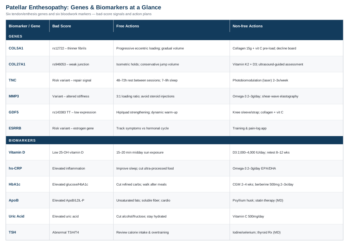
The Genetic Blueprint of Tendon and Enthesis Strength
The patellar enthesis, the specific zone where the patellar tendon fuses into bone at the inferior pole of the kneecap or the tibial tuberosity, is a specialized tissue transition built almost entirely from collagen, proteoglycans, and the enzymes that continuously remodel them. Over the last two decades, researchers led by groups such as Malcolm Collins and Alison September at the University of Cape Town have run candidate-gene studies comparing people with chronic tendinopathy to injury-free controls, and a consistent, if still evolving, picture has emerged. A systematic review of the genetics of tendon injury concluded that the strongest human evidence points to variants in type V collagen, tenascin-C, matrix metalloproteinase-3, and estrogen-related receptor beta, with several other genes showing early but less consistent associations.
It is worth being direct about what this evidence can and cannot tell you. These are common variants found in large portions of the population, not rare mutations, and they shift relative risk rather than guarantee an outcome. Genetic testing for these markers is not yet standard clinical practice, and none of the plans below require a genetic test to be useful, since the underlying nutritional and mechanical strategies benefit most tendons regardless of genotype. Researchers like Ali Torkamani, who has written extensively on interpreting personal genomic data responsibly, and clinicians like Gary Brecka, who popularized turning genetic and biomarker panels into concrete action plans, both make the same point: a risk variant is a reason to pay closer attention, not a diagnosis.
COL5A1: The Collagen Fibril Architect
COL5A1 encodes a chain of type V collagen, a minor but structurally critical collagen that sits at the core of the larger type I collagen fibrils that make up most of your tendon and enthesis tissue. Type V collagen controls how thick and how densely packed those fibrils become during assembly. Certain COL5A1 variants, most studied around the marker rs12722, have been associated with altered tendon stiffness and a higher risk of chronic tendinopathy and ligament injury in multiple cohorts, an association summarized in a review of how gene polymorphisms affect the mechanical properties of human tendon structures. The proposed mechanism is that certain genotypes produce thinner, more loosely packed collagen fibrils, which behave more like an elastic, less load-tolerant structure under repetitive stress.
If the Variant Looks Unfavorable: The Plan Without Supplements
The single most evidence-backed way to compensate for a collagen-structure disadvantage is progressive, tendon-specific mechanical loading, since tendons remodel in direct response to the loads placed on them regardless of starting genotype. A structured eccentric or heavy slow resistance program, the kind validated in a systematic review of randomized trials for patellar tendinopathy, performed on a decline board for 12 weeks, twice daily on training days, is the baseline. Avoid sudden spikes in jumping or sprinting volume, since a genetically softer fibril network tolerates gradual load increases far better than abrupt ones, and build any return to sport around a 10 percent weekly volume increase rule rather than an all-or-nothing comeback.
The Plan With Supplements or Equipment
Collagen protein or gelatin, dosed at 15 grams combined with roughly 200 to 250 mg of vitamin C taken about 60 minutes before loading sessions, has been shown to meaningfully increase markers of collagen synthesis around exercise in the study referenced later in this article. Use it on training days only, not as a daily supplement, since the benefit is tied to the pre-exercise timing rather than constant elevation. Side effects are minimal at these doses, though high-dose vitamin C above 2 grams daily can cause gastrointestinal upset in sensitive individuals, so stay within the studied range. A decline board or a Bromsman-style eccentric overload device makes the loading protocol easier to perform correctly and consistently, which matters more for a structurally weaker fibril network than for an average tendon.
COL27A1: The Lesser-Known Fibril Partner
COL27A1 encodes type XXVII collagen, a fibrillar collagen involved in the mineralization transition zone where soft tendon tissue meets bone, exactly the region implicated in enthesopathy. A case-control study in high-level Croatian athletes found that a protective genotype at the rs946053 marker was significantly overrepresented in tendon-healthy controls compared to athletes with diagnosed tendinopathy, suggesting the less common risk allele may weaken the collagen scaffold specifically at the bone-tendon interface.
If the Variant Looks Unfavorable: The Plan Without Supplements
Because COL27A1's proposed role is specific to the enthesis transition zone, isometric holds at the joint angle where pain typically occurs, held for 30 to 45 seconds for 4 to 5 repetitions, are a reasonable addition before progressing to heavier eccentric work, since isometric loading has been shown in sports medicine practice to reduce pain at the enthesis without adding the shear stress of full range movement. Keep total weekly jumping and deceleration volume conservative during the first 6 to 8 weeks of any return-to-sport plan, and prioritize landing mechanics work with a physiotherapist, since poor deceleration technique concentrates force precisely at this transition zone.
The Plan With Supplements or Equipment
Vitamin K2 (MK-7 form, 90 to 180 mcg daily) and vitamin D together support the mineralization process at bone-tendon interfaces, and while direct enthesis-specific trial data is limited, this combination is reasonable given vitamin D's established role in tendon health discussed later in this article. Cycle vitamin K2 continuously rather than intermittently, since it works through steady-state carboxylation of matrix proteins; side effects are rare but anyone on warfarin or other vitamin K-sensitive anticoagulants should not add K2 without medical supervision. An ultrasound-guided assessment, rather than a piece of home equipment, is the most useful "tool" here, since it can confirm whether the enthesis itself, versus the tendon body, is the primary problem.
TNC: The Tenascin Repair Signal
TNC encodes tenascin-C, an extracellular matrix glycoprotein that is barely present in healthy adult tendon but is strongly upregulated during tendon repair and mechanical stress. Variants in this gene have been associated with tendon injury risk in the same Croatian athlete cohort referenced above, and tenascin-C is thought to influence how well the tissue senses and responds to mechanical load during the repair phase after a flare-up.
If the Variant Looks Unfavorable: The Plan Without Supplements
Since tenascin-C activity is tied to the repair response rather than baseline structure, the practical lever is giving repair phases enough uninterrupted time. Build in a minimum 48 to 72 hour gap between high-load tendon sessions rather than training the same tendon pattern daily, and track pain the morning after loading, not just during it, since delayed-onset tendon pain is a sign the repair window was cut short. Sleep quality also matters more here than for most connective tissue issues, since tissue repair signaling is partly nocturnal; prioritizing 7 to 9 hours consistently is a legitimate part of this plan, not an aside.
The Plan With Supplements or Equipment
There is no strong human trial evidence for a specific supplement targeting tenascin-C expression, so the more defensible option is a photobiomodulation (low-level laser) device, discussed in more detail later in this article, used 2 to 3 times weekly during active flare-ups to support the underlying repair environment. There are no meaningful side effects at standard therapeutic doses beyond mild, transient warmth at the treatment site. Avoid stacking multiple unproven "repair" supplements simultaneously, since doing so makes it impossible to know what, if anything, is actually helping.
MMP3: The Collagen Remodeling Enzyme
MMP3 encodes matrix metalloproteinase-3, an enzyme responsible for breaking down and remodeling collagen and other matrix proteins as part of normal tendon turnover. This is arguably the most directly relevant gene on this list, since a study measuring MMP3 gene variants and patellar tendon properties in an asymptomatic population found that specific genotypes were associated with differences in patellar tendon stiffness and cross-sectional area even before any injury occurred. A separate study found that MMP3 variants were associated with Achilles tendinopathy, with a possible interaction effect alongside COL5A1, meaning the two genes may compound each other's risk.
If the Variant Looks Unfavorable: The Plan Without Supplements
If remodeling enzyme activity runs either too high or too low, the practical response is the same: give the tendon longer, more gradual loading cycles and resist the temptation to progress load every single week. A 3-to-1 loading ratio, three weeks of progressive load followed by one deliberately lighter week, gives the remodeling process time to consolidate before the next stimulus, and this approach is widely used in tendon rehabilitation regardless of genetic background. Avoid corticosteroid injections as a first-line option if you know you carry unfavorable variants here, since exogenous steroids can further disrupt an already imbalanced remodeling enzyme system, weakening tendon structure over time even as they reduce pain short term.
The Plan With Supplements or Equipment
Manganese and copper (found in a standard multivitamin at RDA levels, no need to megadose) are cofactors for the enzymes that cross-link and stabilize new collagen, offering modest theoretical support for a remodeling system under genetic strain. Omega-3 fatty acids (2 to 3 grams daily of combined EPA/DHA) have separately been studied for their role in modulating matrix metalloproteinase activity in connective tissue, and are reasonable to cycle continuously given their broader cardiovascular benefit; the main side effect to watch for is mild GI upset or a fishy aftertaste, manageable by taking them with food. Shear-wave elastography, where available, is a useful piece of diagnostic equipment to periodically check whether tendon stiffness is trending in the right direction, since MMP3-related risk is specifically about tissue mechanical properties.
GDF5: The Enthesis Formation Gene
GDF5, growth differentiation factor 5, is a member of the TGF-beta superfamily with an outsized role in forming synovial joints, cartilage, and the tendon-to-bone attachment during development, which makes it especially relevant to an enthesis-specific condition like this one. A genetic association study of TGF-beta family components and Achilles tendon pathology found that the TT genotype at the rs143383 marker, which is known to reduce GDF5 gene expression, was significantly overrepresented in people with tendon pathology, roughly doubling relative risk in that cohort.
If the Variant Looks Unfavorable: The Plan Without Supplements
Because lower GDF5 expression is linked to reduced structural resilience at the joint and enthesis level generally, not just the tendon body, general joint-supportive strength work around the whole knee, hip abductor and quadriceps strengthening in particular, helps redistribute load away from a potentially more vulnerable enthesis. Warm-up protocols that include a genuine ramp-up in intensity, rather than static stretching alone, are worth prioritizing, since GDF5-related joint tissue may respond better to a gradual mechanical warm-up than to passive stretching before loading.
The Plan With Supplements or Equipment
There is no supplement shown to directly upregulate GDF5 expression in humans, so equipment-based load management is the more honest option here: a knee sleeve or patellar strap during high-load training sessions can modestly reduce strain at the enthesis without masking pain the way medication can, and can be used as needed without meaningful side effects. Collagen and vitamin C dosing, as described under the COL5A1 section, is reasonably applied here too, since it supports the general collagen-building process that a reduced-GDF5 environment may already be handling less efficiently.
ESRRB: The Emerging Estrogen-Pathway Signal
ESRRB, estrogen-related receptor beta, appeared in the same tendon injury systematic review referenced at the start of this section as one of the genes with the strongest current evidence, alongside COL5A1, TNC, and MMP3, though the mechanism connecting this estrogen-signaling gene to tendon structure is less well characterized than the collagen genes above, and human data remains more limited. It is included here specifically because the same review flagged it as a priority area, not because the evidence is as mature as for MMP3 or COL5A1.
If the Variant Looks Unfavorable: The Plan Without Supplements
Given estrogen signaling's known role in connective tissue laxity, tracking whether tendon symptoms fluctuate with hormonal cycles, menstrual cycle phase, perimenopause, or testosterone replacement therapy, is a low-cost, genuinely useful self-experiment, since it can reveal a pattern worth discussing with a physician even without a genetic test in hand. If a clear pattern emerges, adjusting training load intentionally during higher-risk phases, rather than training identically every day of the month or year, is a reasonable, low-risk accommodation.
The Plan With Supplements or Equipment
Because the human evidence for ESRRB is still early, this is one variant where the honest answer is that no specific supplement or equipment protocol is well supported yet, and the more useful action is simply comprehensive symptom tracking (a basic training and pain log app is sufficient equipment) so that if a hormonal-cycle pattern does exist, it becomes visible over 2 to 3 months rather than staying anecdotal.
Reading Your Own Results With a Clear Head
If you decide to look at your own raw genetic data, through a consumer service or a clinician-ordered panel, the approach favored by researchers like Ali Torkamani and popularized in practice by clinicians like Gary Brecka is the same: treat each variant as one input among many, weigh it against your actual symptom history and training load, and change one variable at a time so you can tell what is actually helping. A single unfavorable variant in a gene like COL5A1 is common and, on its own, rarely explains a chronic case; it becomes more informative when it lines up with a biomarker picture that tells the same story, which is exactly where the next section picks up.
Six Blood Markers Worth Pairing With Your Genetic Picture
Genes describe your baseline architecture, but bloodwork describes the metabolic environment your tendon is actually healing in right now, and that environment is far more changeable. Several markers have direct, replicated links to tendon and enthesis health, and unlike most genetic variants, nearly all of them respond meaningfully to intervention within weeks to months.
Vitamin D (25-hydroxyvitamin D)
Vitamin D supports collagen organization and has direct receptors in tenocytes, the cells responsible for tendon maintenance. A study on vitamin D deficiency and distal biceps tendon injury found a significantly higher incidence of tendinopathy in deficient patients compared to matched controls, and vitamin D deficiency has separately been associated with more severe and chronic presentations of tendinopathy at other sites.
How to measure it: a standard 25-OH vitamin D blood draw, widely available through primary care or direct-to-consumer lab services, costing roughly 25 to 60 dollars out of pocket if not covered by insurance.
If the score is bad, the plan without supplements: 15 to 20 minutes of midday sun exposure on bare arms and legs several times weekly, where climate and skin tone allow, is a genuine, cost-free lever, though it is often insufficient alone in northern latitudes or during winter months.
If the score is bad, the plan with supplements or equipment: vitamin D3, dosed at 2,000 to 4,000 IU daily (higher short-term loading doses should be physician-guided), taken continuously rather than cycled, since vitamin D operates on a slow-changing steady state; recheck levels after 8 to 12 weeks. Side effects are rare below 10,000 IU daily but can include hypercalcemia at chronically excessive doses, so retesting rather than guessing is the safer approach.
High-Sensitivity C-Reactive Protein (hs-CRP)
hs-CRP is a general marker of systemic low-grade inflammation. While classic tendinopathy was once considered non-inflammatory, more recent understanding recognizes an inflammatory component in early and flare-up phases, and elevated systemic inflammation can plausibly slow the resolution of a stubborn enthesopathy by keeping the local tissue environment less favorable for repair.
How to measure it: a standard hs-CRP blood test, roughly 15 to 40 dollars, commonly bundled into broader metabolic panels.
If the score is bad, the plan without supplements: addressing the largest drivers of chronic low-grade inflammation, poor sleep, excess visceral fat, and a diet low in fiber and high in ultra-processed food, tends to move hs-CRP more than any single supplement, and should be the first target before adding anything else.
If the score is bad, the plan with supplements or equipment: omega-3 fatty acids (2 to 3 grams daily EPA/DHA combined) have reasonably consistent evidence for modestly lowering hs-CRP over 8 to 12 weeks of continuous use; side effects are generally mild GI upset, and anyone on blood thinners should discuss dosing with a physician first given omega-3's mild antiplatelet effect.
Fasting Glucose and HbA1c
Chronic hyperglycemia is one of the better-established metabolic risk factors for tendinopathy generally. A review on tendinopathy in diabetes mellitus patients found diabetes substantially raises tendinopathy risk, with proposed mechanisms including advanced glycation end-products stiffening collagen and impairing the normal function of tendon repair cells.
How to measure it: fasting glucose and HbA1c are standard, inexpensive blood tests, typically 10 to 30 dollars combined, often included in routine annual bloodwork.
If the score is bad, the plan without supplements: the most reliably effective interventions are also the least novel, reducing added sugar and refined carbohydrate intake, walking after meals to blunt glucose spikes, and building consistent resistance training into the week, all of which improve insulin sensitivity within a matter of weeks.
If the score is bad, the plan with supplements or equipment: a continuous glucose monitor, worn for 2 to 4 weeks as a diagnostic tool rather than a permanent fixture, can reveal which specific foods and habits are driving your glucose highest, allowing far more targeted changes than fasting labs alone; berberine (500 mg, two to three times daily with meals) has trial support for modestly improving glycemic markers and can be cycled in 8 to 12 week blocks with breaks, with GI upset being the most common side effect.
Apolipoprotein B (ApoB) and Lipid Panel
Elevated cholesterol and lipoproteins, particularly ApoB, the marker preferred over standard LDL-C by physicians such as Peter Attia, Thomas Dayspring, and Allan Sniderman because it directly counts the number of atherogenic particles, is also implicated in tendon health. A study on tendon homeostasis in hypercholesterolemia describes how LDL particles accumulate directly within tendon tissue, become oxidized, and trigger local inflammatory changes that can thicken and weaken tendon structure, most classically documented in the Achilles but relevant to load-bearing tendons generally.
How to measure it: an ApoB blood test typically costs 20 to 50 dollars where available, sometimes requiring a specific request beyond a standard lipid panel, which itself runs 15 to 30 dollars.
If the score is bad, the plan without supplements: shifting saturated fat intake toward unsaturated sources, increasing soluble fiber (oats, legumes, psyllium), and adding regular aerobic exercise are the best-supported first steps, typically producing measurable ApoB changes within 6 to 12 weeks.
If the score is bad, the plan with supplements or equipment: psyllium husk (5 to 10 grams daily with water) has reasonable trial support for modestly lowering LDL particle numbers and can be used indefinitely with minimal side effects beyond initial bloating; for markedly elevated ApoB despite lifestyle changes, this is a conversation for a physician regarding statin or other lipid-lowering therapy rather than a supplement decision, since the tendon-specific literature here involves genuinely elevated cholesterol states, not marginal numbers.
Serum Uric Acid
Uric acid is worth tracking specifically for enthesopathy, since monosodium urate crystals are known to deposit directly in and around tendons, including the patellar tendon. A study on hyperuricemia in tendons describes crystal deposition at exactly this site even in people without a formal gout diagnosis, with subclinical inflammation and tissue damage present at elevated uric acid levels well before a gout flare would occur.
How to measure it: a serum uric acid test is inexpensive, typically 10 to 25 dollars, and often included in comprehensive metabolic panels.
If the score is bad, the plan without supplements: reducing alcohol (particularly beer) and fructose-sweetened beverage intake, along with modest reductions in purine-dense foods like organ meats and certain shellfish for those with clearly elevated levels, are the standard first-line dietary changes, alongside adequate hydration.
If the score is bad, the plan with supplements or equipment: vitamin C supplementation (500 mg daily) has modest trial support for lowering serum uric acid over time and can be taken continuously at this dose with minimal side effects; for persistently elevated levels despite lifestyle changes, this warrants a physician discussion about further evaluation rather than self-directed supplementation, since sustained hyperuricemia has implications well beyond tendon health.
Thyroid-Stimulating Hormone (TSH) and Free T4
Thyroid hormone plays a direct role in tenocyte survival and collagen production. A review of thyroid hormone influence on tendon homeostasis describes how thyroid hormones support tenocyte viability and matrix protein secretion, and clinical case reports have documented unexplained tendon pain and even spontaneous tendon rupture as early, easily missed signs of underlying hypothyroidism.
How to measure it: TSH is a standard, low-cost blood test (roughly 15 to 40 dollars), with free T4 as a useful add-on if TSH is abnormal.
If the score is bad, the plan without supplements: subclinical thyroid dysfunction sometimes responds to addressing underlying drivers like significant caloric restriction or overtraining, both of which can suppress thyroid function, so reviewing recent training and nutrition history is a reasonable first step before assuming a primary thyroid problem.
If the score is bad, the plan with supplements or equipment: iodine and selenium status (selenium, 100 to 200 mcg daily, within an upper safe limit) support normal thyroid hormone production and are reasonable to check via diet or a basic multivitamin rather than high-dose isolated supplementation; genuinely abnormal TSH values require physician evaluation and, if indicated, thyroid hormone replacement, which is a prescription matter rather than a supplement decision.
What a Leading Tendon Researcher Told Andrew Huberman That Challenges Standard Advice
Beyond genes and bloodwork, one of the most practically useful deep dives into tendon biology in recent years came from a Huberman Lab podcast conversation with Dr. Keith Baar, a physiologist whose research focuses specifically on how tendons and ligaments adapt to loading. Several points from that conversation directly challenge how patellar tendon issues are typically managed in standard physical therapy, and are worth knowing even if you never listen to the full episode.
Tendons Adapt on a Much Slower Timeline Than Muscle
Muscle can show measurable strength and size changes within two to three weeks of consistent training, but tendon collagen turnover operates on a timeline closer to months, meaning a rehab program judged only by weekly pain checks will consistently look like it is failing before it has had time to actually work.
Collagen Synthesis Has a Narrow Post-Loading Window
Baar's own research, including the vitamin C-enriched gelatin study referenced earlier in this article, found collagen synthesis markers roughly doubled when gelatin and vitamin C were consumed about an hour before intermittent loading exercise, compared to placebo, suggesting there is a specific window in which raw materials most effectively support tendon rebuilding.
Timing Beats Total Daily Intake for Vitamin C and Collagen
The same research suggests it is the proximity of nutrient intake to the loading bout, not simply hitting a daily gram target, that matters most for tendon-specific collagen synthesis, which is a meaningfully different framework than the "just take collagen every morning" advice common in wellness content.
NSAIDs Before Training May Blunt the Very Adaptation You're Training For
Because early tendon adaptation appears to rely on a degree of local signaling that anti-inflammatory drugs can dampen, taking NSAIDs specifically to push through a training session, rather than for occasional pain relief, may work against the long-term repair process even as it masks short-term discomfort.
Heavy Slow Resistance and Isometrics Both Have a Legitimate Place
Rather than treating eccentric-only protocols as the single correct approach, the conversation frames isometric holds as most useful for immediate pain reduction during a flare and heavier slow-tempo resistance work as the better long-term tendon-stiffening stimulus, used sequentially rather than as competing philosophies.
A Stiff-Feeling Tendon Is Not Always a Tendon That Needs Stretching
Baar's research pushes back on the reflex to stretch a tight-feeling tendon, since tendon "tightness" is often a protective, load-related phenomenon rather than a true flexibility deficit, and aggressive stretching of an already irritated enthesis can do more harm than good.
Full Range-of-Motion Loading Builds Resilience Partial Reps Don't
Training through the tendon's full functional range, rather than avoiding end ranges out of caution indefinitely, appears necessary for building tissue that can actually tolerate the demands of jumping and cutting sports, meaning permanent range restriction is a short-term tool, not a long-term strategy.
Blood Flow Restriction Can Let an Irritated Tendon Train Sooner
Using blood flow restriction cuffs allows lighter loads to produce a meaningful training stimulus, which is particularly useful in the early, more painful phase of enthesopathy when heavier direct loading isn't yet tolerated, letting rehab start earlier without the higher mechanical stress of heavy weight.
Connective Tissue Doesn't Get the Same Rest-Day Flexibility as Muscle
Because tendon repair genuinely takes longer than muscle repair, spacing out high-load tendon sessions matters more strictly here than it does for a typical muscle-focused strength program, reinforcing the 48-to-72-hour recovery windows discussed earlier in the genetics section.
Consistency Over Months, Not Weeks, Is What Actually Remodels the Enthesis
The consistent theme across the conversation is that meaningful tendon and enthesis remodeling requires three to six months of consistent, appropriately dosed loading, which reframes a "quick fix" mindset as the actual obstacle to recovery, not the solution to it.
Complementary Approaches With Real Evidence Behind Them
Loading remains the foundation of any patellar enthesopathy plan, but a few complementary approaches have direct human evidence in tendinopathy specifically, and are worth considering as additions rather than replacements.
Low-Level Laser Therapy (Photobiomodulation)
Low-level laser therapy, also called photobiomodulation, uses specific wavelengths of red or near-infrared light applied directly to the skin over an injured tendon, with the proposed mechanism being improved mitochondrial activity and reduced local inflammation in the treated tissue, making it a plausible adjunct for a slow-healing enthesis.
A systematic review and meta-analysis of photobiomodulation for tendinopathy found meaningful improvements in pain and function across the pooled randomized trials, though the authors noted that treatment parameters (wavelength, dose, and session frequency) varied enough across studies that a single standardized protocol cannot yet be firmly recommended.
Realistically, this means seeking out a clinic using a device with published dosing parameters, typically 2 to 3 sessions weekly for 4 to 6 weeks, as an addition to a loading program rather than a substitute for it, and treating it as a reasonable adjunct rather than the primary intervention, given the mixed quality of the evidence base.
Massage Therapy (Deep Transverse Friction)
Deep transverse friction massage is a specific manual technique applied directly across the fibers of an affected tendon, traditionally intended to break down adhesions, stimulate local blood flow, and modulate pain signaling at the treatment site, which is the theoretical basis for using it in patellar tendon cases.
A review of deep transverse friction massage for tendinitis found genuinely mixed results across trials, with some studies showing no significant advantage over other physiotherapy modalities alone, though there was more consistent support for combining it with joint mobilization or other structured interventions rather than using it in isolation.
Given that mixed evidence, the most realistic application is as a short adjunct, a few minutes applied by a trained physiotherapist alongside, not instead of, an eccentric or heavy slow resistance loading program, with expectations set accordingly rather than treating it as a primary fix.
Biofeedback (EMG-Guided Training)
Electromyographic biofeedback uses real-time muscle activity readouts, typically from surface electrodes over the quadriceps, to help a person learn to activate specific muscles more effectively during rehab exercises, which is directly relevant to patellar enthesopathy since poor quadriceps activation and control can shift extra load onto the tendon and its attachment point.
A randomized controlled trial combining patellar taping with EMG biofeedback-guided quadriceps training in athletes with patellofemoral pain found significantly better pain and functional outcomes compared to a sham-taping control group after six weeks, supporting biofeedback as a genuinely useful training aid for improving muscle control around the knee.
In practice, this looks like working with a physiotherapist who has EMG biofeedback equipment during the early phases of a rehab program, using it specifically to correct activation patterns during quadriceps strengthening exercises rather than as a standalone treatment, then phasing it out once correct activation becomes automatic.
Putting It All Together
Patellar enthesopathy rarely has one single cause, which is exactly why it responds poorly to one-size-fits-all advice. A genetic predisposition in a collagen or remodeling gene, an unaddressed vitamin D or metabolic issue, and a training program that ignores how slowly tendons actually adapt can each independently keep a case going, and most chronic, recurring cases involve more than one of these factors stacked together.
The most useful next step is rarely a single dramatic change. Start by getting a basic panel done, vitamin D, hs-CRP, fasting glucose or HbA1c, a lipid panel with ApoB, uric acid, and TSH, since these are inexpensive, widely available, and directly actionable. Pair that with an honest look at your loading history over the past three to six months, since a program that hasn't run long enough or progressed gradually enough will look like a genetic or metabolic problem when it may simply need more time and structure. If you have access to genetic data, review it with the same curiosity, not certainty, that researchers like Ali Torkamani apply to their own findings. From there, the right next conversation is with a sports medicine physician or physiotherapist who can turn your specific numbers, and your specific knee, into a plan built for you rather than for an average case.