This article contains AI generated content.
Patellar Retinaculum Fibrosis: 4 Genes and 7 Biomarkers to Track
If you're reading this, there's a good chance the standard explanations haven't satisfied you. A surgeon or physical therapist told you the tissue around your kneecap has thickened, scarred down, or lost its normal glide — the medial or lateral retinaculum has become fibrotic, often after a dislocation, a lateral release, an ACL reconstruction, or simply months of guarding a painful joint. You were told to stretch, to be patient, to keep moving. That advice isn't wrong. It's just incomplete.
The problem with generic advice is that fibrosis isn't a single, uniform process. Two people with nearly identical surgeries can end up with very different amounts of scar tissue, because the underlying biology — inflammatory tone, collagen turnover, the balance between tissue breakdown and tissue deposition — differs from one person to the next. "Just do your exercises" doesn't tell you why your tissue seems to be laying down scar faster than it should, or what, specifically, you could measure to find out.
This article takes a more specific approach. Instead of general reassurance, it looks at the actual biological markers and genetic variants that researchers studying knee arthrofibrosis have connected to excess scar formation — what they measure, what they may reveal about your own healing pattern, and what a reasonable person can realistically do about each one.
None of this is a cure, and no biomarker panel or gene test will replace a skilled surgeon or physical therapist. But better information tends to produce better decisions. The sections ahead walk through the biomarkers worth tracking, the genetic variants that may nudge your fibrotic risk up or down, a set of ideas from a widely read longevity book that reframes how to think about tissue repair, and a short list of complementary approaches with real supporting evidence. Taken together, they give you something more useful than "wait and see" — a map of where to actually look.
Summary
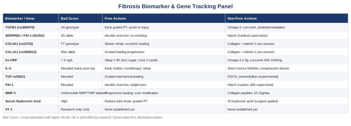
Before you dive in: this article treats patellar retinaculum fibrosis the way a good diagnostic workup would — by asking what's driving the scar tissue rather than just describing it. You'll find seven bloodwork-accessible biomarkers (from a basic hs-CRP most labs already run, to a research-only enzyme called XT-1 that may one day distinguish surgical fibrosis from other scarring), each with what it measures, roughly what it costs, and a two-track plan — one built entirely on habits and rehab timing, one that adds supplements or devices, always with dosing, frequency, and honest side-effect notes. Then it turns to genetics: four variants (in TGFB1, SERPINE1/PAI-1, COL5A1, and COL1A1) that appear repeatedly in fibrosis and connective-tissue-injury research, with a candid look at how much of that evidence actually applies to a human knee versus a heart or a lab dish. A detour through Peter Attia's Outlive pulls out ten ideas that quietly challenge how most orthopedic follow-up care is structured. And a final section covers the complementary therapies — from EMG biofeedback to photobiomodulation — that have actual trial data behind them for knee stiffness and post-surgical recovery, not just plausible theory. The diagram below maps how these pieces connect, from the first inflammatory signal to the stiffness you actually feel.
The 7 Biomarkers Worth Tracking When Your Knee Isn't Healing Like It Should
Knee arthrofibrosis research — most of it done in the context of ACL reconstruction and total knee replacement, since isolated patellar retinaculum fibrosis is rarely studied in its own right — has repeatedly pointed to the same handful of biological signals. A 2022 systematic review of biochemical markers in postsurgical knee arthrofibrosis pulled together this evidence and found that markers tied to the TGF-β signaling pathway showed up most consistently across studies (see the review at PMC9619298). That's the backbone of the list below. None of these are routinely ordered by an orthopedic surgeon during a normal post-op visit, which is part of why this information is hard to come by — but most are obtainable if you know what to ask for.
A quick honest note before the list: these markers are drawn from arthrofibrosis research broadly, since dedicated studies on the patellar retinaculum specifically are scarce. Treat them as the best available proxy, not a retinaculum-specific diagnostic.
1. High-Sensitivity CRP (hs-CRP)
Hs-CRP is the most accessible general marker of systemic inflammation, and it matters here because chronic low-grade inflammation feeds the same IL-6 and TGF-β pathways that drive fibrotic tissue formation. It won't tell you anything about your knee specifically, but it tells you whether your body is running a background inflammatory load that could be slowing normal tissue remodeling everywhere, including at the surgical site. Investors in longevity medicine like Peter Attia and lipidologists like Thomas Dayspring treat hs-CRP as a baseline "is something smoldering" check, not a diagnosis in itself.
How to measure it
A standard blood draw at any lab (Quest, LabCorp, or a direct-to-consumer service) measures hs-CRP for roughly 15 to 30 dollars standalone, or as part of a broader wellness panel. No fasting required.If the score is high: the plan without supplements
Prioritize 7 to 9 hours of sleep, reduce ultra-processed food and added sugar, walk or cycle at an easy pace (zone 2) for roughly 150 minutes a week, address any chronic dental or gum infection, and manage stress. Recheck hs-CRP in 6 to 8 weeks — it should meaningfully improve if lifestyle factors were the driver.If the score is high: the plan with supplements or equipment
Omega-3 fish oil (2 to 3 grams combined EPA/DHA daily) and curcumin (500 to 1,000 mg daily of a bioavailable, piperine-enhanced or phytosome form) both have reasonable evidence for lowering CRP over 8 to 12 weeks. Side effects: fish oil can mildly increase bleeding risk and cause GI upset at higher doses; curcumin can cause GI discomfort and interacts with blood thinners. Reassess every 3 months; stop if hs-CRP normalizes below 1 mg/L and lifestyle factors are stable.2. Interleukin-6 (IL-6)
IL-6 sits upstream of CRP and appears to be one of the most dramatically elevated cytokines in the early post-surgical window in arthrofibrosis models, according to the same systematic review cited above. That timing detail matters: it suggests the first 24 to 72 hours after knee surgery or significant injury may set the inflammatory tone that influences how much scar eventually forms.
How to measure it
IL-6 requires a specialty immunoassay, not a standard primary-care order. Cost runs roughly 70 to 150 dollars through esoteric lab services or a sports medicine and functional medicine practice.If the score is high: the plan without supplements
Early, guided motion after surgery (rather than prolonged immobilization) is consistently associated with better outcomes in rehab protocols. Combine that with structured cryotherapy in the first days post-op, gentle isometric quadriceps activation as early as your surgeon allows, and strict attention to sleep, since sleep restriction independently raises IL-6.If the score is high: the plan with supplements or equipment
Short-course NSAIDs, used only with your surgeon's approval and typically limited to 1 to 2 weeks, can blunt an excessive early inflammatory spike — but prolonged use is controversial because some evidence suggests it may interfere with early tendon and bone healing. Pneumatic compression devices can reduce edema-driven inflammatory load. Omega-3s and curcumin, as above, are reasonable lower-risk additions.3. TGF-β1 (Transforming Growth Factor Beta-1)
TGF-β1 is the closest thing fibrosis research has to a master switch. It drives resident fibroblasts to transform into myofibroblasts, the cells responsible for contracting and depositing the dense collagen that stiffens the retinaculum. It shows up as the central pathway in nearly every review of arthrofibrosis mechanisms, including a widely cited 2019 mechanistic review (see PMC6433953).
How to measure it
Serum TGF-β1 ELISA testing is available mostly through research-oriented or concierge precision-medicine labs, costing roughly 100 to 200 dollars. It is not a mainstream clinical test.If the score is high: the plan without supplements
Controlled, progressive mechanical loading within a pain-free range appears to modulate TGF-β signaling favorably in animal and mechanistic studies, while repeated microtrauma or re-injury reliably retriggers it. Working with a physical therapist on a graded loading progression, rather than pushing hard or resting completely, is the most evidence-aligned strategy here.If the score is high: the plan with supplements or equipment
Human evidence for supplements that safely lower TGF-β1 in joint tissue is thin. Green tea catechins (EGCG) and the prescription drug pentoxifylline have been explored in fibrosis research in other organs, but not established for joint fibrosis — treat these as experimental and discuss with a physician first; pentoxifylline carries GI, headache, and bleeding-risk side effects. Photobiomodulation devices (covered in the complementary approaches section below) have more direct supporting evidence for soft-tissue fibrotic signaling.4. PAI-1 (Plasminogen Activator Inhibitor-1)
PAI-1 blocks the enzyme system that would otherwise break down excess matrix, tipping the balance toward accumulation rather than clearance. Notably, one arthrofibrosis-specific study found PAI-1 protein levels were elevated in fibrotic tissue independent of a person's SERPINE1 gene variant (see PubMed 33816122) — meaning local tissue environment and behavior matter at least as much as genetics here, which is a useful, actionable finding.
How to measure it
A specialty fibrinolysis panel measuring PAI-1 activity or antigen costs roughly 100 to 150 dollars. Morning, fasted sampling is preferred since PAI-1 has a natural diurnal rhythm.If the score is high: the plan without supplements
Regular aerobic exercise is the best-documented way to lower PAI-1. Weight management matters directly, since adipose tissue itself produces PAI-1, and smoking cessation and improved insulin sensitivity both help.If the score is high: the plan with supplements or equipment
Niacin has mixed evidence for lowering PAI-1, mostly from cardiometabolic research rather than joint-specific studies, and carries real side effects (flushing, and at high doses, liver enzyme elevation) — it should only be used under medical supervision. Exercise remains the stronger, safer lever. Recheck every 3 months alongside lifestyle changes.5. MMP-3 and the MMP/TIMP Balance
Matrix metalloproteinases (MMPs) are the enzymes responsible for breaking down excess collagen; their natural inhibitors (TIMPs) keep them in check. When that balance tips too far toward inhibition, fibrotic matrix persists longer than it should. The arthrofibrosis biomarker review noted MMP-related transcription control (via HOX/HOTAIR genes) as a relevant mechanism, and MMP-3 is the most clinically accessible member of this family, already used in rheumatology practice.
How to measure it
An MMP-3 blood test costs roughly 50 to 100 dollars and is available through rheumatology-oriented labs, sometimes bundled into broader joint or autoimmune panels.If the score is low or the balance looks unfavorable: the plan without supplements
Progressive mechanical loading again plays a role, since controlled loading appears to upregulate beneficial remodeling activity. Manual scar mobilization and soft-tissue work (detailed in the complementary approaches section) directly target the same matrix.If the score is low or the balance looks unfavorable: the plan with supplements or equipment
No supplement reliably shifts the MMP/TIMP balance in human joint fibrosis with strong evidence. Collagen peptides (10 to 15 grams daily) are a reasonable, low-risk adjunct supporting overall connective tissue turnover, though the evidence base is stronger for tendon and skin than for fibrotic joint tissue specifically. Side effects are minimal — occasional mild GI upset.6. Serum Hyaluronic Acid (HA)
Serum HA reflects extracellular matrix turnover and synovial tissue activity. It's borrowed conceptually from liver fibrosis panels, where it's an established marker, and applied here as a proxy for how actively your joint's matrix is remodeling.
How to measure it
Cost runs roughly 40 to 80 dollars through specialty or repurposed liver-fibrosis panels; availability varies by lab.If the score is high: the plan without supplements
Reduce repetitive joint strain and work with a physical therapist on graded, not excessive, loading.If the score is high: the plan with supplements or equipment
Intra-articular hyaluronic acid injections (viscosupplementation) have evidence in osteoarthritis but not specifically in arthrofibrosis — this is a conversation to have with an orthopedic surgeon, not a self-directed choice. Oral HA supplements have weak evidence for joint fibrosis specifically and are optional at best.7. XT-1 (Xylosyltransferase-1) — An Emerging Marker Worth Watching
XT-1 is the enzyme that initiates proteoglycan synthesis in connective tissue, and it's included here mainly for transparency: the same systematic biomarker review found early evidence that XT-1 may distinguish surgical arthrofibrosis from other types of fibrosis, which would make it more specific to exactly this problem than anything else on this list.
How to measure it
Currently research-only. It is not commercially available for routine patient testing, so there's no realistic way to order it outside of a clinical study.The honest plan
There isn't one yet — no protocol targets XT-1 directly, with or without supplements. It's listed here so you know what to watch for as this research matures, and so you can ask a sports medicine research center whether any local trials are enrolling.Genetics tell a related but different part of this story — not what's happening in your tissue right now, but why your baseline tendency toward scar formation might differ from someone else's.
What Your Genes May Be Telling You About How You Scar
Genetic research specific to patellar retinaculum fibrosis essentially doesn't exist as its own field. What does exist is a body of work on genes tied to fibrosis in other tissues and to connective tissue injury risk generally, some of it well replicated, some of it inconsistent. Ali Torkamani, a genomics researcher known for large-scale precision medicine work, and Gary Brecka, who has popularized broad epigenetic and methylation-focused panels through consumer health programs, are reasonable starting points if you want to go deeper into personal genetic testing — but it's worth being clear that neither has published research specific to knee fibrosis. Their value here is more in the general framework: test broadly, treat genotype as a probability rather than a verdict, and prioritize the biomarkers that reflect what your genes are actually doing over the genes themselves.
TGFB1 (rs1800470)
This variant has been linked to increased myocardial fibrosis risk in heart transplant recipients carrying the AA genotype, with a notably strong effect size in that population (see PMC8807540). That's cardiac tissue, not knee tissue, so the extrapolation to joint fibrosis is reasonable but unproven — TGF-β1's fibrotic role is fairly universal across tissue types, which is why it's included, but the evidence is indirect.
If the gene looks unfavorable: the plan without supplements
Lean harder into early, guided physical therapy after any knee surgery or significant injury, avoid repeat trauma, and track the hs-CRP and IL-6 biomarkers above more closely than someone without this variant might need to.If the gene looks unfavorable: the plan with supplements or equipment
The same anti-inflammatory approach outlined for hs-CRP and IL-6 (omega-3s, curcumin) applies, plus consideration of photobiomodulation during rehab. There's no way to "turn off" this gene at a consumer level — the goal is managing its downstream effects, not the variant itself.SERPINE1 / PAI-1 (rs1799889, the 4G/5G variant)
The 4G allele is generally associated with higher PAI-1 production. But the arthrofibrosis-specific study referenced earlier found that actual PAI-1 protein elevation in fibrotic tissue didn't track cleanly with this genotype — a useful and honest finding that tells you the measured biomarker (PAI-1 levels) is more informative here than the genotype alone.
If the gene looks unfavorable: the plan without supplements
Regular aerobic exercise, weight management, and not smoking — the same levers that lower PAI-1 protein levels regardless of genotype.If the gene looks unfavorable: the plan with supplements or equipment
Niacin, cautiously and under medical supervision, given its side-effect profile. Given that this study found genotype doesn't reliably predict protein levels, it's more useful to simply test PAI-1 directly than to act on the gene result in isolation.COL5A1 (rs12722)
A systematic review and meta-analysis found the TT genotype associated with significantly higher risk of tendon and ligament soft-tissue injuries, particularly in Caucasian populations (see PMC5880610), though more recent research in elite athletes has produced mixed results — worth flagging as evidence that's solid at the population level but not perfectly predictive for any one individual.
If the gene looks unfavorable: the plan without supplements
A slower, more conservative rehab progression after knee surgery, with emphasis on eccentric loading protocols under a physical therapist's supervision, and resisting the urge to rush a return-to-sport timeline.If the gene looks unfavorable: the plan with supplements or equipment
Collagen peptides (roughly 15 grams) combined with 50 to 100 mg of vitamin C, taken 30 to 60 minutes before rehab or loading sessions, has some human evidence for enhancing collagen synthesis around the loading window. Side effects are minimal; cycle it around training sessions rather than using it continuously all day.COL1A1 (rs1800012)
A separate meta-analysis found this variant associated with tendon and ligament injury risk in a similar way to COL5A1 (see PMC5432363), reinforcing the idea that structural collagen gene variants may predispose some people to both injury and, plausibly, altered healing and scarring patterns afterward.
If the gene looks unfavorable: the plan without supplements
Same graded-loading approach as COL5A1 — the practical response to both genes converges on the same rehab philosophy.If the gene looks unfavorable: the plan with supplements or equipment
The same collagen-plus-vitamin-C protocol applies. Watch for mild GI upset or, rarely, an allergic reaction if you're sensitive to bovine or marine collagen sources.Genetics and biomarkers explain the biology. The next section steps back and looks at how one influential longevity thinker argues we should approach tissue repair and joint durability in general.
Ten Ideas From Outlive That Reframe How to Think About a Stiff, Scarred Knee
Peter Attia's Outlive: The Science and Art of Longevity isn't about knees specifically, but its central argument — that most of medicine treats problems reactively, long after the window for prevention has closed — maps directly onto how arthrofibrosis is usually managed: wait for stiffness to become a problem, then refer to physical therapy. Here are ten ideas from the book worth pulling into how you think about your own recovery.
1. Medicine 3.0 versus Medicine 2.0
Attia's core distinction is between medicine that waits for disease to declare itself (2.0) and medicine that acts on risk markers years before a problem is undeniable (3.0). Applied here: tracking hs-CRP or IL-6 after a knee injury, rather than waiting to see if stiffness develops, is a 3.0 approach to a 2.0 problem.2. The Centenarian Decathlon
Attia asks readers to define the specific physical tasks they want to still be able to do decades from now, then train backward from those goals. For retinaculum fibrosis, this means defining concrete functional targets — full-depth squatting, kneeling, stairs without compensation — rather than a vague goal of "less stiffness."3. Zone 2 Training as a Metabolic Foundation
Attia emphasizes low-intensity aerobic work for mitochondrial health and systemic inflammation control, which connects directly to the CRP and IL-6 discussion above — this isn't just a cardiovascular recommendation, it's an anti-inflammatory one.4. Strength and VO2 Max as the Strongest Predictors of Functional Independence
Attia argues these two measures predict long-term function better than almost any other metric. For a knee recovering from fibrosis, quadriceps and hip strength aren't just rehab checkboxes — they're the load-bearing insurance against future stiffness and reinjury.5. Inflammaging as a Root Driver of Tissue Dysfunction
The book leans on the concept of chronic low-grade inflammation accelerating tissue decline broadly, a mechanism well documented in aging research (see PMC10248980) — the same biological thread running through the hs-CRP and IL-6 sections above.6. Objective Markers Over Subjective Feeling
Attia's repeated refrain is to test rather than guess. This is the entire premise of this article's biomarker approach — "my knee feels tight" is a starting point, not a data point.7. Sleep as a Repair Mechanism, Not a Luxury
The book treats sleep as one of the highest-leverage inputs for tissue repair and inflammation control, directly reinforcing the sleep recommendations under both hs-CRP and IL-6 above.8. Skepticism Toward Reflexive Pharmacological Fixes
Attia is cautious about leaning on medications like NSAIDs as a default rather than addressing root causes — echoing the caution given earlier about short-course-only NSAID use around fibrotic healing.9. Stability and Movement Quality, Not Just Raw Strength
Attia stresses that injury prevention depends as much on movement quality and joint stability as on strength alone — relevant to protecting a healing retinaculum from the repeated microtrauma that retriggers TGF-β signaling.10. Patience With Biological Timelines
Perhaps the most directly applicable idea: connective tissue remodels on a timeline of months, not weeks, far slower than muscle. Attia's broader argument about playing a long game applies almost literally to fibrotic tissue recovery.These reframes are about mindset and timelines. The next section gets more concrete, covering complementary therapies with actual clinical trial evidence behind them.
Complementary Approaches Worth Adding to Your Recovery Plan
The following five approaches were selected because each has meaningful human evidence tied to knee stiffness, post-surgical scar tissue, or closely related joint conditions — not because they're popular in general wellness circles. Evidence quality varies, and that's noted honestly for each.
Biofeedback (EMG Biofeedback)
Electromyographic biofeedback gives you real-time visual or audio feedback on muscle activation, which is directly relevant after knee surgery because arthrogenic muscle inhibition — the nervous system's reflexive shutdown of the quadriceps around a swollen or scarred joint — is extremely common and can itself prolong stiffness by discouraging normal joint motion.
A randomized controlled trial in patients recovering from ACL reconstruction found that adding EMG biofeedback for the vastus medialis to standard rehabilitation improved knee extension range and strength compared to standard rehabilitation alone (see PMC3582470).
Realistically, this means asking your physical therapist whether they have EMG biofeedback equipment, or renting a portable unit for home use, typically for 15 to 20 minute sessions several times a week during the early rehab phase, tapering off once normal quadriceps activation returns without visual cues.
Low-Level Laser Therapy / Photobiomodulation
Photobiomodulation uses specific wavelengths of red or near-infrared light to influence cellular activity in soft tissue, and there is a plausible mechanistic link to modulating inflammatory and fibrotic signaling, alongside more established evidence in joint pain and function.
A systematic review and meta-analysis of low-level laser therapy combined with exercise for knee osteoarthritis found meaningful improvements in pain, range of motion, and function compared to exercise alone (see PubMed 36576096). This evidence is from osteoarthritis, not arthrofibrosis specifically, so treat it as a plausible extension rather than direct proof.
In practice, this looks like 10 to 24 sessions of near-infrared light (in the 780–860 nm or 904 nm range, per treatment guidelines) applied directly over the retinaculum and surrounding tissue, usually delivered by a physical therapist or sports medicine clinic, occasionally supplemented with a home-use device between sessions.
Massage Therapy and Manual Scar Mobilization
Manual therapy directly targets the mechanical side of fibrosis — breaking up tethered scar tissue and restoring normal tissue glide around the retinaculum and surrounding structures, which is a fundamentally different mechanism from anything biochemical listed above.
A systematic review of soft tissue therapy for postoperative knee conditions found consistent improvements in functional outcomes, and in one small cohort of patients who had failed to respond to standard rehabilitation, targeted soft-tissue treatment improved knee flexion deficits by 35 degrees and flexion contractures by 12 degrees (see PMC8704673).
Realistically, this means starting gentle scar mobilization once the surgical incision is fully healed (usually 3 to 6 weeks post-op, confirmed with your surgeon), progressing to instrument-assisted or manual soft tissue work with a trained therapist, done 1 to 2 times weekly during active fibrosis remodeling, tapering as mobility normalizes.
Tai Chi
Tai chi combines slow, controlled range-of-motion work with balance and proprioceptive training, which is relevant to a fibrotic knee both for regaining lost motion gently and for rebuilding the movement confidence that guarding and stiffness often erode.
A large randomized trial comparing tai chi to standard physical therapy for knee osteoarthritis found tai chi produced benefits comparable to a full course of physical therapy over 12 weeks of twice-weekly group sessions (see PubMed 27183035). Again, this evidence is from osteoarthritis rather than post-surgical fibrosis, so the extrapolation is reasonable but not proven for this exact condition.
In practice, this looks like a beginner tai chi class (many are offered through physical therapy clinics or community centers) twice a week, used as a low-impact complement to formal rehab rather than a replacement for it, particularly useful once acute swelling has resolved and gentle range-of-motion work is appropriate.
Yoga
Yoga's slow, sustained stretching combined with strength-through-range-of-motion work addresses both the flexibility and the strength deficits that commonly accompany a fibrotic, stiff knee.
A 2024 systematic review and meta-analysis of yoga for knee osteoarthritis, covering multiple randomized trials, found significant improvements in pain, stiffness, and physical function compared to control groups (see PubMed 38753745).
Realistically, this means a gentle, knee-modified yoga class (many studios and physical therapists offer these), 2 to 3 sessions a week, avoiding deep knee flexion poses until your surgeon or therapist confirms your tissue can tolerate that range, and treating it as a mobility supplement to strength training rather than a substitute for it.
Turning This Into a Plan
Patellar retinaculum fibrosis isn't one thing — it's the downstream result of inflammatory signals, a specific growth factor pathway, a matrix-remodeling balance, and in some people, a genetic tendency that nudges all of the above in one direction. The biomarkers in this article give you a way to see which of those pieces is actually active in your case rather than guessing. The genetics give context, not a verdict — and in at least one case (PAI-1), the research itself shows the measured protein matters more than the gene. The Attia-inspired mindset shift and the complementary therapies round out a plan that treats your knee as something to actively manage, not passively wait out.
The concrete next step is smaller than it might feel: pick one or two of the biomarkers above that map to your specific history — recent surgery, slow rehab progress, family history of poor healing — and ask your doctor or a sports medicine practice about ordering them. Bring this list to that conversation. Track your range of motion alongside whatever markers you check. And keep a qualified surgeon or physical therapist in the loop on every change you make, especially anything involving supplements, devices, or accelerated loading. Better data, used carefully, is how you move from managing symptoms to actually understanding what your knee is doing.
Musculoskeletal: Joint Conditions Tendon & Ligament Conditions
Autoimmune: Connective Tissue Conditions