This article contains AI generated content.
· UpdatedPatellar Sleeve Fracture - 6 Genes And 7 Biomarkers To Track
Introduction
A patellar sleeve fracture is not the kind of injury that announces itself with obvious warning signs. It happens when the cartilaginous sleeve surrounding the lower pole of the kneecap tears away — most often in a child or teenager during a jump, a sudden sprint, or a deceleration — and the X-ray may look deceptively subtle while the damage is anything but. What makes it harder to process is that most young patients who experience this are otherwise healthy and active, with no prior indication that this particular structure was vulnerable. That disconnect is part of what makes the injury so disorienting, both for families and for the athletes themselves.
The typical clinical response is appropriate as far as it goes: confirm the injury, decide between conservative management and surgical fixation, immobilize, then move through physiotherapy toward a return-to-sport timeline. But that framework stops short of asking a deeper question — why did this fracture happen, and what does the healing environment actually look like underneath the surface? Two children with the same injury, the same surgeon, and the same rehabilitation program can have strikingly different outcomes. Some return fully. Others struggle with persistent pain, delayed union, or cartilage complications that emerge months later. The difference often lies in factors that are measurable but rarely measured: bone mineral quality, cartilage integrity, the efficiency of anabolic repair signals, and the background level of inflammation that either supports or quietly undermines healing.
This is where biomarkers and genetics offer something that standard orthopedic care does not. A targeted panel of blood and urine tests can reveal whether a young patient's healing environment is biochemically favorable — or whether a vitamin D deficiency, low IGF-1, or chronic low-grade inflammation is working against the repair process in real time. Genetic variants in collagen genes, vitamin D receptor function, and cartilage remodeling enzymes can explain susceptibility and guide a more personalized recovery strategy.
The biomarker section that follows is the most practical starting point — concrete, testable, and actionable within weeks. The genetics section goes one layer deeper, for those who want to understand predisposition at a biological level and build a prevention strategy that lasts beyond the immediate recovery. Neither section promises a cure or a guaranteed outcome. But better information reliably leads to better decisions, and in pediatric orthopedic recovery, early and well-informed decisions compound significantly over time.
7 Biomarkers Worth Tracking During Recovery
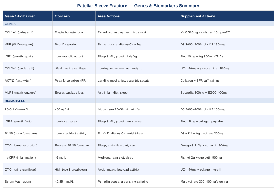
Most blood panels ordered after a patellar sleeve fracture are focused on ruling out infection or anemia. They reveal little about whether bone is mineralizing efficiently, whether cartilage breakdown is outpacing repair, or whether inflammatory signaling is disrupting the healing cascade. The seven markers below address those gaps directly. They are organized from the most accessible and affordable to the most specialized, with a clear plan for each unfavorable result — both with and without supplementation.
1. 25-OH Vitamin D
Why it matters
Vitamin D is essential for calcium absorption, bone mineralization, and the regulation of the growth plate — the very structures centrally involved in a patellar sleeve fracture. In children and adolescents, even mild deficiency disrupts the quality of newly forming bone and slows the calcification of cartilaginous tissue. Research consistently links low vitamin D status to increased fracture risk and impaired fracture healing in pediatric populations. Vitamin D also plays a key role in immune modulation, meaning it directly influences how cleanly and completely the inflammatory phase of healing resolves. A child who fractures with a vitamin D level of 18 ng/mL is healing in a fundamentally different biochemical environment than one at 55 ng/mL.
How to measure it
Serum 25-hydroxyvitamin D (25-OH-D) is the standard test and can be ordered by a general practitioner or purchased directly through direct-to-consumer lab services. Cost: $30–80 USD. Optimal range is generally considered 40–60 ng/mL (100–150 nmol/L). Values below 30 ng/mL indicate insufficiency; below 20 ng/mL, deficiency. The NIH Office of Dietary Supplements vitamin D fact sheet provides detailed reference ranges for children and adolescents.
If the score is bad, the plan without supplements
Increase daily sun exposure to 15–30 minutes of midday sun on arms and legs (without sunscreen in that window), adjusted for skin tone and latitude. Prioritize dietary vitamin D: wild-caught salmon provides 600–1000 IU per serving; egg yolks, UV-exposed mushrooms, and fortified foods contribute smaller amounts. Eliminate the main depletion factors: excess indoor time, heavy sunscreen use year-round, and high body fat percentage (which sequesters vitamin D in adipose tissue). This approach typically yields modest improvements of 5–10 ng/mL over 3–6 months and is rarely sufficient on its own to correct frank deficiency.
If the score is bad, the plan with supplements or equipment
Vitamin D3 (cholecalciferol) 2000–4000 IU/day for a child or adolescent with insufficiency, always combined with Vitamin K2 (MK-7 form) at 100–150 mcg/day to direct calcium appropriately toward bone rather than soft tissue and arterial walls. Take both with the fattiest meal of the day for optimal absorption. Retest at 3 months to adjust dose. Cycling: not required — D3 and K2 can be taken year-round, monitored twice per year. Side effects: toxicity below 5000 IU/day is rare but possible in individuals with granulomatous disease; monitor via retesting. Symptoms of excess (fatigue, excessive thirst, nausea) are the clinical signal to stop and recheck.
2. IGF-1 (Insulin-Like Growth Factor 1)
Why it matters
IGF-1 is the primary anabolic signal driving bone and cartilage repair. Produced mainly in the liver in response to growth hormone, it stimulates osteoblast activity and chondrocyte proliferation — the cells that rebuild bone and cartilage respectively. During adolescent fracture healing, adequate IGF-1 is particularly critical because the growth plates are still active and tissue remodeling is happening at an accelerated rate. Children with low IGF-1 for their age — even without a formal growth disorder — may heal more slowly, with less complete regeneration of the osteochondral interface. This biomarker is rarely checked after pediatric orthopedic injury, which is precisely why it slips through the standard care framework.
How to measure it
Serum IGF-1 is available from most labs as a routine blood test. Cost: $60–150 USD. Reference ranges are tightly age- and sex-dependent; a result in the lower quartile for age should prompt further investigation, ideally with a pediatric endocrinologist who can contextualize it against growth velocity and clinical history.
If the score is bad, the plan without supplements
IGF-1 is powerfully influenced by three lifestyle variables: sleep quality and quantity (most growth hormone — which drives hepatic IGF-1 production — is released during slow-wave sleep; 8–9 hours is the pediatric minimum), protein intake (1.2–1.6g/kg body weight per day using whole food sources), and resistance loading (as cleared by the surgeon). Eliminating ultra-processed foods and excess sugar load improves the IGF-1/insulin ratio by reducing compensatory hyperinsulinemia. These changes can raise IGF-1 meaningfully within 8–12 weeks.
If the score is bad, the plan with supplements or equipment
Zinc bisglycinate 15–25 mg/day supports the growth hormone secretion that drives IGF-1 production. Take with food to reduce nausea. Cycling: 8 weeks on, 2 weeks off to avoid copper depletion with prolonged use (check copper at 6 months if continuous use is planned). Hydrolyzed collagen peptides 10–15g/day — specifically the glycine, proline, and hydroxyproline content — provide the amino acid building blocks for osteochondral repair; combining with Vitamin C 500 mg at the same time improves conversion efficiency. No cycling required for collagen. Side effects: zinc at excess doses depletes copper; collagen — occasional mild GI discomfort at high doses.
3. P1NP (Procollagen Type 1 N-Terminal Propeptide)
Why it matters
P1NP is the most sensitive specific marker of bone formation currently available for clinical use and is endorsed by the International Osteoporosis Foundation as a first-line bone formation marker. When osteoblasts synthesize new type I collagen — the structural scaffold of bone — P1NP is released as a byproduct into the circulation. Elevated P1NP during fracture healing is a favorable sign; unexpectedly low P1NP in the recovery window suggests impaired osteoblast activity. Tracking it at baseline and again 6–8 weeks into recovery provides a direct window into whether the bone-building process is running at the right speed. This context is invisible to standard imaging.
How to measure it
Blood test (serum or plasma), available through specialty labs and academic medical centers. Cost: $60–120 USD. Not routinely ordered in pediatric fractures, but increasingly accessible through functional medicine panels and direct-to-consumer labs. A baseline measurement at the time of initial management, with a follow-up at 6–8 weeks, is most informative.
If the score is bad, the plan without supplements
Low P1NP in a recovering patient suggests the bone-building environment needs structural support. The most powerful non-supplemental intervention is correcting vitamin D deficiency first — D3 is one of the strongest activators of osteoblast function through VDR signaling. Adequate dietary calcium (dairy, fortified plant milks, leafy greens) is the next priority. Once surgical clearance allows, even gentle low-level weight-bearing (standing, walking, or hydrotherapy) stimulates osteoblast recruitment through mechanotransduction.
If the score is bad, the plan with supplements or equipment
Vitamin D3 + K2 (as above) combined with magnesium glycinate 200–300 mg/day — magnesium is both a cofactor in osteoblast enzymatic activity and necessary for converting vitamin D into its active form. Orthosilicic acid (bioavailable silicon) 10 mg/day supports collagen cross-linking in bone matrix; the research base is still building but human studies show effects on bone formation markers. Recheck P1NP at 8–12 weeks. Cycling: not required for magnesium or K2 at these doses; silicon: standard research protocol is 3 months on. Side effects: magnesium — loose stools above 400 mg elemental/day; titrate up slowly.
4. CTX-I (C-Terminal Telopeptide of Type I Collagen)
Why it matters
Where P1NP measures bone formation, CTX-I measures bone resorption — the breakdown of existing bone by osteoclasts. In healthy fracture healing, resorption and formation are tightly coupled: old or damaged tissue is removed and new matrix is laid down in its place. But in the presence of excess systemic inflammation, high cortisol, corticosteroid use, or nutritional deficiency, resorption can outpace formation and net bone loss occurs even during what appears to be the recovery phase. Monitoring the P1NP-to-CTX-I ratio gives a more complete picture of whether remodeling is productive or destructive. A useful principle from metabolic bone medicine, endorsed by clinicians like Thomas Dayspring for cardiovascular analogues, is that isolated data points mislead — ratios reveal.
How to measure it
Fasting morning blood draw (CTX-I is strongly diurnal, peaking in the early morning hours, and must be standardized for meaningful comparisons over time). Cost: $60–120 USD. Specialty labs; can be collected in the same blood draw as P1NP.
If the score is bad, the plan without supplements
High CTX-I (excess resorption) is commonly driven by inflammation, elevated cortisol, or inadequate calcium/vitamin D intake. Address sleep quality first — cortisol rises measurably with poor sleep within 48 hours and directly activates osteoclasts. Shift the diet toward an anti-inflammatory Mediterranean pattern. Within surgical clearance, low-level mechanical loading stimulates the mechanosensory pathways that favor formation over resorption.
If the score is bad, the plan with supplements or equipment
Omega-3 fatty acids (EPA + DHA combined) 2–3g/day have solid evidence for reducing osteoclast activity through suppression of inflammatory prostaglandins. Take with meals; no cycling required. Curcumin with piperine 500 mg twice daily reduces NF-κB-driven inflammatory signaling that recruits osteoclasts. Cycling: 8 weeks on, 2 weeks off. Side effects: omega-3 at high doses may modestly prolong clotting time — discuss with surgeon in the early postoperative period; curcumin at high doses may modestly reduce iron absorption — separate by 2 hours from iron-rich meals.
5. High-Sensitivity CRP (hs-CRP)
Why it matters
Systemic inflammation is one of the most under-recognized barriers to clean fracture healing. High-sensitivity CRP is an affordable, widely available marker of background inflammatory activity. Values above 1 mg/L suggest meaningful inflammatory load; above 3 mg/L is defined as high risk in cardiovascular research and is equally concerning in a bone-healing context. Elevated CRP reflects circulating inflammatory cytokines — IL-6, TNF-α — that directly inhibit osteoblast function, disrupt IGF-1 signaling, and accelerate cartilage matrix breakdown. In a pediatric patient with persistently elevated hs-CRP during recovery, the cellular environment is working against the repair process at every level. Identifying this is not academic — it changes the intervention.
How to measure it
Standard blood test, widely available through any laboratory. Cost: $15–50 USD. Should ideally be drawn at least two weeks after the acute injury phase — when the initial post-traumatic inflammatory surge has partially resolved — to get a meaningful reading of background inflammation rather than the expected injury response.
If the score is bad, the plan without supplements
An anti-inflammatory dietary pattern is the single most consistent and evidence-supported CRP-lowering intervention available without supplements. Mediterranean-pattern eating — emphasis on oily fish, extra-virgin olive oil, colorful vegetables, legumes, nuts — consistently reduces hs-CRP in pediatric as well as adult populations in randomized trials. Sleep quality matters enormously: sleep deprivation raises CRP within 48–72 hours in controlled experiments. Reducing body weight toward a healthy range has a powerful additional effect.
If the score is bad, the plan with supplements or equipment
Fish oil (EPA + DHA) 2g/day — among the best-studied CRP-lowering nutrients. Quercetin 500 mg/day has multiple RCT-level evidence for reducing hs-CRP, particularly when combined with bromelain 100 mg for enhanced absorption. Cycling: quercetin 8 weeks on, 2 weeks off; fish oil can be used continuously. Side effects: fish oil — fishy aftertaste (enteric-coated form minimizes this); quercetin at high doses — occasional headache; discuss with prescribing physician given concurrent medications in a surgical patient.
6. Urinary CTX-II (Cartilage Degradation Marker)
Why it matters
This is the most specialized marker on the list, but also the most directly relevant to the specific pathology of a patellar sleeve fracture. The injury occurs at the osteochondral junction — the interface between bone and hyaline cartilage — and the cartilaginous sleeve itself is largely composed of type II collagen. CTX-II (C-terminal crosslinking telopeptide of type II collagen) is released into urine as cartilage breaks down. Elevated CTX-II in the weeks and months after recovery suggests that the cartilaginous component of the repair is struggling even when anatomical union appears complete on imaging. This marker is routinely used in osteoarthritis research and is underused in pediatric sports medicine — it can serve as an early warning system for the cartilage complications that occasionally emerge long after the fracture itself has been declared healed.
How to measure it
Second-morning urine collection, adjusted for creatinine to normalize for hydration variation. Cost: $80–200 USD. Available through specialty labs. Pediatric reference ranges are less standardized than adult values — interpretation should involve a sports medicine or pediatric rheumatology specialist. Tracking trend over time (3-month intervals) is more informative than a single measurement.
If the score is bad, the plan without supplements
Avoid premature return to high-impact loading — running, jumping, cutting movements — until both clinical and, ideally, imaging criteria support it. Low-impact movement that loads cartilage beneficially without exceeding its adaptive capacity (cycling, hydrotherapy, controlled resistance work) is the appropriate activity mode during elevated CTX-II periods. Maintaining a healthy body weight reduces cumulative cartilage stress regardless of other interventions.
If the score is bad, the plan with supplements or equipment
Undenatured type II collagen (UC-II) 40 mg/day on an empty stomach — mechanism involves oral tolerance, reducing immune-mediated cartilage degradation. RCT evidence includes significant CTX-II reductions in joint conditions. Cycling: 3 months on, 1 month off. Hydrolyzed collagen peptides (type II specific) 10g/day + Vitamin C 500 mg taken together at the same time as physical activity for synergistic effect. No cycling required. Side effects: both are very well tolerated; UC-II — occasional mild GI discomfort; collagen — minimal at standard doses. For those with shellfish allergies, confirm collagen sourcing (prefer marine or bovine-derived, non-shellfish).
7. Serum Magnesium
Why it matters
Magnesium participates in more than 300 enzymatic reactions in the body, including those governing bone mineralization, vitamin D activation, muscle contraction, and inflammatory regulation. Deficiency is common in children who eat highly processed diets and in adolescent athletes who lose it through sweat. Its relevance to patellar sleeve recovery is threefold: it is required to enzymatically activate vitamin D into its usable form; it is co-deposited with calcium in bone crystal structure and is necessary for optimal bone density; and it regulates normal quadriceps and patellar tendon muscle mechanics — which directly affect the load environment at the repair site during rehabilitation. The NIH Office of Dietary Supplements magnesium fact sheet outlines reference ranges and dietary sources in detail.
How to measure it
Serum magnesium is included in most comprehensive metabolic panels. Cost: $15–40 USD. Important caveat: serum magnesium reflects only approximately 1% of total body magnesium and may appear normal even when intracellular stores are meaningfully depleted. If deficiency is clinically suspected but serum is borderline (below 0.85 mmol/L), a 24-hour urinary magnesium test is more sensitive and is the preferred functional assessment.
If the score is bad, the plan without supplements
Dietary sources: pumpkin seeds, dark leafy greens (spinach, Swiss chard), black beans, almonds, and dark chocolate are among the richest sources per serving. Reduce the major dietary depletion drivers: excess caffeine (blocks magnesium absorption), highly processed foods, and excess sugar. This dietary approach can meaningfully correct borderline deficiency over 4–8 weeks, particularly when gut absorption is healthy.
If the score is bad, the plan with supplements or equipment
Magnesium glycinate or magnesium malate 300–400 mg elemental magnesium/day — these forms have superior bioavailability compared to magnesium oxide and are significantly gentler on the digestive tract. Take in the evening (mild sedative effect supports recovery sleep). Cycling: not required — continuous use at these doses is generally safe and appropriate. Avoid magnesium citrate if stools are already loose. Recheck serum magnesium at 6–8 weeks to confirm trend. Side effects: loose stools above 400 mg elemental magnesium/day; titrate up over 2 weeks starting at 150 mg to minimize this.
The Genetics Behind Patellar Sleeve Vulnerability
Biomarkers tell you what the healing environment looks like right now. Genetics tells you something different — why this injury happened at all, and what structural tendencies will persist through every future training cycle. Understanding the genetic dimension is not about determinism; it is about calibration. Two adolescents with identical training loads may face very different injury risks because their collagen quality, vitamin D responsiveness, or cartilage remodeling activity differs at the gene level. Researchers like Ali Torkamani at Scripps Research and practitioners like Gary Brecka have helped translate this kind of genomic data into practical, personalized intervention frameworks. The six variants below are the most directly relevant to patellar sleeve fracture risk and recovery.
COL1A1 — The Bone and Tendon Scaffold Gene
What it affects: COL1A1 encodes the alpha-1 chain of type I collagen, the structural protein that makes up approximately 90% of bone's organic matrix and is also the dominant collagen in tendons and ligaments. The Sp1 binding site polymorphism (rs1800012) — the most studied variant — has been linked in multiple population studies to reduced bone matrix tensile strength, increased fracture susceptibility, and slower connective tissue repair after injury. Carriers of the T-allele at this site tend to produce collagen fibers with reduced cross-linking density, mechanically more fragile under the kind of sudden eccentric load that produces an avulsion fracture.
How to test: Consumer genomic platforms (23andMe, AncestryDNA) or sport-specific genetic panels (DNAFit, SelfDecode). Alternatively, serum P1NP and CTX-I together give an indirect functional picture of collagen turnover quality without genetic sequencing — a useful first step when genomic testing is not accessible.
If the gene is bad, the plan without supplements
Prioritize progressive mechanical loading throughout rehabilitation — controlled tension on collagen fibers stimulates lysyl oxidase, an enzyme responsible for collagen cross-linking and therefore tensile strength. This is not an argument for aggressive loading; it is an argument for a carefully periodized return-to-activity protocol rather than prolonged passive rest. Sports technique review is equally important: landing mechanics that distribute force through the hip and ankle (rather than concentrating it at the knee) reduce peak patellar tendon load, which matters more for a COL1A1 T-allele carrier than for a genetically average peer.
If the score is bad, the plan with supplements or equipment
Vitamin C 500 mg + collagen peptides 15g taken together 30–45 minutes before a physical therapy session — this timing protocol, established in research by Shaw et al. published in the American Journal of Clinical Nutrition, exploits the window when collagen synthesis is most responsive to amino acid availability and ascorbate co-factor action. Lysine 1000 mg/day supports hydroxylation steps in collagen cross-linking. Cycling: collagen peptides 3 months on, 1 month off; vitamin C is safe continuously at 500 mg; lysine has no established cycling protocol at this dose. Side effects: very high-dose vitamin C (above 2g/day) causes loose stools; 500 mg is well tolerated in virtually all patients.
VDR — The Vitamin D Receptor Gene
What it affects: Even when circulating vitamin D levels are adequate, the biological response depends on the Vitamin D Receptor gene. Several VDR polymorphisms — particularly FokI (rs2228570), BsmI (rs1544410), and TaqI (rs731236) — affect the binding affinity and transcriptional efficiency of the vitamin D receptor in osteoblasts, immune cells, and cartilage. Individuals with less efficient VDR variants may require higher circulating vitamin D levels to generate the same bone-protective signal, meaning a level of 35 ng/mL that is sufficient for one person may be functionally inadequate for another with an unfavorable receptor genotype. This is one reason why tracking both vitamin D level and bone formation markers together is more informative than either alone.
How to test: Consumer genomics platforms. Functionally, comparing vitamin D blood levels against clinical outcomes (bone turnover markers, inflammatory markers) over supplementation cycles provides indirect evidence of receptor efficiency without genetic sequencing.
If the gene is bad, the plan without supplements
Maximize endogenous vitamin D synthesis through consistent midday sun exposure and dietary vitamin D sources (oily fish, eggs, UV-exposed mushrooms). Focus equally on the downstream cofactors that the VDR pathway depends on: dietary calcium from whole foods, and magnesium (which is enzymatically required for vitamin D activation regardless of receptor function). This nutritional infrastructure reduces the demand on the receptor signaling system itself.
If the score is bad, the plan with supplements or equipment
Those with suspected VDR inefficiency typically benefit from maintaining serum 25-OH-D at the higher end of the optimal range — 55–65 ng/mL — rather than the lower end. This may require 3000–5000 IU/day of D3 plus K2 (MK-7) 150–200 mcg/day, monitored via blood testing every 3 months until a stable level is achieved. A broad-spectrum mineral supplement ensuring calcium, magnesium, zinc, and boron availability will support the downstream signaling that the VDR is trying to activate. Cycling: not required. Side effects: as above; monitor for hypercalcemia symptoms at doses above 4000 IU/day.
IGF1 Gene — Anabolic Repair Capacity
What it affects: The IGF1 gene contains a promoter region microsatellite polymorphism — variations in the number of CA repeats — that affects baseline IGF-1 production capacity. The 192-bp allele has been associated with higher IGF-1 levels across multiple cohorts; individuals with non-192 alleles may have structurally lower IGF-1 for their age, impairing anabolic recovery from orthopedic injuries throughout life. Since patellar sleeve fractures occur during the peak growth years when IGF-1 production should be at its biological maximum, a genetically limited IGF-1 axis compounds the biological challenge of repair.
How to test: Serum IGF-1 measurement (see biomarkers section) is the most accessible indirect indicator of functional IGF-1 status. Genetic testing through specialty wellness genomics panels.
If the gene is bad, the plan without supplements
Protect the three primary drivers of IGF-1 production: sleep quality and duration (growth hormone — and therefore IGF-1 — is primarily released during slow-wave sleep; 8–9 hours in adolescents is non-negotiable during recovery), adequate protein intake (1.4–1.6g/kg/day from whole food sources), and resistance loading (as cleared post-surgery — even light eccentric work restores GH pulse frequency). Reducing ultra-processed food intake improves the GH/IGF-1 axis by reducing hyperinsulinemia, which competes with GH signaling.
If the score is bad, the plan with supplements or equipment
Zinc 20–25 mg/day + magnesium 300 mg/day in the evening (the ZMA combination has published support for supporting the GH/IGF-1 axis in athletes, with effects particularly clear when baseline deficiencies exist). Cycling: zinc 8 weeks on, 2 weeks off to protect copper balance; magnesium can be continuous. For older adolescents under specialist guidance only: arginine 2–3g/day has preliminary evidence for stimulating GH pulse amplitude; this should not be used without pediatric nutritionist involvement due to the complexity of GH axis modulation in growing patients. Side effects: zinc excess depletes copper over time; arginine at high doses causes GI discomfort; monitor serum IGF-1 to track response.
COL2A1 — Cartilage Collagen Integrity
What it affects: COL2A1 encodes type II collagen, the primary structural protein of hyaline cartilage. Because patellar sleeve fractures occur exactly at the osteochondral interface, the quality of the cartilaginous sleeve — determined in part by COL2A1 variants — influences both susceptibility to the initial avulsion and the quality of cartilage repair afterward. Variants that reduce type II collagen fibril organization or cross-linking density may make the sleeve more prone to separation under sudden eccentric load, and may result in less mechanically competent cartilage matrix during the repair process. This gene is less commonly tested in consumer panels but is included in clinical genetics workups for cartilage-related conditions.
How to test: Specialty or clinical genetics panel. Functionally, urinary CTX-II tracking (see biomarkers section) provides the most accessible indirect signal of type II collagen degradation activity without sequencing.
If the gene is bad, the plan without supplements
Prioritize low-impact movement (swimming, cycling, aquatic therapy) throughout recovery to provide beneficial compressive loading to cartilage without the peak impact forces that accelerate type II collagen breakdown. Maintain a healthy body weight — excess mechanical load on the joint is among the strongest drivers of cartilage degradation regardless of genetic background. Early return to high-impact sport should be delayed beyond standard timelines in those with confirmed COL2A1 vulnerability.
If the score is bad, the plan with supplements or equipment
UC-II (undenatured type II collagen) 40 mg/day on an empty stomach in the morning — the mechanism here is oral tolerance induction, reducing immune-mediated cartilage attack. RCT evidence from joint conditions shows significant reductions in CTX-II with UC-II supplementation. Glucosamine sulfate 1500 mg/day supports cartilage matrix synthesis through chondrocyte stimulation; the evidence base spans multiple randomized trials. Cycling: 3 months on, 1 month off for both. Side effects: UC-II is very well tolerated; glucosamine — rare shellfish allergy cross-reactivity (use bovine or fermented plant-derived sources if shellfish allergy is present).
ACTN3 — The Explosive Force Gene
What it affects: ACTN3 encodes alpha-actinin-3, a structural protein found exclusively in fast-twitch (type IIx) muscle fibers. The R577X polymorphism (rs1815739) — among the most widely studied sports genetics variants — results in a complete absence of functional alpha-actinin-3 protein in XX homozygotes. RR genotype carriers express high levels of this protein and are associated with greater peak muscle force generation and explosive power output. There is a mechanically plausible and increasingly supported case that RR athletes generate the sudden quadriceps force spikes — during explosive jumps and landings — that create the peak patellar tendon tension capable of producing a sleeve avulsion. Understanding this polymorphism informs how an athlete trains and moves, not whether they should compete.
How to test: Widely included in consumer genetics panels (23andMe, AncestryDNA, DNAFit, Athletigen).
If the gene is bad, the plan without supplements
RR carriers specifically benefit from landing mechanics training: learning to distribute impact forces across the hip and ankle rather than concentrating them at the knee. Video-based landing analysis by a sports physiotherapist is a worthwhile investment for high-level young athletes who carry this variant and have already experienced a patellar sleeve injury. Eccentric quadriceps strengthening via Spanish squat variations and Nordic-style hamstring curls (introduced progressively after surgical clearance) builds tendon stiffness and attenuates shock transmission at the patellar tendon insertion.
If the score is bad, the plan with supplements or equipment
No supplement changes ACTN3 genotype, but supporting the connective tissue that absorbs the forces generated by fast-twitch fibers is productive and actionable. Collagen peptides 15g + Vitamin C 500 mg before each training or physiotherapy session (Shaw et al. timing protocol). In later-stage rehabilitation, blood flow restriction (BFR) training using a BFR cuff device ($40–150 for entry-level equipment, $300+ for clinical-grade cuffs) allows significant adaptive stimulus to the quadriceps at very low joint load — particularly valuable for RR carriers who need connective tissue adaptation without the joint stress that comes with high-load conventional training. Always use BFR under the supervision of a qualified clinician until protocols are established.
MMP3 — The Matrix Remodeling Gene
What it affects: Matrix metalloproteinase 3 (MMP3) is an enzyme that breaks down extracellular matrix components including type II collagen, fibronectin, and proteoglycans — the molecular architecture of cartilage and connective tissue. The 5A/6A promoter polymorphism (rs3025058) affects transcriptional activity: 5A allele carriers express substantially more MMP3 than 6A carriers under the same inflammatory stimulus. In the context of patellar sleeve recovery, elevated MMP3 activity may explain why some individuals develop persistent joint surface degradation despite anatomically successful fracture healing — MMP3 upregulated by post-injury inflammation continues breaking down cartilage matrix long after the mechanical injury has resolved.
How to test: Specialty genetics panels. Serum MMP-3 can also be measured directly as a biomarker ($80–150 USD) to assess activity level without genetic testing — a useful option when sequencing is not accessible.
If the gene is bad, the plan without supplements
The primary driver of MMP3 upregulation is systemic inflammation. Everything that reduces background inflammation — Mediterranean diet, sleep optimization, stress management, resolution of gut dysbiosis — reduces MMP3 activity independent of genetics. Avoiding repetitive high-impact loading until cartilage healing is imaging-confirmed is a critical mechanical corollary: MMP3 is upregulated by both inflammatory cytokines and mechanical overload, and an MMP3 5A carrier is sensitive to both inputs simultaneously.
If the score is bad, the plan with supplements or equipment
Boswellia serrata extract (AKBA fraction) 100–200 mg/day inhibits MMP-3 expression via suppression of the 5-LOX inflammatory pathway; RCT evidence in joint conditions includes significant reductions in MMP-3 serum levels. Cycling: 8 weeks on, 2 weeks off. EGCG (epigallocatechin gallate) from green tea extract 400 mg/day suppresses MMP-3 transcription in cartilage tissue in controlled research; take with food. Side effects: Boswellia — rare GI discomfort; EGCG — at doses above 800 mg/day and with prolonged continuous use, hepatic enzyme elevation has been reported in case studies; monitor liver function at 6 months if using long-term. At 400 mg/day, this risk is minimal but worth noting.
What "Outlive" by Peter Attia Teaches About Bone Health and Recovery
Outlive: The Science and Art of Longevity by Peter Attia is built around a central argument: that modern medicine excels at treating disease once it has arrived but almost entirely neglects the biology of prevention and functional preservation. While the book is not written about pediatric fractures, its treatment of bone health, biomarkers, and repair biology directly challenges assumptions embedded in standard orthopedic care. These ten points are the most immediately relevant for anyone navigating a patellar sleeve fracture.
1. Peak Bone Density Is Built in a Narrow Developmental Window
Attia is explicit that peak bone mineral density is achieved in the late teens to early twenties — and that the trajectory established during this window persists for decades. A child recovering from a patellar sleeve fracture is in this exact window. How bone heals and mineralizes during this period is not just about the current injury; it shapes the structural quality of the skeleton for the next fifty years. Standard care that focuses exclusively on anatomical union and return to sport misses the broader opportunity to actively optimize bone mineral density during this specific developmental phase.
2. The Standard DXA Scan Is Incomplete
The dual-energy X-ray absorptiometry (DXA) scan is the default bone density measurement, but Attia argues it captures only a fraction of what matters structurally. Trabecular Bone Score (TBS), which assesses three-dimensional bone microarchitecture rather than just density, and quantitative CT provide additional information that better predicts fracture risk — particularly in younger patients whose overall density may appear normal while microstructural organization is still maturing or already impaired.
3. Protein Intake for Bone Healing Is Consistently Underemphasized
Attia directly addresses the outdated belief that high protein intake is harmful to bone health. The current evidence runs in the opposite direction: adequate to high protein intake is associated with better bone formation, better fracture outcomes, and higher P1NP levels. Growing children recovering from fracture require consistent amino acid availability for collagen synthesis, and the standard rehabilitation plan rarely includes specific nutritional targets for protein — a gap that matters more than most practitioners acknowledge.
4. Zone 2 Exercise Has Bone-Relevant Anti-Inflammatory Effects
Low-intensity sustained aerobic exercise — what Attia calls Zone 2, performed at approximately 60–70% of maximum heart rate — improves mitochondrial efficiency, reduces systemic inflammatory load, and improves insulin sensitivity. For a child medically cleared for limited activity during fracture recovery, gentle cycling or swimming at this intensity is not merely "keeping fit" — it is actively improving the cellular environment within which bone repair is occurring, by reducing the CRP and IL-6 levels that impair that process.
5. IGF-1 Is the Critical Repair and Longevity Signal
Attia discusses IGF-1 in depth as a central longevity marker, noting that optimal — not maximal — IGF-1 is associated with the best repair and health outcomes. During fracture recovery, ensuring IGF-1 is in the favorable range through sleep quality, protein intake, and resistance loading (as cleared) is one of the most direct interventions available. He also notes that IGF-1 is rarely tested in general clinical practice, despite being one of the most informative single-marker windows into anabolic repair capacity.
6. Chronic Inflammation Is the Hidden Antagonist of Every Repair Process
One of Attia's most repeated arguments throughout the book is that chronic, low-grade inflammation — detectable via hs-CRP and related markers — disrupts virtually every repair and maintenance process in the body. The mechanism he cites operates at the level of cytokine signaling: elevated IL-6 and TNF-α suppress osteoblast recruitment and inhibit the IGF-1 receptor response in cartilage. His framework makes tracking hs-CRP during fracture recovery not an optional add-on but a core element of understanding the repair environment.
7. Supplements Should Be Approached With Pharmacological Rigor
Attia's supplement philosophy is disciplined: he treats each compound with the same analytical framework he applies to a pharmaceutical drug — mechanism, dose, bioavailability, duration, and interaction profile. He specifically endorses Vitamin D and omega-3 fatty acids as having both strong mechanistic rationale and meaningful epidemiological and clinical evidence for bone and anti-inflammatory applications, while dismissing most other marketed supplements as lacking comparable evidence quality. This calibration is genuinely useful when navigating a supplement market that inflates most claims.
8. Sleep Disruption Directly Suppresses Tissue Repair
Attia cites the research demonstrating that growth hormone is primarily secreted during slow-wave sleep, making sleep quality a direct variable in IGF-1 production and tissue repair rate. Disrupting slow-wave sleep — through blue light exposure before bed, irregular sleep timing, or untreated sleep apnea — measurably suppresses the hormonal environment that drives repair. For a recovering adolescent, consistent sleep hygiene is not a lifestyle preference — it is a therapeutic variable that affects bone healing as directly as any supplement.
9. Early Biomarker Monitoring Changes the Long-Term Trajectory
Rather than waiting for problems to become symptomatic, Attia's preventive framework calls for proactive biomarker tracking to identify unfavorable trends before they become clinical complications. Applied to patellar sleeve recovery: do not wait until a young athlete develops cartilage symptoms to begin tracking CTX-II, and do not wait until healing seems slow to check vitamin D. Early data creates the ability to intervene early, and in pediatric recovery — where the biological window for adaptation is still open — the return on early intervention is compounded.
10. Resistance Training Is the Most Powerful Single Intervention for Musculoskeletal Health
Attia's most consistent message across the entire book is that progressive resistance training — particularly eccentric loading protocols — is the most evidence-supported tool for maintaining and restoring musculoskeletal function. For a patellar sleeve recovery, this translates into a physiotherapy program that eventually includes eccentric quadriceps loading (Spanish squats, decline squats), hip and glute strengthening, and a carefully graduated return to plyometrics. The alternative — prolonged rest that allows surrounding tissue to atrophy — creates secondary mechanical problems that outlast the original injury.
Recovery Approaches Worth Considering Alongside Standard Care
The following modalities each have meaningful published evidence in the context of musculoskeletal healing, post-fracture pain, or pediatric recovery. They are presented as thoughtful additions to an orthopedic care plan — not as replacements for it, and not as cure claims.
Low-Level Laser Therapy (Photobiomodulation)
Low-level laser therapy uses specific wavelengths of near-infrared and red light (typically 630–1000 nm) to stimulate mitochondrial function in target tissue. The mechanism relevant to fracture healing involves photobiomodulation of cytochrome c oxidase in bone and cartilage cells, increasing ATP production, reducing oxidative stress, and promoting osteoblast and fibroblast activity. Multiple systematic reviews have examined its effect on bone consolidation, with the most consistent findings in the 810–830 nm wavelength range applied at 4–8 J/cm² energy density, 3–5 times per week. A search of the published literature on PubMed reflects a growing body of human clinical data supporting LLLT-accelerated fracture healing.
Clinical sessions with a physiotherapist or sports medicine practitioner using medical-grade LLLT equipment are the gold standard, particularly in the early postoperative period. A typical protocol is 10–15 minutes applied over the fracture site, 3–5 times per week, for 6–8 weeks. Consumer-grade red light panels (Joovv, Mito Red) represent a home-use alternative once the acute phase has resolved. Cost: clinical sessions $50–150 per session; home devices $200–800. Always confirm with the treating orthopedic surgeon before beginning — standard contraindications include direct application over growth plates in some protocols, an important caveat in pediatric patients.
Massage Therapy for Surrounding Musculature
After a patellar sleeve fracture, the surrounding musculature — particularly the quadriceps, vastus medialis, and hip flexors — undergoes significant atrophy and altered tension patterns due to immobilization and pain-related inhibition. This disrupted muscle balance affects joint mechanics throughout rehabilitation and can increase the load through the healing patellar tendon during the return-to-activity phase. Manual massage therapy, applied by a qualified physiotherapist or sports massage therapist, helps maintain tissue extensibility, reduce compensatory muscle guarding, and support lymphatic drainage in the immobilized limb — all of which contribute to a more favorable rehabilitation environment. Research in the Journal of Orthopaedic and Sports Physical Therapy has demonstrated that soft-tissue massage combined with exercise is superior to exercise alone in reducing quadriceps inhibition after knee injury.
Swedish effleurage and petrissage techniques to the quadriceps and posterior thigh, with specific attention to the distal quadriceps above the knee (avoiding the fracture site), are appropriate once the acute phase has resolved. One 30–45 minute session per week with a therapist experienced in sports or pediatric rehabilitation is a reasonable starting frequency. Self-massage using a foam roller on the midthigh (stopping well above the patella) can supplement professional sessions at minimal cost. Cost: $70–130 per professional session; foam roller $25–50. Communicate the complete surgical and fracture history before any session so the therapist avoids contraindicated techniques near the repair.
Mindfulness Meditation and Stress Reduction
Mindfulness-Based Stress Reduction (MBSR) is a structured 8-week program combining mindfulness meditation, body scan, and gentle movement practices. Its relevance to fracture recovery operates on two levels: it addresses the anxiety and psychological distress that are common — and rarely treated — in young athletes facing enforced inactivity, and it has measurable effects on inflammatory biomarkers. A randomized controlled trial published in Psychoneuroendocrinology found that MBSR practice reduced salivary cortisol and serum CRP over 8 weeks compared to control conditions. Given that both elevated cortisol and elevated CRP impair the bone-healing environment through the mechanisms described earlier in this article, the biochemical case for a stress-reduction practice during fracture recovery is not merely anecdotal.
For children and adolescents, abbreviated mindfulness programs (4-week adaptations with shorter daily sessions of 10–15 minutes) have shown similar anxiolytic and anti-inflammatory effects to the full 8-week adult format. The most accessible entry point is diaphragmatic breathing practiced twice daily: 4 counts in, 4 counts hold, 6 counts out, for 10 minutes per session. Apps such as Headspace for Teens or Calm provide age-appropriate structured guidance. Formal MBSR programs are available through many hospital-affiliated wellness centers. Cost: guided apps $10–15 per month; formal programs $300–600 for the 8-week course. There are no contraindications, making this a zero-risk parallel intervention to standard orthopedic care.
Conclusion
A patellar sleeve fracture is a specific injury at a specific moment in a young person's development, and how it heals is shaped by factors that clinical imaging alone cannot capture. The biomarkers covered in this article are measurable, interpretable, and actionable — they provide a direct window into the biochemical conditions that either support or undermine the healing process in real time. The genetic variants add a longer-range understanding of why certain tissue structures are structurally vulnerable, and what can be done differently going forward.
The most useful next step is not to implement everything simultaneously. Start with the three most accessible and affordable markers: vitamin D, hs-CRP, and serum magnesium. These three tests cost less than $150 combined, can be ordered through a general practitioner or direct-to-consumer lab service, and will immediately reveal whether the foundational healing environment is adequate. Bring the results to the treating orthopedic surgeon or sports medicine physician, and use them as the basis for a more specific conversation about recovery support. For those who want to go deeper, the genetic panel and the cartilage-specific markers offer the next layer of detail.
Better information does not guarantee a perfect outcome. But it reliably leads to better questions — and in pediatric recovery, better questions asked early tend to produce better decisions throughout.