This article contains AI generated content.
· UpdatedPatellar Stress Fracture Genes and Biomarkers — 5 Genes and 6 Biomarkers to Track
Introduction
A patellar stress fracture is one of those injuries that catches people off guard. You trained consistently, you weren't doing anything reckless, and yet the knee started hurting in a way that wouldn't resolve with standard rest protocols. When imaging finally confirms a stress fracture of the patella, the next question — the one most clinicians don't answer well — is: why did this happen to you, and what does your body actually need to heal and not repeat the experience?
The generic advice you'll encounter covers the basics: reduce load, protect the knee, gradually reintroduce activity. That framework isn't wrong, but it's incomplete in a way that matters. Two athletes with nearly identical training loads can have vastly different stress fracture risk based on their bone turnover rate, hormonal status, nutritional sufficiency, and even the collagen architecture encoded in their genes. Treating everyone the same means some people recover well while others spiral into repeated injury cycles with no clear explanation.
What actually differentiates people in terms of fracture susceptibility and healing speed often comes down to measurable biology — biomarkers that can be checked with a standard blood panel or a slightly more advanced test, and genetic variants that shape how effectively your body builds, maintains, and repairs bone. These aren't obscure variables. They're trackable, and in many cases, addressable.
This article takes a closer look at both layers. The first and primary focus is on six key biomarkers — the most clinically relevant, most actionable measurements you can get today — with practical guidance on what to do if any of them come back unfavorable. The second layer explores five genetic variants increasingly linked to stress fracture risk, with concrete strategies for compensating when the underlying predisposition isn't working in your favor. Taken together, these perspectives offer something a standard X-ray cannot: a roadmap toward understanding and correcting the biology behind your injury.
6 Biomarkers That Reveal Why Your Patella Isn't Healing
Bone is a dynamic tissue. It is constantly being broken down and rebuilt through a process called remodeling, regulated by hormones, nutrients, mechanical signals, and inflammatory status. A stress fracture means the breakdown side temporarily outpaced the building side — and identifying which factors drove that imbalance is where biomarkers become genuinely useful.
The six markers below represent a scientifically grounded, practically accessible panel. Some can be ordered through your primary care physician; others may require a sports medicine or integrative medicine specialist. Costs reflect approximate US ranges and vary by lab and insurance.
Biomarker 1 — 25-Hydroxyvitamin D
Vitamin D is arguably the single most important biomarker to check after a stress fracture, and also the most commonly deficient in both athletes and the general population. Its active form regulates calcium absorption in the gut, supports phosphorus metabolism, modulates bone mineralization, and plays a role in muscle function that affects how forces are transmitted through the patella.
Low vitamin D status does not just reduce bone mineral density over time — it actively impairs the acute healing response after injury. Research consistently shows that athletes with serum 25-OH-D below 30 ng/mL face significantly elevated stress fracture risk compared to those with levels above 40 ng/mL. A landmark military study published in the Journal of Bone and Mineral Research (Lappe et al., 2008) found that calcium and vitamin D supplementation reduced stress fracture incidence in female Navy recruits by 20 percent.
How to Measure It
A standard serum 25-hydroxyvitamin D blood test (25-OH-D) ordered through any lab. Cost: $30–$80 without insurance. Optimal range for bone health and fracture prevention is generally considered 40–60 ng/mL. Levels below 30 are clinically deficient; levels between 30–40 are suboptimal for athletes under high mechanical load.
If the Score Is Bad, the Plan Without Supplements
Prioritize midday sun exposure (when UV-B is present) for 20–30 minutes on large skin surfaces like arms and legs, five days per week. Dietary sources with meaningful vitamin D content include wild-caught fatty fish (salmon, sardines, mackerel), pasture-raised egg yolks, and beef liver. These alone rarely correct a clinical deficiency but support maintenance once levels are restored.
If the Score Is Bad, the Plan With Supplements or Equipment
Vitamin D3 is far more effective than D2 for raising serum levels. A typical repletion protocol for deficiency runs 5,000–10,000 IU D3 per day for 8–12 weeks, followed by reassessment and maintenance dosing of 2,000–5,000 IU. Critical co-factor: always pair D3 with vitamin K2 (MK-7 form, 100–200 mcg/day), which directs calcium into bone rather than soft tissue. Magnesium glycinate (300–400 mg/day) is also essential, as magnesium is required to convert vitamin D into its active form. Recheck levels after 90 days; avoid exceeding 100 ng/mL without physician oversight. No established cycling needed at standard doses; high-dose protocols (above 10,000 IU) should be supervised.
Biomarker 2 — P1NP (Procollagen Type 1 N-Terminal Propeptide)
P1NP is the most sensitive serum marker of bone formation available. When osteoblasts — the cells responsible for building new bone — synthesize type 1 collagen, P1NP is released as a byproduct into the bloodstream. Its level reflects how actively your body is producing bone matrix.
After a patellar stress fracture, ideally P1NP should be rising as part of the healing response. If it remains low relative to markers of bone breakdown, it signals that the anabolic side of remodeling is blunted — often due to insufficient protein intake, low IGF-1, hormonal suppression, or caloric restriction.
How to Measure It
A fasting blood draw (morning preferred). Cost: $50–$150 through specialty or functional medicine labs; not always available through standard panels, but increasingly offered. Reference ranges differ by age and sex; for premenopausal women a normal range is approximately 19–83 ng/mL, and for men 22–87 ng/mL, though sports medicine physicians increasingly prefer the upper half of normal for injured athletes.
If the Score Is Bad, the Plan Without Supplements
Protein intake is the primary lever. Bone matrix is primarily type 1 collagen, which requires adequate dietary protein, particularly glycine and proline. Target at least 1.6–2.2 grams of protein per kilogram of body weight daily during recovery, prioritizing whole food sources: animal proteins, eggs, bone broth (high glycine content). Adequate caloric intake overall is essential — energy restriction directly suppresses bone formation markers. Resistance training even within the limitations of the injury (upper body, contralateral limb) supports systemic anabolic signaling.
If the Score Is Bad, the Plan With Supplements or Equipment
Collagen peptide supplementation (10–15 grams, taken 30–60 minutes before activity or physical therapy) with vitamin C (50–200 mg alongside the dose) has shown in research by Shaw et al. (2017) to increase collagen synthesis markers. Creatine monohydrate (3–5 grams/day, no loading necessary) supports phosphocreatine availability in bone-forming cells. Zinc supplementation (15–30 mg/day with food) supports osteoblast activity; cycle off every 8–12 weeks and ensure copper intake (1–2 mg/day) is maintained alongside zinc to avoid copper depletion.
Biomarker 3 — CTX (C-Terminal Telopeptide of Type 1 Collagen)
CTX is the mirror image of P1NP — a marker of bone resorption. When osteoclasts break down old bone collagen, CTX fragments are released into blood and urine. Elevated CTX signals accelerated bone breakdown, which in the context of a stress fracture can mean the body is removing damaged bone tissue faster than it can replace it.
The clinical picture comes from reading P1NP and CTX together. A high CTX with low P1NP is the most concerning pattern — net bone loss. High CTX with appropriately high P1NP may actually reflect active remodeling and healing, which is more reassuring. The ratio and trend over repeated measurements matter more than any single reading.
How to Measure It
Serum CTX (also called beta-CTX or CrossLaps), fasting blood draw. Timing is critical: CTX is highest in the morning and falls significantly after eating. Always draw fasting before 10 AM for reproducible results. Cost: $50–$120. Reference ranges for premenopausal women: typically below 0.57 ng/mL; for men under 50: below 0.30 ng/mL. Postmenopausal women have higher baseline values.
If the Score Is Bad, the Plan Without Supplements
The most powerful non-supplemental intervention to reduce excess bone resorption is mechanical loading appropriate to the stage of healing. Weight-bearing activity, even partial, signals bone to retain density. Additionally, reducing cortisol through sleep optimization (7–9 hours, consistent schedule) directly lowers osteoclast activity — elevated cortisol is one of the strongest drivers of elevated CTX. Anti-inflammatory nutrition patterns (reducing ultra-processed foods, refined carbohydrates, and omega-6 seed oils) also attenuate the cytokine signaling that upregulates bone resorption.
If the Score Is Bad, the Plan With Supplements or Equipment
Omega-3 fatty acids (EPA+DHA, 2–3 grams/day) have demonstrated modest but meaningful reductions in bone resorption markers in clinical trials. Strontium citrate (not ranelate — the pharmaceutical form) at 340–680 mg/day has shown some evidence for shifting the resorption-formation balance, though evidence in stress fractures specifically is limited. Whole-body vibration platforms (20–40 Hz, 10–20 minutes/day) have shown reductions in CTX in multiple randomized controlled trials in post-menopausal women and may be relevant during the non-weight-bearing phases of stress fracture recovery.
Biomarker 4 — PTH (Parathyroid Hormone)
Parathyroid hormone is produced by the parathyroid glands in response to falling serum calcium. Its job is to raise calcium — partly by pulling it from bones. Chronically elevated PTH means the body is in a state of sustained calcium mobilization from skeletal tissue, a situation incompatible with optimal fracture healing.
Secondary hyperparathyroidism (elevated PTH due to vitamin D deficiency or chronic low calcium intake) is far more common than most people realize. In athletes managing high training loads, inadequate calcium and low vitamin D create the perfect conditions for elevated PTH-mediated bone resorption — a factor that compounds stress fracture risk and delays healing.
How to Measure It
Intact PTH (iPTH) blood test, ideally alongside calcium and vitamin D. Cost: $30–$80. Optimal range: 10–55 pg/mL. Values above 65 pg/mL with normal calcium strongly suggest secondary hyperparathyroidism driven by vitamin D or calcium insufficiency. High PTH with high calcium warrants further endocrine investigation.
If the Score Is Bad, the Plan Without Supplements
Increase dietary calcium from whole food sources: dairy (if tolerated), tinned sardines and salmon with bones, kale, bok choy, tofu made with calcium sulfate. Target at least 1,000–1,200 mg/day from food. Pair calcium-rich meals with vitamin D sources. Reducing caffeine excess and alcohol (both increase urinary calcium excretion) helps retain the calcium you consume.
If the Score Is Bad, the Plan With Supplements or Equipment
Calcium supplementation should be taken in divided doses (no more than 500 mg per sitting for adequate absorption), as calcium citrate rather than carbonate, which absorbs better regardless of stomach acid levels. Total supplemental calcium should not exceed 500–1,000 mg/day; prioritize food sources for the remainder. Simultaneously correcting vitamin D (as above) is the most reliable way to normalize secondary elevated PTH — the PTH elevation typically resolves within 8–12 weeks of adequate D3 repletion. Avoid standalone high-dose calcium supplementation without adequate K2, as unguided calcium can deposit in arterial walls.
Biomarker 5 — Ferritin and Full Iron Panel
Iron deficiency is one of the most underappreciated contributors to stress fracture risk — particularly in female athletes, endurance athletes, and anyone eating a primarily plant-based diet. The connection is multi-layered: iron is required for oxygen delivery to bone tissue (through hemoglobin and myoglobin), for collagen hydroxylation enzymes (prolyl hydroxylase is iron-dependent), and for normal immune function relevant to the inflammatory phase of fracture healing.
Low ferritin — often present even before frank anemia develops — is associated with significantly elevated stress fracture risk in military and athletic populations. A 2012 study published in Medicine & Science in Sports & Exercise found that iron-deficient, non-anemic female military recruits had substantially higher rates of stress fractures than iron-replete controls.
How to Measure It
Request a full iron panel: serum ferritin, serum iron, total iron binding capacity (TIBC), and transferrin saturation. Ferritin alone can be falsely elevated during acute inflammation (it is an acute-phase reactant), so the full panel gives a clearer picture. Cost: $40–$100 for the full panel. Optimal ferritin for athletes: 50–100 ng/mL for women, 70–150 ng/mL for men. Levels below 30 ng/mL warrant intervention regardless of hemoglobin.
If the Score Is Bad, the Plan Without Supplements
Increase heme iron from animal sources (red meat, organ meat, shellfish — particularly oysters), which absorbs 2–3× more efficiently than plant-based non-heme iron. Consume vitamin C alongside iron-rich meals to enhance non-heme absorption. Separate iron-rich meals from calcium-rich foods and coffee/tea, which significantly inhibit absorption. Cooking with cast iron cookware adds small but meaningful amounts of elemental iron, particularly to acidic foods.
If the Score Is Bad, the Plan With Supplements or Equipment
Iron bisglycinate is the preferred supplemental form — gentler on the GI tract than ferrous sulfate and better absorbed. Typical dose: 25–36 mg elemental iron/day taken on an empty stomach with vitamin C. Iron supplementation should only be done under confirmed laboratory deficiency — excess iron is harmful and not benign. Recheck ferritin at 90 days. Alternate-day dosing (rather than daily) has shown comparable iron repletion in some trials while reducing GI side effects. Do not cycle on and off; maintain therapeutic dosing until targets are reached and sustained.
Biomarker 6 — Sex Hormones: Estradiol, Testosterone, and SHBG
The connection between sex hormones and bone density is well-established and directly relevant to patellar stress fractures. Estrogen — in both women and men — suppresses osteoclast activity, preserving bone density. Testosterone in men has direct anabolic effects on bone and indirect effects through conversion to estrogen. Low estrogen is one of the most potent drivers of bone fragility in athletes of both sexes.
In female athletes, the suppression of estrogen through the mechanisms of Relative Energy Deficiency in Sport (RED-S — formerly called the Female Athlete Triad) is particularly dangerous. Even athletes with normal body weight can develop functional hypothalamic amenorrhea if caloric availability relative to training load is insufficient, leading to estrogen levels comparable to postmenopause. In male athletes, overtraining syndrome, very low body fat, or other stressors can suppress testosterone to levels that meaningfully impair bone turnover.
How to Measure It
Serum estradiol (E2), total testosterone, free testosterone, and sex hormone-binding globulin (SHBG). In women, testing should be done on days 2–5 of the menstrual cycle for accurate baseline estradiol; in men, morning blood draw is standard (testosterone follows a diurnal pattern). Cost: $80–$200 for the full panel. For athletic bone health, estradiol in women should be above 50 pg/mL; total testosterone in men above 400–500 ng/dL; SHBG elevation can suppress free hormone availability even when total levels appear adequate.
If the Score Is Bad, the Plan Without Supplements
The most impactful intervention for suppressed sex hormones in athletes is energy availability restoration. Increasing caloric intake to match training load — particularly carbohydrates, which have the greatest effect on suppressing cortisol and LH/FSH — often restores hormonal function within 1–3 months. Reducing training volume temporarily (by 20–30%) while maintaining strength work can break the pattern. Adequate fat intake (minimum 20–25% of calories) is critical, as fat is the precursor for steroid hormone synthesis.
If the Score Is Bad, the Plan With Supplements or Equipment
Zinc (15–25 mg/day) and magnesium (300–400 mg/day as glycinate) are foundational for testosterone production and LH signaling. Ashwagandha (Withania somnifera, 300–600 mg/day of root extract, standardized to withanolides) has demonstrated in randomized trials — including a 2019 study published in Medicine — meaningful increases in testosterone and reductions in cortisol in men under physical stress. In women with exercise-induced amenorrhea, the primary intervention is nutritional; hormone replacement therapy may be appropriate under physician guidance if recovery does not normalize menses. Do not use testosterone boosters or anabolic agents without medical supervision.
The Genetic Layer: 5 Variants That Influence Your Risk
Biomarkers show you the current state of your bone biology. Genetics explain why that state exists. For some people, bone fragility isn't primarily about training mistakes or nutritional gaps — it's shaped by inherited variants in genes governing collagen structure, vitamin D metabolism, estrogen signaling, and muscle force transmission. Understanding your genetic predispositions doesn't change the variants you carry, but it sharpens the interventions you should prioritize.
Direct-to-consumer genomic testing (23andMe, AncestryDNA) provides raw data that can be interpreted through tools like Genetic Genie or Rhonda Patrick's nutrigenomics frameworks, or through a clinical geneticist. For deeper clinical analysis, whole-exome sequencing or sports genetics panels from specialized providers are increasingly available.
Gene 1 — COL1A1 (Collagen Type 1 Alpha 1)
Type 1 collagen is the primary structural protein of bone matrix. COL1A1 encodes one of the two alpha chains that form the collagen triple helix. The most studied variant is the Sp1 polymorphism (rs1800012), a G-to-T substitution in the first intron. Carriers of the T allele — particularly TT homozygotes — produce collagen with altered binding characteristics, associated with lower bone mineral density and significantly higher risk of stress fractures across multiple athletic and military cohorts.
Research published in Medicine & Science in Sports & Exercise by Bennell et al. found that COL1A1 Sp1 polymorphism was an independent risk factor for stress fractures in athletes, with TT homozygotes having two to three times the risk of GG homozygotes.
If the Gene Is Bad, the Plan Without Supplements
Increase dietary collagen precursors through food: bone broth consumed regularly (2–4 cups/week minimum), generous intake of animal proteins rich in glycine (skin, connective tissue, slow-cooked meats). Mechanical loading that is progressive and well-periodized — avoiding abrupt spikes in training volume — remains the most powerful stimulus for adaptive collagen remodeling. Prioritize recovery periods between high-load sessions.
If the Score Is Bad, the Plan With Supplements or Equipment
Hydrolyzed collagen peptides (10–15 grams/day) with vitamin C (100 mg) taken 45–60 minutes before mechanical loading sessions have shown in human trials (Shaw et al., published in American Journal of Clinical Nutrition) to increase collagen synthesis markers. Silicon (from orthosilicic acid sources or horsetail extract) is a less commonly discussed but mechanistically relevant mineral for collagen crosslinking — doses of 10–25 mg of bioavailable silicon/day appear safe. No standard cycling protocol; most interventions are intended as ongoing maintenance given the genetic predisposition.
Gene 2 — VDR (Vitamin D Receptor)
The vitamin D receptor mediates virtually all of vitamin D's genomic effects — bone remodeling, calcium absorption, immune modulation, and muscle function. Several VDR polymorphisms are linked to bone outcomes: the most studied include BsmI (rs1544410), ApaI (rs7975232), TaqI (rs731236), and FokI (rs2228570). Certain combinations of these variants (particularly the "BBAA" or "bbAA" haplotypes) are associated with lower bone mineral density and impaired response to vitamin D supplementation.
The clinical implication is significant: a person with unfavorable VDR variants may need substantially higher vitamin D supplementation to achieve the same serum 25-OH-D response and bone protection as someone with favorable variants.
If the Gene Is Bad, the Plan Without Supplements
Maximize sun exposure as described above, but recognize that VDR variants impair the downstream cellular response to vitamin D — so sun exposure alone may be insufficient. Focus heavily on the co-nutrients that modulate VDR signaling: magnesium (required for VDR activation), zinc (co-regulates VDR transcription), and adequate dietary fat (vitamin D is fat-soluble and requires bile acids for absorption). Resistance training has been shown to upregulate VDR expression in muscle and bone tissue independently of serum levels.
If the Score Is Bad, the Plan With Supplements or Equipment
With unfavorable VDR polymorphisms, standard vitamin D dosing may be insufficient. Work with a physician to target serum 25-OH-D at the higher end of optimal (50–70 ng/mL) rather than settling for 30 ng/mL. Some practitioners use calcitriol (the active 1,25-D form) in specific cases where conversion is genetically impaired, though this requires medical supervision and careful monitoring. Regular reassessment every 90 days with full bone turnover markers is more important in this population.
Gene 3 — LRP5 (Low-Density Lipoprotein Receptor-Related Protein 5)
LRP5 is a co-receptor in the Wnt signaling pathway, which is the primary anabolic signaling pathway for bone formation. Activating mutations in LRP5 cause extremely high bone density; loss-of-function mutations cause severe osteoporosis. Common polymorphisms (including rs3736228, Ala1330Val) are associated with modest reductions in bone mineral density and elevated fracture risk across large population studies.
Individuals with suboptimal LRP5 function have a blunted anabolic bone response to mechanical loading — they don't build as much bone per unit of exercise as those with favorable variants.
If the Gene Is Bad, the Plan Without Supplements
Since Wnt signaling is the primary pathway affected, mechanical loading is the most powerful intervention — and one that acts through Wnt. Specifically, high-impact loading (jumping, plyometrics within injury-safe limits, vibration) upregulates Wnt signaling and can partially compensate for blunted LRP5 function. This means that during and after recovery, progressive impact loading should be a deliberate priority rather than optional. Reduce prolonged sitting and sedentary behavior, as mechanical unloading downregulates Wnt even further in those with LRP5 variants.
If the Score Is Bad, the Plan With Supplements or Equipment
Sclerostin is a protein that naturally inhibits LRP5/Wnt signaling; certain nutrients downregulate sclerostin expression. Strontium citrate (340 mg/day) activates LRP5/6 through a calcium-sensing receptor pathway and has been used as an adjunct to support bone formation signaling. Whole-body vibration (25–50 Hz, 10–20 minutes/day) activates Wnt signaling independently of LRP5 and may partially bypass the genetic blunting. Bone-stimulating electrical devices (pulsed electromagnetic field therapy, PEMF) have some clinical evidence for upregulating Wnt-dependent bone formation in fracture nonunion contexts.
Gene 4 — ESR1 (Estrogen Receptor Alpha)
Estrogen exerts its bone-protective effects by binding to estrogen receptor alpha, encoded by ESR1. The most studied polymorphisms are PvuII (rs2234693) and XbaI (rs9340799). Certain ESR1 variants impair the receptor's sensitivity to estrogen, meaning that even with adequate serum estradiol, the bone-protective signaling downstream is attenuated.
This is particularly relevant for female athletes who appear to have normal hormonal profiles by blood test but continue to experience bone fragility — the problem may not be estrogen levels per se, but estrogen sensitivity at the receptor level.
If the Gene Is Bad, the Plan Without Supplements
Dietary phytoestrogens (isoflavones from fermented soy products, lignans from flaxseed) bind to estrogen receptors and may partially compensate for reduced receptor sensitivity — though evidence is modest. The priority without supplementation is ensuring all other bone-health inputs are maximized, since the estrogen-receptor pathway's reduced contribution needs to be offset by other signaling pathways (mechanical loading, PTH pulsatility, IGF-1). Consistent sleep, which supports GH-mediated bone anabolism, becomes more critical.
If the Score Is Bad, the Plan With Supplements or Equipment
Fermented soy isoflavones (specifically genistein and daidzein, 40–80 mg/day from fermented sources such as tempeh or natto) have shown modest BMD benefits in postmenopausal women with certain ESR1 variants. DIM (diindolylmethane, from cruciferous vegetables or as a supplement, 100–200 mg/day) helps modulate estrogen metabolism toward more favorable forms. For women with confirmed low estradiol alongside ESR1 variants, bioidentical estrogen under physician guidance may be appropriate — the receptor variant context should inform that clinical conversation.
Gene 5 — ACTN3 (Alpha-Actinin-3)
ACTN3 encodes alpha-actinin-3, a structural protein found exclusively in fast-twitch (type IIx) muscle fibers. The R577X variant (rs1815739) creates a premature stop codon, resulting in complete absence of alpha-actinin-3 in approximately 18% of the global population (XX homozygotes). While this variant is associated with endurance performance advantages, it also significantly alters force transmission mechanics in the musculoskeletal system.
The connection to patellar stress fractures lies in biomechanics: the quadriceps muscle group is the primary force generator loading the patella. Without functional alpha-actinin-3, fast-twitch fiber force characteristics change, potentially altering the pattern and magnitude of forces transmitted through the patella during high-load activities. A study by Yang et al. (2003) established the foundational understanding of ACTN3 and athletic phenotype, though stress fracture-specific research in this gene is still early stage.
If the Gene Is Bad, the Plan Without Supplements
Training strategy matters: XX homozygotes respond better to higher training volumes at moderate intensity rather than explosive, high-load sessions — which aligns with their muscle fiber composition. Quadriceps strengthening protocols emphasizing full range of motion, eccentric loading, and VMO (vastus medialis oblique) development are particularly important to distribute patellar load more evenly. Gait analysis and biomechanical screening may identify aberrant loading patterns that a physical therapist can correct with targeted neuromuscular training.
If the Score Is Bad, the Plan With Supplements or Equipment
Creatine monohydrate (3–5 grams/day) is especially relevant for XX homozygotes, as creatine partially compensates for the fast-twitch fiber power deficit. Beta-alanine (3.2–6.4 grams/day, split into smaller doses to minimize paresthesia) supports carnosine levels in fast-twitch fibers and may modulate force-fatigue curves relevant to patellar loading. Custom orthotics or patellar taping (McConnell technique) used under sports physiotherapy guidance can reduce peak compressive forces on the patella during training, providing external compensation for genetically altered force transmission.
What Bone Health Research Tells Us That Most Doctors Miss
Peter Attia's Outlive: The Science and Art of Longevity dedicates significant attention to bone density as a critical longevity marker — one he argues is consistently undervalued in conventional medicine until catastrophic fractures occur. The book synthesizes decades of research into a practical framework that challenges several assumptions common in standard clinical care. For anyone dealing with a stress fracture, these are among the most actionable insights.
Bone Density Trajectories Are Set Younger Than You Think
Peak bone mass is largely established by the mid-20s. How you load bone, nourish it, and manage hormonal health in your teens and 20s has lasting consequences. A patellar stress fracture in a young adult may be the first visible symptom of a bone-building deficit that began years earlier.
DEXA Scans Are an Underused Tool in Athletes
Attia advocates for baseline DXA (dual-energy X-ray absorptiometry) scanning as a routine health measure, not just for postmenopausal women. An athlete who has sustained a stress fracture with no compelling training error has a strong case for DXA scanning of the lumbar spine and hip to assess BMD and T/Z-scores — findings that would change the intervention strategy considerably.
Protein Is as Important as Calcium for Bone
The conventional narrative focuses on calcium. The emerging evidence, which Attia emphasizes, shows that protein — specifically adequate leucine, glycine, and proline — is equally critical for bone matrix integrity. Bone is not just mineral; it is mineral embedded in a collagen framework that requires continuous protein synthesis to maintain.
Muscle and Bone Are Deeply Coupled
Bone responds to mechanical signals from muscle contractions. Muscle atrophy — from illness, disuse, or under-eating — directly accelerates bone loss through reduced mechanical stimulation and reduced IGF-1 production. This is why even during stress fracture recovery, maintaining as much muscle mass as possible through non-aggravating exercise is not optional — it is bone-protective.
Resistance Training Outperforms Cardio for Bone
High-impact and resistance training stimulate bone formation far more effectively than steady-state cardio. For fracture-prone athletes whose sport is primarily aerobic (distance running, cycling), supplemental resistance training is not a nice-to-have — it is a structural necessity for bone health.
Cortisol Is a Silent Bone Thief
Chronically elevated cortisol — from under-recovery, life stress, or overtraining — suppresses osteoblast activity and increases osteoclast activity simultaneously. Attia notes this as one of the most damaging and underappreciated physiological patterns for long-term skeletal health. Managing recovery quality is as important as the training stimulus itself.
The DXA Z-Score Is More Useful Than the T-Score for Athletes
T-scores compare to a young healthy adult reference population; Z-scores compare to age- and sex-matched peers. For young athletes, a Z-score below -1.0 is a significant finding that warrants investigation and intervention, even if the T-score appears unremarkable.
Low-Carbohydrate Diets May Impair Bone Healing
Though often promoted for body composition, severely carbohydrate-restricted diets in athletes with high training volumes can suppress LH/FSH, reduce IGF-1, and elevate cortisol — all of which impair bone remodeling. This is particularly relevant during stress fracture recovery, where anabolic hormonal conditions are needed.
Sleep Is When Bone Repairs
Growth hormone — the primary driver of overnight bone repair — is secreted in pulses during deep sleep. Consistently poor or shortened sleep is, mechanistically, directly harmful to stress fracture healing. Seven to nine hours of quality sleep is not a lifestyle preference in this context; it is a clinical priority.
Bone Health Is Systemic, Not Isolated
Attia's overarching framework treats bone as part of an integrated system. Metabolic health, hormonal balance, gut absorption, nutritional status, and training periodization all converge on the skeleton. Addressing only the local fracture site while ignoring systemic biology explains why many athletes experience recurrent stress fractures despite apparent rest and recovery.
Complementary Approaches With Meaningful Evidence
Beyond biomarker optimization and genetic compensation strategies, a few evidence-informed modalities have clinical relevance for patellar stress fracture recovery — either for supporting bone healing, managing pain during rehabilitation, or reducing factors that impair the healing environment.
Low-Level Laser Therapy (Photobiomodulation)
Photobiomodulation (PBM) involves the application of low-level red or near-infrared light (typically 630–1000 nm wavelength) to tissue. At the cellular level, PBM activates cytochrome c oxidase in mitochondria, increasing ATP production and reducing oxidative stress — effects that accelerate tissue repair and modulate the inflammatory environment in bone and surrounding soft tissue. For stress fractures, the mechanism is particularly relevant during the early and mid-healing phases when cellular energy and inflammatory resolution determine the rate of callus formation.
A randomized controlled trial published in Photomedicine and Laser Surgery (Sella et al.) examined the effects of low-level laser therapy on stress fractures and found accelerated healing and earlier return to activity in the treatment group compared to controls. A broader meta-analysis on PBM and bone repair (Pires Oliveira et al., published in Photomedicine and Laser Surgery) confirmed pro-osteogenic effects across multiple study types, though most evidence comes from smaller trials and animal models.
In practice, PBM for patellar stress fractures involves targeted treatment to the anterior knee 3–5 sessions per week during active healing phases. Devices should deliver 2–4 J/cm² at the tissue level; laser wavelengths of 810–904 nm penetrate bone more effectively than superficial red light. Physiotherapy clinics and sports medicine centers typically offer this; home devices (Joovv, PlatinumLED, and similar) provide near-infrared exposure but with lower power output than clinical units. Treatment should begin only after fracture is confirmed to be non-displaced and healing, and be coordinated with the treating physician.
Massage Therapy
Massage therapy is not a direct intervention for the patellar fracture itself, but its role in stress fracture management is meaningful: it targets the secondary musculoskeletal dysfunction that develops around the injury. During patellar stress fracture recovery, altered gait, compensatory loading patterns, and relative immobility create tension and dysfunction in the quadriceps, IT band, hip flexors, and calf complex. Unresolved soft-tissue restrictions impair rehabilitation and alter the biomechanics that predisposed to the fracture in the first place.
A systematic review published in the Journal of Athletic Training (2016) on soft tissue mobilization in musculoskeletal conditions supports its role in reducing pain, improving range of motion, and facilitating functional recovery — while direct patellar stress fracture trials are limited. The rationale for inclusion is primarily clinical rather than specific-trial-based.
A practical protocol involves working with a licensed massage therapist or physical therapist skilled in sports massage: weekly or twice-weekly sessions focusing on the quadriceps (particularly the distal VMO), patellar retinaculum, and hip musculature during the non-weight-bearing phase, transitioning to more functional soft-tissue work as load is reintroduced. The patella itself and the fracture site should not be directly compressed or mobilized until cleared by the treating physician; work focuses on the surrounding tissues.
Pulsed Electromagnetic Field Therapy (PEMF)
PEMF therapy applies low-frequency electromagnetic pulses to bone tissue, stimulating the electrical environment that naturally occurs under mechanical loading. Bone is piezoelectric — it generates electrical signals under compression — and these signals regulate osteoblast activity. PEMF mimics this process, providing a mechanical-equivalent signal during periods when actual loading is contraindicated.
The clinical evidence for PEMF in fracture healing is more substantial than for many complementary modalities. A Cochrane-referenced analysis as well as multiple randomized trials — including work published in the Journal of Orthopaedic Research — have demonstrated accelerated fracture healing and improved bone density outcomes with PEMF use. The evidence is strongest for delayed-union fractures and non-displaced stress fractures, both clinically relevant to the patellar context.
Application involves using an FDA-cleared PEMF device targeted over the anterior knee for 30–60 minutes daily, or as specified by the manufacturer protocol. Clinical units (such as the OrthoLogic Physio-Stim or similar bone-growth stimulators) are prescribed for fracture healing and may be covered by insurance in some jurisdictions. Consumer-grade whole-body PEMF mats (Bemer, HealthyLine) provide lower therapeutic specificity but may support systemic healing as adjuncts. Coordinate use with your orthopedic specialist, as PEMF is most effective in the subacute and mid-healing phase rather than the very acute inflammatory phase.
Conclusion
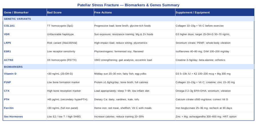
A patellar stress fracture is not a random event. Behind it lies a specific interaction of mechanical load, bone remodeling capacity, nutritional status, hormonal environment, and often, inherited predispositions that shaped your bone biology long before the injury occurred. The six biomarkers outlined here — vitamin D, P1NP, CTX, PTH, ferritin, and sex hormones — give you a measurable window into that biology. The five genetic variants, from COL1A1 to ACTN3, help explain why some people are more vulnerable and which interventions to prioritize.
The next smart step is to bring this information into a concrete conversation with a sports medicine physician, physiatrist, or functional medicine provider who is willing to run a targeted panel and interpret the results in the context of your training and history. Testing, not guessing, is what turns a generalized recovery plan into one that is genuinely calibrated to your biology — and that is the difference between recovering once and understanding how to stay well long-term.