This article contains AI generated content.
Patellar Stress Reaction: 5 Genes And 7 Biomarkers To Track
Anterior knee pain that flares every time you resume running, jumping, or squatting under load has a particular way of wearing people down. You rest, it settles. You return to training, it comes back within a few weeks, sometimes in almost the exact same spot on the front of the knee. An MRI eventually shows a stress reaction in the patella, a form of bone-marrow irritation that sits one step before a true stress fracture, and the standard advice is to offload the joint, reduce mileage, and wait.
That advice is not wrong, but it is incomplete. Two athletes with the same MRI grade, the same training load, and the same physiotherapy program can recover at very different speeds, and one of them may re-injure within a season while the other does not. Generic rest-and-strengthen protocols treat the bone as a passive structure that simply needs less stress. They rarely ask why this particular bone failed to keep up with a training load that plenty of other athletes tolerate without issue.
That "why" usually lives in biochemistry: how efficiently the body absorbs and uses calcium, whether bone is being broken down faster than it is rebuilt, whether hormone levels support bone formation, and whether energy availability is high enough for repair to happen at all. Some of it also lives in genetics, in the collagen and receptor genes that shape how strong and how adaptable a person's bone and connective tissue are in the first place.
None of this replaces a sports medicine evaluation or an MRI. But looking at the right blood work, and understanding what genetic tendencies might be working against you, turns a vague "rest it and hope" plan into a targeted one. The rest of this article walks through the laboratory markers worth checking, the genetic variants worth knowing about, a training philosophy that challenges the default rest-first mindset, and a short list of complementary approaches with real supporting evidence.
Summary
A patellar stress reaction is rarely just a training-load problem. It is often the visible result of an invisible mismatch between the mechanical demand placed on bone and the biological resources available to repair it, resources that show up in blood work long before they show up on an MRI. Vitamin D status, bone turnover markers, sex hormones, and iron stores can each independently explain why one runner's bone remodels fast enough to keep up with training while another's falls behind and starts to break down. Underneath those numbers, a handful of collagen and bone-density genes set the baseline strength and adaptability of the tissue itself, which is why two people with identical training loads can have completely different outcomes. This article breaks down the seven biomarkers most worth tracking, the five genes most likely to be involved, a bone-loading philosophy from a well-known longevity physician that pushes back against reflexive rest, and a short set of complementary techniques with genuine supporting evidence.
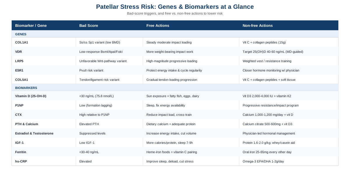
Seven Biomarkers That Explain Why Bone Isn't Keeping Up With Load
A stress reaction happens when the rate of bone breakdown (resorption) outpaces the rate of bone rebuilding (formation) at a specific, repeatedly loaded site. That balance is influenced by nutrient status, hormone levels, inflammation, and overall energy availability. The seven markers below are the ones sports medicine and endocrinology research keeps coming back to when explaining who gets bone stress injuries and who doesn't.
25-Hydroxyvitamin D
Vitamin D governs how much calcium the gut actually absorbs, which makes it the single most cited nutritional risk factor for bone stress injuries in athletes. Low 25(OH)D levels have been directly linked to stress fractures in both collegiate and adolescent athletes, and one prospective study found that a 25(OH)D level below roughly 75.8 nmol/L (about 30 ng/mL) meaningfully raised stress fracture risk, according to research summarized in a review of adolescent stress fractures linked to vitamin D insufficiency.
How to measure it: a standard 25-hydroxyvitamin D blood test, available through a physician or most direct-to-consumer lab services. Cost typically runs $30 to $80 out of pocket, and it is frequently bundled into broader athlete panels.
If the score is low, the plan without supplements: increase sensible, unprotected sun exposure (roughly 15 to 20 minutes midday, several times a week, skin-type dependent) and add vitamin D-rich foods such as fatty fish, egg yolks, and fortified dairy. Re-test after 8 to 12 weeks, since sunlight-driven changes are slow and seasonal.
If the score is low, the plan with supplements: vitamin D3 (cholecalciferol), typically 2,000 to 4,000 IU daily, paired with vitamin K2 and adequate dietary calcium to support proper mineral deposition rather than soft-tissue calcification. One pilot trial of high-risk collegiate athletes used a similar approach and reported fewer stress fractures over the study period, described in Vitamin D3 Supplementation and Stress Fractures in High-Risk Collegiate Athletes. Re-check levels at 3 months; doses above 4,000 IU/day for extended periods should be supervised, since excess vitamin D can raise blood calcium and, paradoxically, stress the kidneys rather than the bone.
Bone Turnover Markers: P1NP and CTX
P1NP (procollagen type 1 N-terminal propeptide) reflects how much new bone is being formed, while CTX (C-terminal telopeptide of type I collagen) reflects how much existing bone is being broken down. Measured together, they show whether a bone is in a net-building or net-losing state well before that shows up on imaging, which is why they are considered reference markers for fracture risk assessment, as outlined in guidance on standardizing CTX-I and P1NP measurement and supported by a large meta-analysis of bone turnover markers and fracture prediction.
How to measure it: a morning fasting blood draw, since both markers have daytime and meal-related variability. This is a specialty test, usually $100 to $250, ordered through a sports medicine physician, endocrinologist, or a specialty lab rather than a typical consumer panel.
If the ratio looks unfavorable (CTX high relative to P1NP), the plan without equipment or supplements: reduce total impact load temporarily (cross-training in a pool or on a bike), prioritize sleep, since most bone remodeling activity happens overnight, and address any underlying low energy availability, since under-eating relative to training load is one of the strongest drivers of a resorption-dominant state.
If the ratio looks unfavorable, the plan with supplements or equipment: a structured, progressive resistance and impact-loading program (detailed further in the training section below) is the single most evidence-backed way to shift the balance toward formation, since mechanical loading is what tells bone to rebuild. Calcium (1,000 to 1,200 mg/day from food where possible) and vitamin D support the raw materials for that rebuilding. Re-test bone turnover markers no sooner than 3 to 6 months, since bone remodels slowly and short-interval testing mostly captures noise.
Parathyroid Hormone and Calcium
Parathyroid hormone (PTH) is the body's calcium thermostat: when blood calcium drops, PTH rises and pulls calcium out of bone to compensate. Chronically elevated PTH, often from inadequate calcium or vitamin D intake, means bone is quietly being sacrificed to keep blood calcium stable, which is a mechanism directly implicated in reduced bone density in athletes.
How to measure it: intact PTH and total or ionized calcium, drawn together, roughly $40 to $90 combined through most labs.
If PTH is elevated, the plan without supplements: increase dietary calcium from dairy, leafy greens, canned fish with bones, or fortified plant milks to the 1,000 to 1,200 mg/day range, and confirm adequate protein intake, since protein supports the calcium-binding matrix of bone.
If PTH is elevated, the plan with supplements: calcium citrate or calcium carbonate (500 to 600 mg per dose, since absorption drops at higher single doses) taken with meals, alongside vitamin D3 to support absorption. Avoid combining large calcium doses with iron supplements at the same time, since they compete for absorption; space them by several hours. Re-test PTH and calcium after correcting vitamin D first, since low vitamin D is the most common upstream driver.
Estradiol and Testosterone
Sex hormones are directly bone-protective: estradiol restrains bone resorption in both women and men, and testosterone supports bone formation and muscle mass that offloads bone. Athletes with low energy availability, whether from intentional or unintentional under-eating, often show suppressed estradiol (in women) or suppressed testosterone (in men), which is one of the clearest biological pathways to a bone stress injury. A recent literature review of biomarkers used to diagnose relative energy deficiency in sport confirms that low estradiol, low progesterone, and reduced testosterone are consistently associated with low energy availability states and impaired bone health.
How to measure it: estradiol and/or total and free testosterone via blood draw, roughly $50 to $150 depending on the panel. For menstruating women, timing relative to the cycle matters and should be discussed with a provider; missed or irregular periods are themselves a red flag worth reporting even before lab work.
If levels are low, the plan without supplements or equipment: the first-line fix is almost always increasing energy intake relative to training load, particularly carbohydrate around workouts, since low energy availability is the most common reversible cause. Reducing training volume temporarily while intake catches up is often necessary, and menstrual irregularities in female athletes should prompt a conversation with a physician rather than a wait-and-see approach.
If levels are low, the plan with supplements or equipment: there is no supplement shortcut for hormone-suppressing energy deficits; supplements do not fix a caloric problem. Where a clinical hormonal deficiency is confirmed by a physician (rather than assumed), medical management is a physician-led decision, not a self-directed supplement plan. This is one of the few areas in this article where professional oversight, not a home protocol, is the responsible next step.
IGF-1
Insulin-like growth factor 1 (IGF-1) is bone-trophic, meaning it directly supports osteoblast activity and new bone formation. It drops in states of low energy availability and chronic under-fueling, which is part of why RED-S is so damaging to bone health over time, as described in the same RED-S biomarker review.
How to measure it: a fasting IGF-1 blood test, roughly $80 to $150, best interpreted alongside overall energy intake rather than in isolation.
If IGF-1 is low, the plan without supplements: increase total caloric and protein intake, particularly around training sessions, and prioritize 7 to 9 hours of sleep, since growth hormone (which drives IGF-1 production) is released predominantly during deep sleep.
If IGF-1 is low, the plan with supplements or equipment: adequate protein (roughly 1.6 to 2.0 g/kg body weight per day) from food is the most reliable lever; a whey or casein supplement can help close a gap if whole-food intake is inconsistent, taken once daily post-training or before bed, with no evidence of benefit from exceeding normal protein targets. There is no well-supported case for IGF-1-boosting supplements outside of addressing the underlying energy deficit.
Ferritin and Iron Studies
Iron supports collagen synthesis and oxygen delivery to healing tissue, and low ferritin is common in endurance athletes, especially menstruating women. While the direct causal chain from low iron to bone stress injury is still being worked out, low ferritin clusters with the same low-energy-availability picture that damages bone, and a recent systematic review of iron deficiency in female athletes confirms both the high prevalence and the performance and recovery consequences of going untreated.
How to measure it: a ferritin and iron panel (ferritin, serum iron, transferrin saturation), typically $40 to $90. Ferritin is an inflammation-sensitive marker, so a recent illness or hard training block can temporarily distort it.
If ferritin is low (commonly cited thresholds are below 30 to 40 ng/mL for female endurance athletes), the plan without supplements: increase heme-iron food sources (red meat, poultry, fish) and pair plant iron sources with vitamin C to improve absorption; avoid drinking coffee or tea immediately with iron-rich meals, since polyphenols block absorption.
If ferritin is low, the plan with supplements: oral iron (ferrous sulfate or a gentler bisglycinate form), commonly 25 to 65 mg of elemental iron every other day rather than daily, since alternate-day dosing improves absorption and reduces gastrointestinal side effects according to modern iron-repletion research. Common side effects include constipation and stomach upset; take with food if needed, away from calcium and dairy. Re-test ferritin at 8 to 12 weeks; iron supplementation should be guided by an actual deficiency, not taken preventively, since excess iron carries its own long-term health risks.
hs-CRP
High-sensitivity C-reactive protein is a general inflammation marker. It won't diagnose a stress reaction, but a persistently elevated hs-CRP during a rehab period can flag systemic inflammation, illness, or overtraining that is competing with the bone's ability to repair itself.
How to measure it: a simple, widely available blood test, usually $15 to $40, often included in routine metabolic panels.
If hs-CRP is elevated, the plan without supplements: review sleep quality, training load, and life stress, since all three independently raise systemic inflammation; a short deload week is often more useful than any single intervention.
If hs-CRP is elevated, the plan with supplements or equipment: omega-3 fatty acids (roughly 1 to 2 g EPA/DHA combined daily) have modest, well-tolerated anti-inflammatory evidence in general populations; take with a meal to reduce fishy aftertaste and mild GI upset. This is a supporting measure, not a substitute for addressing training load, sleep, or an underlying illness.
What Your Genes May Be Doing In The Background
Blood work captures what's happening right now, but some of the variation in who develops a patellar stress reaction, and who recovers quickly versus repeatedly re-injures, is written into collagen and bone-density genes that never change. Researchers like Ali Torkamani, whose work on genomic risk interpretation popularized reading these variants for everyday athletes, and Gary Brecka, known for translating functional genomic markers into practical protocols, have both pushed the idea that knowing your baseline collagen and bone genetics changes how aggressively you should manage the modifiable side (vitamin D, loading, calcium) rather than assuming a one-size-fits-all recovery timeline. The evidence behind each gene below ranges from well-replicated to early-stage, and that distinction matters for how much weight to put on any single result.
COL1A1 (Collagen Type I Alpha 1)
COL1A1 codes for the main structural collagen in bone. A well-studied variant at a binding site for the Sp1 transcription factor (commonly reported as "Ss" or "ss" genotypes) is associated with lower bone mineral density and a higher fracture risk, an association strong and consistent enough to be confirmed in a meta-analysis of over 7,800 participants. Mechanistic work suggests the effect on fracture risk is larger than what bone density alone would predict, pointing to a separate effect on bone quality, as shown in research on how this polymorphism affects bone density and quality. This is one of the better-replicated gene-bone associations in the literature, though most of the underlying data comes from osteoporosis and fracture cohorts rather than athletic stress injuries specifically.
If the gene variant looks unfavorable, the plan without supplements or equipment: treat vitamin D and calcium intake as non-negotiable rather than optional, since a weaker collagen scaffold has less margin for nutritional shortfalls; prioritize consistent, moderate impact loading (walking, light jogging, jump-rope) year-round rather than large seasonal swings in training volume, which is harder on a less resilient matrix.
If the gene variant looks unfavorable, the plan with supplements or equipment: vitamin C (which is required for collagen cross-linking) from food or a modest 500 mg/day supplement, and collagen peptides (roughly 15 g/day, ideally taken 30 to 60 minutes before loading exercise) have some early evidence for supporting connective tissue, though evidence in bone specifically is thinner than in tendon. There are no notable side effects at these doses; the main caution is not substituting supplements for addressing training load and vitamin D status.
VDR (Vitamin D Receptor)
The VDR gene doesn't change how much vitamin D you have, it changes how effectively your cells respond to the vitamin D that's there. Certain VDR genotypes (variants at sites called BsmI, ApaI, and FokI are the most studied) have been linked to differences in bone density, circulating osteocalcin, and PTH in healthy adolescents, as shown in research on VDR polymorphism and bone density in adolescent girls, and other work has found that the bone density benefit of weight-bearing exercise itself varies by VDR genotype in young athletes. In practice, this means two people can have identical vitamin D blood levels and get different amounts of bone benefit from that same level.
If the gene variant looks unfavorable, the plan without supplements or equipment: lean harder on weight-bearing, impact-based exercise rather than relying on vitamin D correction alone, since the research suggests mechanical loading and vitamin D receptor activity interact, and consistent impact training may partly offset a less responsive receptor.
If the gene variant looks unfavorable, the plan with supplements or equipment: aim for the higher end of a normal 25(OH)D range (40 to 60 ng/mL rather than the bottom of "sufficient") under a physician's guidance, on the logic that a less efficient receptor may need more circulating substrate to achieve the same effect; re-test every 3 to 4 months rather than assuming correction is permanent, since this is a compensation strategy, not a fix to the receptor itself.
LRP5 (LDL Receptor-Related Protein 5)
LRP5 is a central player in the Wnt signaling pathway that governs osteoblast activity, the cells responsible for building new bone. Genome-wide association studies have repeatedly identified LRP5 as one of the strongest single genetic loci for bone mineral density and fracture risk, with a Bayesian meta-analysis confirming associations between specific LRP5 variants and bone density across multiple studies. Rare, severe LRP5 mutations cause dramatic bone disease in either direction (very low or very high bone mass); the common variants relevant to most people have a much smaller, incremental effect.
If the gene variant looks unfavorable, the plan without supplements or equipment: since LRP5 acts through mechanical loading signals in the Wnt pathway, progressive, high-magnitude loading (not just cardio) is the most direct lever available without medication; this is discussed further in the loading-focused strategy below.
If the gene variant looks unfavorable, the plan with supplements or equipment: there is no supplement that reliably activates Wnt signaling in a way current evidence supports for healthy adults; the most defensible option is equipment-based, such as a weighted vest for daily walking or a home setup for basic resistance training, used to consistently apply the mechanical stimulus the pathway responds to. No meaningful side effects, aside from the same overuse risk that applies to any progressive loading program if increased too quickly.
ESR1 (Estrogen Receptor Alpha)
ESR1 codes for the estrogen receptor that mediates estrogen's protective effect on bone. The PvuII variant in particular has been associated with hip fracture risk in a meta-analysis of ESR1 polymorphisms and fracture risk, with the effect present in men as well as women, since men also rely on estrogen (converted from testosterone) for bone protection. The evidence is described by the researchers themselves as still limited by small sample sizes in individual studies, so this is a moderate-confidence association rather than a settled one.
If the gene variant looks unfavorable, the plan without supplements or equipment: protecting energy availability and, in women, menstrual regularity becomes even more important, since a receptor that is already less efficient at using estrogen has less room to also cope with low estrogen levels from under-fueling.
If the gene variant looks unfavorable, the plan with supplements or equipment: there is no supplement that meaningfully changes estrogen receptor sensitivity. The practical response is tighter monitoring of the estradiol/testosterone biomarker above, and a lower threshold for involving a physician if menstrual cycles become irregular or testosterone trends low, rather than attempting to self-manage a receptor-level issue.
COL5A1 (Collagen Type V Alpha 1)
COL5A1 regulates the diameter and assembly of collagen fibers in tendons and ligaments, and by extension the periosteum and connective tissue surrounding bone. Certain COL5A1 variants are consistently associated with tendon and ligament injury risk in a meta-analysis of COL5A1 polymorphisms and tendon-ligament injuries in Caucasian populations. This gene is more directly tied to soft tissue than to bone mineral density itself, but because the patella is a heavily tendon-loaded bone (via the quadriceps and patellar tendons), a less favorable COL5A1 profile may partly explain why the surrounding soft tissue transmits more strain to the bone underneath it.
If the gene variant looks unfavorable, the plan without supplements or equipment: build in a longer, more gradual tendon-loading progression before increasing running or jumping volume, since less adaptable collagen fibers typically need more time under controlled load to remodel safely; isometric quadriceps holds (progressing gradually in duration and load) are a low-risk starting point.
If the gene variant looks unfavorable, the plan with supplements or equipment: the same vitamin C plus collagen peptide approach described under COL1A1 has the most direct rationale here, since it targets collagen synthesis broadly; a foam roller or massage tool used for short sessions (a few minutes, a few times a week) can support soft-tissue quality around the patella as a low-cost complement, without replacing an actual loading progression.
The Bone-Loading Idea That Pushes Back Against "Just Rest It"
Peter Attia's book Outlive spends a significant portion of its musculoskeletal argument on a theme that runs counter to how many stress injuries are still managed by default: that bone and connective tissue, much like cardiovascular fitness, respond to progressive mechanical stress rather than to prolonged avoidance of it. That framework is directly relevant to a patellar stress reaction, where the instinctive plan is often extended, unstructured rest rather than a graded return to loading. Below are ten of the book's most load-bearing ideas, adapted to this specific context.
The "Marginal Decade" Framing
Attia argues that most people lose function not gradually but in a compressed final stretch of life, and that the structural capacity you build decades earlier determines how much you have left to lose. Applied here, a stress reaction in your 20s or 30s is an early signal worth taking seriously, not a nuisance to push through and forget.
Bone Density Has A Ceiling Age
Peak bone mass is typically reached by the late twenties, after which the trend is gradually downward. This is a strong argument for treating a stress reaction as a prompt to build bone reserve now, through loading and nutrition, rather than assuming it will simply resolve on its own.
Total Avoidance Of Load Is Its Own Risk
The book is explicit that prolonged, complete offloading (extended non-weight-bearing rest, in the extreme) can itself reduce bone density, since bone remodels in response to the absence of stress just as it does to its presence. The practical implication for a stress reaction is that the right question isn't "rest or not," it's "how much load, at what intensity, right now."
Mechanotransduction Rebuilds Bone
Bone responds to mechanical strain through a signaling process (mechanotransduction) that triggers osteoblast activity, essentially the biological basis of Wolff's Law. Controlled, incrementally increasing load is what tells bone to lay down new tissue at the exact site under strain, which is the rationale behind graded return-to-run and return-to-jump protocols rather than open-ended rest.
Muscle Is Protective Armor For Bone
A recurring point in the book is that muscle mass and strength act as shock absorbers that reduce the force transmitted directly to bone and joints. For a patellar stress reaction, quadriceps and hip strength work isn't cosmetic, it's load management.
Stability Training Reduces Injury Risk From Repetitive Single-Limb Loading
Running and jumping are single-limb-dominant activities, and Attia emphasizes stability and balance training as an underrated injury-prevention lever, since poor single-leg control changes how force travels through the knee on every stride or landing.
Zone 2 Training Supports The Recovery Environment
Aerobic base training (sustained, easy-effort cardio) is framed less as a performance tool and more as a way to improve mitochondrial efficiency and tissue oxygenation broadly, which plausibly supports the metabolic environment tissue repair depends on, even though this specific link to bone healing is more theoretical than proven.
Objective Testing Beats Waiting For Injury
A core argument of the book is that most people only discover a structural weakness after it becomes an injury. Applied to bone stress injuries, this favors periodic strength testing, gait analysis, or even a baseline DEXA scan for athletes with a prior stress injury, rather than waiting for the next flare-up to reveal the same weakness again.
Protein Timing Around Training Supports Repair
Attia is a proponent of distributing adequate protein intake across the day, with particular attention around training sessions, on the logic that tissue repair, bone included, is substrate-dependent and benefits from a steady rather than sporadic amino acid supply.
Sleep Is A Limiting Factor, Not A Luxury
Sleep is presented as one of the most underrated recovery tools available, given its role in growth hormone release and tissue repair broadly. For a bone that is actively trying to remodel, inconsistent or insufficient sleep is a direct constraint on that process, not a separate wellness issue.
The Goal Is A Graded Return, Not A Cleared-Or-Not Binary
Perhaps the most practically useful shift in mindset is moving away from treating recovery as a single "cleared to return" moment and toward a structured, symptom-monitored progression of load, which is closer to how modern bone stress injury rehabilitation protocols are actually built.
Complementary Approaches Worth Considering
None of the following replace load management, nutrition, or a sports medicine evaluation, and the evidence for each is drawn mostly from closely related conditions (patellar tendinopathy, patellofemoral pain) rather than bone stress reactions specifically, since dedicated trials on patellar bone stress reactions are scarce. They are worth knowing about as add-ons, not centerpieces.
Photobiomodulation (Low-Level Laser Therapy)
Photobiomodulation uses low-level red or near-infrared light, theorized to support mitochondrial activity and reduce local inflammation in irritated tendon and bone tissue. It is relevant here because the anterior knee is a common target for this therapy in exactly the kind of overuse presentations that overlap with patellar stress reactions.
A systematic review and meta-analysis of photobiomodulation for tendinopathy found that when combined with exercise, it produced greater pain reduction and functional improvement than exercise alone, though the review is also candid that overall evidence quality remains mixed, with some individual trials showing no clear benefit.
Realistically, this is best used as an adjunct alongside, not instead of, a structured loading program, typically applied two to three times per week over several weeks under the guidance of a physical therapist with access to the equipment; it is not something to rely on as a standalone fix, and expectations should stay modest given the mixed trial quality.
Biofeedback-Guided Gait Retraining
Biofeedback in this context means using real-time visual or auditory feedback (from motion sensors or video) to help a runner consciously adjust their movement pattern, most often hip and knee alignment during stance phase. It's relevant because altered hip mechanics are a well-documented contributor to abnormal patellofemoral loading, the same joint region implicated in patellar stress reactions.
The foundational study here, real-time gait retraining on hip kinematics, pain and function in patellofemoral pain syndrome, found that runners who received real-time feedback on hip adduction during treadmill running showed improved mechanics and reduced pain over eight sessions, with feedback gradually withdrawn as the new pattern became automatic.
Applying this practically means working with a physical therapist or running coach who has motion-capture or even simple mirror/video feedback tools, over a structured multi-week block (the original protocol used eight sessions), rather than attempting to self-diagnose a gait fault; overcorrecting a movement pattern without professional guidance can shift stress to a different structure rather than resolving it.
Massage Therapy
Massage therapy applied to the quadriceps, IT band, and surrounding soft tissue is often used to reduce muscular tension that may be increasing tensile load on the patellar tendon and, by extension, the underlying bone. The rationale is mechanical rather than purely relaxation-based: tighter quadriceps and lateral structures can alter patellar tracking and loading patterns.
Evidence specific to bone stress reactions is limited, and most supporting research comes from broader overuse-injury and tendinopathy populations rather than controlled trials on patellar stress reactions themselves, so this should be considered a plausible, low-risk adjunct rather than a well-proven intervention.
In practice, this looks like regular sessions (weekly to biweekly during a rehab phase) focused on the quadriceps and lateral thigh structures, ideally coordinated with, not substituted for, the loading and strengthening work a physical therapist prescribes; it carries minimal risk beyond routine post-massage soreness.
Conclusion
A patellar stress reaction that keeps coming back is rarely explained by training load alone. Vitamin D status, bone turnover balance, sex hormones, iron stores, and inflammation each tell part of the story, and inherited collagen and bone-density genes set the baseline that biomarkers are working against. None of these pieces is a diagnosis on its own, and none of them replaces an actual clinical evaluation, but together they turn "rest and hope it doesn't come back" into a plan built around your own biology.
The most useful next step is usually the most boring one: get the core panel checked (vitamin D, calcium and PTH, and a sex hormone panel at minimum), track how your symptoms respond as you reintroduce load in a structured, graded way, and bring both the numbers and the pattern of pain to a sports medicine physician or physical therapist who can put them in context. Genetics can inform how cautious or aggressive to be with that plan, but the biomarkers and how your knee actually responds to load are what should drive the decisions in the meantime.