This article contains AI generated content.
· UpdatedPatellofemoral Arthritis Genes and Biomarkers – 4 Genes and 7 Biomarkers to Track
Introduction
If you have been told you have patellofemoral arthritis, you have probably received the standard advice: lose weight, strengthen your quadriceps, avoid stairs when flared, and take anti-inflammatories as needed. That advice is not wrong, but for a large number of people it simply is not enough. Pain persists, function declines, and the underlying process continues quietly — often because nothing is being tracked and nothing is being corrected at a deeper level.
Patellofemoral arthritis involves the gradual breakdown of cartilage between the kneecap and the femur. Unlike tibiofemoral osteoarthritis, which tends to get more research attention, the patellofemoral compartment is biomechanically unique: it is under enormous compressive load during everyday activities like sitting, climbing, or squatting, and it is highly sensitive to subtle changes in alignment, inflammation, and cartilage metabolism. Generic protocols miss these nuances entirely.
What makes this condition especially worth investigating at a molecular level is that two people with the same X-ray finding can have radically different trajectories — one stabilizes, the other deteriorates. That difference is rarely random. It reflects differences in their inflammatory signaling, their cartilage turnover rate, their metabolic state, and in some cases their genetic predispositions. Tracking the right numbers can reveal which of those factors are actively driving harm in your specific situation.
This article takes a more targeted approach. The first section covers the seven most useful biomarkers to track if you have patellofemoral arthritis — what they measure, how to get them tested, what a concerning result looks like, and what to do about it, with and without supplementation. A second section covers the four most relevant gene variants identified in osteoarthritis research, along with practical responses for each. A third section summarizes key insights from Peter Attia's Outlive, which provides an unusually rigorous framework for thinking about musculoskeletal longevity. Finally, a section on complementary approaches covers the modalities with the clearest clinical evidence for this specific condition. Better information genuinely does lead to better decisions — and this article is designed to give you that information.
7 Biomarkers to Track for Patellofemoral Arthritis
Most people with joint pain never get beyond a basic inflammatory panel, if they get any blood work at all. But several biomarkers — some inexpensive and widely available, others more specialized — can give you a much sharper picture of what is driving your condition and how fast it may be progressing. Here are the seven most clinically meaningful ones for patellofemoral arthritis.
1. High-Sensitivity C-Reactive Protein (hs-CRP)
Why it matters: hs-CRP is the most accessible systemic inflammation marker, and chronic low-grade inflammation is now understood to be a central driver of cartilage degradation in osteoarthritis — not just a consequence of it. In patellofemoral arthritis specifically, elevated hs-CRP correlates with accelerated synovial inflammation and worse pain scores. The distinction between standard CRP and high-sensitivity CRP matters: the hs version detects inflammation in the range that predicts metabolic and musculoskeletal risk, typically below 10 mg/L where standard CRP reads zero.
How to measure it: A simple blood draw, available at any standard lab. Cost typically ranges from $10–$40 depending on your country and whether ordered by a physician or via direct-to-consumer services. Target: below 1.0 mg/L. Above 3.0 mg/L is associated with significantly elevated disease activity.
If the score is bad — without supplements: Reducing inflammatory load through diet is the single most evidence-backed free intervention. This means eliminating refined seed oils (soybean, corn, sunflower), reducing ultra-processed carbohydrates, prioritizing omega-3 rich whole foods (fatty fish, walnuts, flaxseed), and addressing sleep quality aggressively. Regular zone-2 cardiovascular exercise (3–5 sessions per week of 30–45 minutes at conversational intensity) has been shown in multiple RCTs to reduce hs-CRP independent of weight loss. Losing visceral fat, even 5–10% of body weight, produces dramatic CRP reductions.
If the score is bad — with supplements and equipment: Fish oil at 2–4g EPA+DHA per day is one of the best-studied anti-inflammatory interventions, with meta-analyses consistently showing hs-CRP reduction. Use it for at least 12 weeks before reassessing. Frequency: daily, indefinite. Side effects: fishy breath, loose stools at high doses — take with meals. Curcumin with piperine (500–1000mg/day in a phospholipid form like Meriva or BCM-95) has shown CRP reduction in OA patients in several RCTs. Cycle: 8–12 weeks on, reassess, can be used continuously at lower doses. Side effects: GI upset at high doses; avoid with blood thinners at high doses.
2. Cartilage Oligomeric Matrix Protein (COMP)
Why it matters: COMP is a glycoprotein released into the bloodstream when cartilage is under excessive mechanical stress or is actively degenerating. Serum COMP levels are among the most specific available markers of cartilage damage in the knee. Importantly for patellofemoral arthritis, COMP rises with compressive loading abnormalities — meaning it can flag mechanical problems before imaging changes become obvious. It is also used in some research settings to monitor response to treatment.
How to measure it: Serum COMP requires a specialized lab; it is not on standard panels. Cost is typically $80–$200 and usually requires a physician's order. Reference ranges vary by lab, but elevated values above approximately 12–15 U/L in middle-aged adults are generally considered significant. Some direct-to-consumer longevity clinics include it in premium panels.
If the score is bad — without supplements: The most direct free intervention is reducing compressive loading on the patellofemoral joint. This means substituting high-impact activities (running, jumping, stair climbing) with low-impact alternatives (swimming, cycling at a comfortable seat height, water aerobics) while still maintaining quadriceps and VMO strength. Taping techniques (McConnell taping) and patellar tracking braces can redistribute load acutely. Losing excess body weight reduces compressive forces significantly: every kilogram lost removes roughly 3–4kg of patellofemoral load during stair descent.
If the score is bad — with supplements and equipment: Unloader braces designed specifically for the patellofemoral joint (not generic knee sleeves) can reduce compressive load during daily activities and are worth the investment if COMP remains elevated despite activity modification. Collagen peptides (10–15g/day with vitamin C, taken 30–60 minutes before exercise) have shown promising results in supporting cartilage matrix integrity in several RCTs. Frequency: daily with pre-exercise timing. Cycle: ongoing; evidence suggests benefits build over 3–6 months. Side effects: minimal; source from quality suppliers to avoid contaminants.
3. CTX-II (C-Terminal Cross-Linking Telopeptide of Type II Collagen)
Why it matters: CTX-II is a urine-based biomarker that reflects the breakdown rate of type II collagen — the primary structural collagen in joint cartilage. Elevated CTX-II indicates that cartilage is being degraded faster than it can be repaired, making it one of the most direct available measures of cartilage catabolism. Several prospective studies have shown that elevated urinary CTX-II predicts radiographic progression of knee OA over time, independent of pain levels.
How to measure it: Measured in a first-morning urine sample, typically by ELISA. Cost ranges from $100–$250 at specialty labs. Results are usually expressed as ng/mmol creatinine. Values above the 75th percentile of age/sex norms are clinically significant. Some integrative medicine and longevity-focused clinics now include CTX-II in their osteoarthritis monitoring panels.
If the score is bad — without supplements: The most evidence-backed non-supplement approach to reducing CTX-II is correcting biomechanical loading patterns. This includes gait retraining (with a physical therapist if possible) to reduce knee valgus and patellar maltracking, and prioritizing low-impact strengthening over high-impact loading. Adequate sleep is underrated here: cartilage regeneration occurs primarily during slow-wave sleep, and sleep deprivation increases catabolic signaling systemically.
If the score is bad — with supplements and equipment: Undenatured type II collagen (UC-II, 40mg/day) — distinct from hydrolyzed collagen peptides — has shown significant reductions in joint pain and functional improvement in knee OA in RCTs, and operates through an oral tolerance mechanism that may modulate cartilage autoimmunity. Frequency: daily on an empty stomach. Cycle: minimum 12 weeks to assess; often used indefinitely. Side effects: rare; mild GI in some individuals. Avocado-soybean unsaponifiables (ASU, 300mg/day) have specific evidence for reducing CTX-II in knee OA clinical trials. Cycle: 12 weeks on, 4 weeks off. Side effects: generally well tolerated; avoid in soy allergies.
4. Vitamin D (25-OH-D)
Why it matters: Vitamin D receptor is expressed in chondrocytes, synovial cells, and osteoblasts. Deficiency disrupts the regulation of both cartilage metabolism and subchondral bone remodeling — a process that is intimately connected to patellofemoral arthritis progression. Low 25-OH-D levels have been associated in epidemiological studies with greater cartilage loss, lower muscle strength (particularly in the quadriceps), and increased systemic inflammation. Peter Attia consistently places optimal vitamin D in his longevity biomarker stack, targeting 40–60 ng/mL rather than the basic sufficiency threshold of 20 ng/mL.
How to measure it: Standard blood test, available everywhere. Cost: $20–$60. Optimal target for joint health is broadly considered to be 40–60 ng/mL (100–150 nmol/L). Below 30 ng/mL is deficient; below 20 ng/mL represents significant clinical deficiency.
If the score is bad — without supplements: Safe sun exposure remains the most natural route: 10–20 minutes of midday sun on arms and legs (without sunscreen) three to four times per week can meaningfully raise levels during spring and summer months in temperate climates. Dietary sources (fatty fish, egg yolks, fortified foods) contribute modestly. For most people with genuine deficiency, sun and diet alone are insufficient to reach optimal levels.
If the score is bad — with supplements and equipment: Vitamin D3 supplementation (2,000–5,000 IU/day) is one of the safest and most cost-effective interventions available. Always co-supplement with vitamin K2 (100–200mcg MK-7 form) to direct calcium appropriately and avoid arterial calcification risk. Frequency: daily with a fat-containing meal. Cycle: indefinite, with retesting every 3–6 months to avoid toxicity (which begins above approximately 100–120 ng/mL). Side effects: none at physiological doses; hypercalcemia is theoretically possible only at very high chronic doses without K2.
5. Interleukin-6 (IL-6)
Why it matters: IL-6 is a key pro-inflammatory cytokine that drives synovial inflammation in osteoarthritis. In the patellofemoral joint, elevated IL-6 accelerates chondrocyte apoptosis and suppresses cartilage matrix synthesis. Serum IL-6 also correlates with pain sensitization through central mechanisms — meaning it may contribute not just to joint damage but to the amplification of pain signals in the nervous system. Tracking it separately from hs-CRP provides complementary information: CRP is downstream of IL-6, so IL-6 can be elevated when CRP appears borderline.
How to measure it: Serum IL-6 is available at specialty and reference labs. Cost: approximately $50–$150. Normal levels in healthy adults are typically below 7 pg/mL. Chronic low-grade elevation in the range of 2–7 pg/mL is increasingly recognized as clinically meaningful.
If the score is bad — without supplements: The most powerful free interventions for IL-6 are resistance exercise (paradoxically, acute IL-6 rises after exercise but chronic training reduces baseline levels), sleep optimization (even partial sleep restriction significantly elevates IL-6 the following day), and stress reduction (psychological stress elevates IL-6 through the HPA axis). Reducing visceral adiposity is critical: adipose tissue, particularly visceral fat, is a major IL-6 secretion site.
If the score is bad — with supplements and equipment: Magnesium glycinate or malate (300–400mg elemental magnesium/day) has shown moderate anti-inflammatory effects including IL-6 reduction in deficient individuals. Tart cherry extract or fresh tart cherry juice has demonstrated IL-6 reduction in several exercise and OA studies. Frequency: daily. Cycle: can be used continuously. Side effects: magnesium — loose stools if dose is too high; tart cherry — high in natural sugars if using juice, watch glycemic load.
6. Leptin
Why it matters: Leptin is a hormone produced by adipose tissue that has recently been identified as a significant driver of osteoarthritis pathology — particularly in the patellofemoral compartment. Leptin receptors are expressed in chondrocytes, and elevated leptin levels directly stimulate the production of matrix-degrading enzymes (MMPs), prostaglandins, and nitric oxide within cartilage. There is a notable sex difference: women tend to have higher leptin-to-adiponectin ratios, which may partly explain their higher patellofemoral OA prevalence. Elevated leptin is both a metabolic signal and a direct cartilage toxin.
How to measure it: Fasting serum leptin, available at most labs. Cost: $30–$80. Optimal fasting leptin is generally considered below 10 ng/mL for men and below 15 ng/mL for women, though these thresholds are debated. High leptin despite a normal BMI can indicate leptin resistance — a more complex situation requiring adiponectin co-testing.
If the score is bad — without supplements: Leptin is almost entirely governed by adipose tissue mass and insulin sensitivity. Reducing fasting insulin through dietary carbohydrate modification and time-restricted eating (a 14–16 hour overnight fast) lowers leptin over weeks to months. Intermittent fasting protocols have consistently shown leptin reduction in RCTs. High-intensity interval training (HIIT, 2–3 sessions per week) improves leptin sensitivity and reduces adipose volume.
If the score is bad — with supplements and equipment: Berberine (500mg two to three times daily with meals) reduces insulin resistance and leptin levels in several RCTs involving metabolic syndrome. Cycle: 8 weeks on, 4 weeks off (to avoid gut microbiome disruption). Side effects: significant GI effects in some; do not combine with metformin without medical supervision. Alpha-lipoic acid (600mg/day) has shown leptin-lowering effects in combination with caloric restriction. Frequency: daily. Side effects: mild GI; possible biotin depletion with long-term use.
7. Matrix Metalloproteinase-3 (MMP-3)
Why it matters: MMP-3 (stromelysin-1) is an enzyme that degrades collagen and proteoglycans in cartilage and synovial tissue. Elevated serum MMP-3 is one of the most specific available markers of active joint tissue breakdown, and it is measurable before significant structural damage appears on MRI. In patellofemoral arthritis, elevated MMP-3 signals that the remodeling balance has tipped toward destruction — a state that is potentially reversible with targeted interventions but will worsen silently without them.
How to measure it: Serum MMP-3 can be ordered through specialty labs and some integrative medicine clinics. Cost: approximately $80–$180. Normal adult values are approximately 17–59 ng/mL; levels above 60–80 ng/mL in a joint-pain context are generally considered clinically elevated.
If the score is bad — without supplements: Mechanical unloading strategies (activity modification, patellar bracing, gait retraining) directly reduce the stimuli for MMP-3 secretion by reducing abnormal compressive and shear forces on the cartilage matrix. Dietary approaches that reduce insulin and inflammatory cytokine load (which are upstream regulators of MMP secretion) will also lower MMP-3 over time. Cold water immersion or contrast therapy post-exercise has emerging evidence for reducing post-exercise MMP elevations.
If the score is bad — with supplements and equipment: Doxycycline at subantimicrobial doses is actually the most rigorously studied MMP inhibitor for OA, but requires a physician's prescription and is not a first-line choice due to side effects with long-term use. More accessible: Boswellia serrata extract (AKBA standardized, 100–250mg/day) has shown direct MMP-3 inhibition in synovial tissue in human clinical trials. Cycle: 8–12 weeks on, 4 weeks off. Side effects: mild GI; rare hepatic effects at high doses. Green tea extract (EGCG, 400–800mg/day) has demonstrated MMP inhibition in cartilage cell studies and small clinical trials.
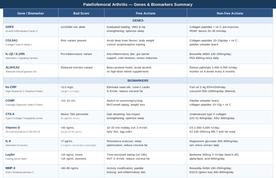
Summary Table: Genes and Biomarkers at a Glance
The following table summarizes all genes and biomarkers covered in this article, including target values and the primary free and paid interventions.
4 Key Genes Linked to Patellofemoral Arthritis
Genetics do not determine destiny in osteoarthritis, but they can significantly shift your baseline risk and the rate of progression. Understanding which gene variants may be active in your biology allows you to prioritize the interventions that matter most for your specific profile. Genetic testing (through services like 23andMe, combined with interpretation tools, or through clinical genetic panels) is now accessible enough to make this actionable.
GDF5 – Growth Differentiation Factor 5
What it does: GDF5 encodes a signaling protein critical for joint formation, cartilage repair, and the maintenance of chondrocyte viability. The rs143384 variant in the GDF5 promoter region is one of the most robustly replicated genetic risk factors for knee osteoarthritis in the world, with association confirmed across European, Asian, and other populations. The risk allele reduces GDF5 expression, leading to reduced cartilage repair capacity and thinner articular cartilage, particularly in the patellofemoral compartment. Research published in Nature Genetics was among the first to establish this association with functional cartilage consequences.
If the gene variant is present — plan without supplements: Since GDF5 governs repair signaling, the most important free intervention is ensuring that cartilage repair stimuli are present. This means regular, graduated mechanical loading — not rest. Chondrocytes require appropriate compression to synthesize matrix proteins, but excessive or uneven loading suppresses GDF5 signaling. A physical therapist-guided exercise program emphasizing proper patellar tracking, VMO activation, and hip abductor strengthening is essential. Sleep quality is non-negotiable: GDF5 signaling interacts with growth hormone pathways that are most active during deep sleep.
If the gene variant is present — plan with supplements or equipment: Collagen peptides with vitamin C (taken pre-exercise) provide the raw substrate that GDF5-mediated signaling uses to build cartilage matrix. Bioelectrical stimulation devices designed for cartilage repair (such as pulsed electromagnetic field therapy, PEMF) have shown modest but real effects on cartilage anabolism in GDF5-deficient tissue in preclinical models and are increasingly available as consumer devices. Frequency: PEMF 20–30 minutes daily. Cycle: 8–12 weeks on, ongoing as maintenance. Side effects: PEMF is generally considered safe; avoid with pacemakers.
COL9A1 – Collagen Type IX Alpha-1
What it does: Type IX collagen acts as a cross-linking molecule between type II collagen fibrils in articular cartilage, providing tensile resistance and structural integrity. Variants in COL9A1 have been associated with earlier onset and more rapid progression of knee OA, partly because they reduce the mechanical resilience of the cartilage matrix. A cartilage that is structurally weaker at the collagen fibril level is more vulnerable to the compressive and shear forces that patellofemoral arthritis generates.
If the gene variant is present — plan without supplements: Reducing peak compressive loading on the patellofemoral joint is the highest priority. Avoid deep knee flexion under load (deep squats, lunges below 90 degrees, stair climbing with heavy loads). Optimize body weight — the patellofemoral joint sustains 3–8 times body weight during squatting. A patellofemoral unloader brace during high-demand activities can directly reduce the mechanical stress that exploits this collagen vulnerability. Proprioceptive training (balance board, single-leg stance work) reduces cumulative micro-trauma.
If the gene variant is present — plan with supplements or equipment: Hydrolyzed collagen peptides (10–15g/day) provide building blocks that may partially compensate for reduced intrinsic collagen quality. Vitamin C (500–1000mg/day) is essential as a cofactor in collagen hydroxylation — without adequate vitamin C, new collagen is structurally inferior. Frequency: daily, with exercise timing. Cycle: indefinite; these are substrate-level supports. Side effects: minimal at these doses. Silica supplementation (from horsetail extract, orthosilicic acid) is sometimes used to support connective tissue synthesis; evidence is early but mechanistically plausible.
IL-1β / IL1RN – Interleukin-1 Signaling Variants
What it does: The IL-1 gene cluster, including IL-1β and its natural receptor antagonist IL-1RN, controls one of the most potent inflammatory pathways in joint biology. IL-1β directly suppresses cartilage matrix synthesis and stimulates MMP production. Variants that increase IL-1β signaling or reduce IL-1RN activity create a pro-inflammatory milieu in the joint. People with these variants often have elevated synovial inflammation even in the absence of obvious injury, and they tend to respond less well to conservative management alone.
If the gene variant is present — plan without supplements: Dietary intervention is particularly important for IL-1 variants because the IL-1 pathway is strongly activated by dietary-derived endotoxin (lipopolysaccharide from gut bacteria, elevated by high-fat processed meals). An anti-inflammatory dietary pattern — emphasizing whole foods, soluble fiber, polyphenols, and fermented foods — directly modulates IL-1 signaling through gut barrier integrity. Cold exposure (cold showers, 2–3 minutes at end of shower) has been studied as a means of reducing systemic IL-1 cytokine activity via norepinephrine elevation.
If the gene variant is present — plan with supplements or equipment: Boswellia serrata (AKBA fraction, 100–200mg/day) specifically inhibits 5-LOX, a key enzyme in the arachidonic acid pathway upstream of IL-1 activity. A 2014 RCT published in Phytomedicine showed significant pain and function improvement in knee OA at 8 weeks. Cycle: 12 weeks on, 4 weeks off. Side effects: GI discomfort at high doses; theoretical interaction with blood thinners. Palmitoylethanolamide (PEA, 600mg twice daily) modulates mast cell activation and IL-1 signaling and has shown analgesic effects in musculoskeletal pain RCTs. Cycle: 8 weeks minimum to assess; can be used indefinitely. Side effects: excellent safety profile; few reported adverse effects.
ALDH1A2 – Aldehyde Dehydrogenase 1 Family Member A2
What it does: ALDH1A2 encodes an enzyme responsible for retinoic acid synthesis. Retinoic acid plays a critical regulatory role in chondrocyte differentiation, cartilage homeostasis, and the suppression of hypertrophic cartilage states. Reduced ALDH1A2 function leads to abnormal chondrocyte maturation and is associated with a more rapid progression toward the bone-on-bone remodeling pattern seen in advanced OA. This gene's role is less widely known clinically, but it represents an important area of emerging OA genomics research.
If the gene variant is present — plan without supplements: Dietary retinoic acid precursors become more important when endogenous synthesis is impaired. Regular consumption of beta-carotene-rich foods (sweet potato, carrots, leafy greens) provides substrate for retinoic acid production through alternate pathways. Avoiding alcohol is particularly important: alcohol directly inhibits ALDH enzymes, compounding the functional deficit. Avoiding high-dose vitamin A supplementation (retinol form) paradoxically risks disrupting retinoic acid signaling balance.
If the gene variant is present — plan with supplements or equipment: Low-dose retinol supplementation (as retinyl palmitate, not mega-dose) at approximately 2,500–5,000 IU/day provides preformed vitamin A that bypasses the ALDH1A2-dependent conversion step. This is not about high doses but ensuring the downstream retinoic acid pool is not depleted. Frequency: daily with a fat-containing meal. Cycle: ongoing at moderate doses; monitor with periodic vitamin A blood levels if supplementing for more than 6 months. Side effects: toxicity risk only at doses above 10,000 IU/day chronically; this is a key reason to avoid high-dose retinol supplements sold for "anti-aging."
Key Insights from Outlive by Peter Attia
Peter Attia's Outlive: The Science and Art of Longevity (2023) does not focus exclusively on joint disease, but it contains arguably the most rigorous framework available for thinking about the biomarkers that govern long-term musculoskeletal function. What makes it worth summarizing here is that Attia treats conditions like osteoarthritis not as inevitable aging outcomes but as metabolic and inflammatory processes that can be tracked, modeled, and meaningfully slowed. His approach challenges the fatalism that many patients — and some physicians — bring to OA management.
1. Zone 2 Cardio Is the Single Most Powerful Anti-Inflammatory Tool
Attia argues that consistent zone-2 aerobic training (conversational pace, 45–60 minutes, 4–5 times per week) is more important than almost any supplement for reducing systemic inflammatory burden. It improves mitochondrial efficiency in muscle, reduces visceral fat, and lowers IL-6 and hs-CRP over months. For patellofemoral arthritis patients, cycling and swimming are ideal zone-2 vehicles.
2. ApoB Is More Important Than LDL for Tracking Inflammatory Risk
Attia considers ApoB (apolipoprotein B) the most important cardiovascular and systemic inflammatory marker — more useful than total LDL because it captures the number of atherogenic particles. Elevated ApoB correlates with higher systemic inflammation and metabolic dysfunction, which in turn accelerates OA. He targets ApoB below 60–80 mg/dL for high-risk patients.
3. Muscle Mass Is a Critical Longevity Organ
Attia frames muscle as the primary defense against metabolic disease. This matters directly for patellofemoral OA: the quadriceps and VMO are the primary force absorbers across the patellofemoral joint. Without adequate muscle mass, every step transmits disproportionate force to cartilage. He advocates for resistance training 3–4 times per week as non-negotiable.
4. Blood Glucose Variability Drives More Inflammation Than Average Glucose
Using continuous glucose monitors (CGMs), Attia demonstrates that it is glucose spikes — not just average glucose — that drive glycation, oxidative stress, and inflammatory cytokine cascades. For OA patients, reducing glucose variability through meal composition and post-meal walking may reduce systemic inflammatory signals independently of medication.
5. Sleep Architecture Is a Repair Window — Not Just Rest
Attia dedicates significant attention to sleep as a biological repair process. During slow-wave sleep, growth hormone secretion peaks — and GH is one of the primary upstream regulators of cartilage anabolic activity. Optimizing sleep (consistent schedule, dark room, no alcohol within 3 hours) is framed not as lifestyle advice but as a physiological intervention with measurable biomarker consequences.
6. Visceral Adiposity Is More Dangerous Than BMI Suggests
Attia emphasizes that visceral fat — not subcutaneous fat — is the primary source of inflammatory cytokines (IL-6, TNF-α, leptin). Many normal-weight individuals carry sufficient visceral fat to sustain a chronic inflammatory state. DEXA scans or waist-to-hip ratios provide better estimates of visceral fat than BMI alone.
7. Protein Intake Is Almost Universally Too Low
For muscle maintenance and cartilage matrix support, Attia recommends 1.6–2.2g of protein per kilogram of ideal body weight per day — far above typical dietary guidelines. Inadequate protein accelerates sarcopenia, weakens periarticular muscle, and reduces substrate for cartilage collagen synthesis. Most OA patients are significantly below this threshold.
8. VO2 Max Predicts Musculoskeletal Resilience
Attia presents VO2 max as one of the strongest predictors of long-term functional independence, including the ability to manage joint disease without disability. It can be improved at any age through structured aerobic training and predicts the capacity of muscles to sustain cartilage-protective contraction patterns over decades.
9. Insulin Resistance Is Often Subclinical but Measurable
He advocates tracking fasting insulin alongside glucose to identify insulin resistance years before diabetes or pre-diabetes appears. Elevated fasting insulin (above 8–10 µIU/mL) correlates with elevated leptin, visceral fat, and inflammatory cytokines — all active drivers of OA acceleration.
10. The "Centenarian Decathlon" Framework
Attia's most practical framework: define what physical tasks you want to be capable of at 80 or 90, then train backward from those demands. For someone with patellofemoral arthritis, this might mean being able to walk stairs unassisted, hike at moderate intensity, or carry groceries — and then structuring today's training to build the specific strength and proprioception required, rather than treating exercise as symptom management.
Complementary Approaches with Clinical Evidence
The following modalities have meaningful human clinical evidence for knee osteoarthritis and patellofemoral pain and are worth integrating alongside the biomarker and genetic strategies above.
Tai Chi
Tai chi is a slow, flowing movement practice rooted in Chinese martial arts that combines joint mobility, postural control, proprioception training, and mindful movement. For patellofemoral arthritis, these properties are particularly relevant: the practice builds VMO and hip abductor strength, improves patellar tracking through better alignment habits, and reduces pain through both mechanical and central sensitization pathways.
A landmark randomized controlled trial published in the New England Journal of Medicine in 2016 compared tai chi to standard physical therapy in 204 adults with symptomatic knee OA and found equivalent pain reduction and functional improvement at 12 weeks, with benefits persisting at 52 weeks in the tai chi group (Wang et al., NEJM 2016). This is one of the most methodologically rigorous trials in complementary medicine for OA.
A realistic protocol for patellofemoral arthritis involves joining a beginner tai chi class (or following a structured online program) two to three times per week for at least 12 weeks. The key is consistency over intensity — tai chi's benefits are cumulative and depend on movement pattern habituation, not effort. Modified forms avoiding very deep knee flexion are appropriate during flares.
Low-Level Laser Therapy (Photobiomodulation)
Low-level laser therapy (LLLT) — also called photobiomodulation — uses specific wavelengths of red and near-infrared light to stimulate cellular energy production (via cytochrome c oxidase in mitochondria), reduce local inflammation, and promote tissue repair in cartilage and synovium. For patellofemoral arthritis, its appeal lies in its ability to target superficial joint tissue (the patellofemoral joint sits close to the skin surface) with minimal side effects.
A 2010 systematic review and meta-analysis in The Lancet covering 22 RCTs of LLLT for knee OA found significant short-term pain reduction and functional improvement, particularly with wavelengths in the 780–860nm range and at adequate doses (Bjordal et al., Lancet 2009). The effect is dose-dependent — too low has no effect, too high may be inhibitory.
For practical application, clinic-based LLLT is available from sports medicine and physiotherapy practices. Consumer-grade near-infrared devices (panels or targeted devices delivering 630–850nm at 50–200mW) are increasingly available for home use. A typical protocol is 5–10 minutes over the anterior knee, 3–5 times per week, for 4–8 weeks. Results vary — LLLT is a useful adjunct, not a standalone treatment.
Yoga
Yoga offers a combination of stretching, progressive strengthening, proprioceptive training, and breathwork that addresses several of the biomechanical factors that worsen patellofemoral arthritis. Hip flexor and iliotibial band tightness — both common contributors to patellar maltracking — are directly addressed in most yoga styles. Yoga also has a well-established effect on cortisol reduction, which is relevant given the role of chronic stress in elevating IL-6.
A systematic review and meta-analysis of yoga in knee OA published in Rheumatology International found significant improvements in pain, stiffness, and physical function compared to controls across multiple RCTs, with the most consistent benefits appearing after 8–12 weeks of regular practice. Iyengar yoga, which uses props to maintain alignment without excessive joint stress, is particularly well-suited to patients with active patellofemoral symptoms.
A practical approach for patellofemoral arthritis involves two to three 45-minute sessions per week, avoiding deep knee flexion poses (hero pose, lotus) during symptomatic periods. Chair-assisted modifications make most yoga sequences accessible regardless of pain level. A teacher with experience in therapeutic or adaptive yoga is preferable to unguided online classes, at least initially.
Mindfulness Meditation and MBSR
Mindfulness-based stress reduction (MBSR) is an 8-week structured program combining mindfulness meditation, body scan practice, and gentle movement developed by Jon Kabat-Zinn at the University of Massachusetts. Its relevance to patellofemoral arthritis goes beyond stress management: chronic pain involves central sensitization, a state in which the nervous system amplifies pain signals independent of local tissue damage. MBSR directly addresses this through neuroplastic mechanisms.
A 2014 RCT by Zautra et al. in Pain found that MBSR significantly reduced pain catastrophizing, improved mood, and reduced pain interference in knee OA patients compared to a health education control. Multiple meta-analyses of mindfulness in chronic musculoskeletal pain confirm moderate, consistent effect sizes for pain and disability outcomes.
The most accessible starting point is the 8-week MBSR program, available in-person at hospitals and wellness centers or online through platforms like Palouse Mindfulness (free, structured). Commitment to daily practice of 20–40 minutes is required for clinical benefit. The investment is purely in time — there are no costs if using free online resources — making it an exceptionally high value-to-cost intervention for patients managing chronic joint pain.
Conclusion
Patellofemoral arthritis is not a condition you simply manage while it worsens. For a meaningful number of people, it is a condition with identifiable, measurable, and partially correctable drivers — elevated inflammatory cytokines, accelerated cartilage catabolism, metabolic dysfunction, and in some cases genetic variants that shift the biological baseline. None of these are destiny, but none will improve without targeted attention.
The clearest next step is to start measuring. Requesting an hs-CRP and vitamin D level costs very little and can be done at any standard lab. If those results are concerning, COMP and CTX-II provide the next level of precision. Genetic testing, if accessible, can guide which biological pathways deserve the most investment. And complementary practices like tai chi and MBSR cost nothing beyond time, yet carry randomized controlled trial evidence behind them.
Work with a physician who is willing to interpret these markers in context, not just flag them against narrow reference ranges. The goal is not to treat numbers — it is to reduce the biological processes that are actively degrading your joint, and give your body the conditions it needs to repair as much as possible. That is a realistic, grounded aim, and it starts with knowing your actual numbers.
Musculoskeletal: Bone Conditions Joint Conditions
Autoimmune: Inflammatory Conditions Connective Tissue Conditions