This article contains AI generated content.
Patellofemoral Capsule Sprain Genes And Biomarkers - 6 Genes And 5 Biomarkers To Track
Introduction
If you've dealt with a patellofemoral capsule sprain, you already know the frustrating part isn't the initial injury — it's the aftermath. The swelling calms down, the pain becomes manageable, and yet the knee still feels unreliable on stairs, on uneven ground, or halfway through a run. Standard advice loops back to the same three things: rest, strengthen the quadriceps, and give it time. That advice isn't wrong, but it's built for an average knee, and your knee has a specific history, a specific structure, and possibly a specific genetic and biochemical profile that makes it behave differently than the textbook case.
Generic rehab protocols assume every capsule is made of the same tissue in the same proportions, healing at the same rate, under the same biochemical conditions. In reality, collagen composition, inflammation regulation, and tissue remodeling capacity vary meaningfully between people — and some of that variation is written into genes that have been studied specifically in the context of ligament and capsule injuries. Other parts of the picture show up not in DNA but in blood work: vitamin D status, glycation markers, inflammatory tone — all things that shape how well soft tissue repairs itself and how resistant it is to re-injury.
This article takes a more granular approach. Instead of repeating "strengthen your VMO and be patient," it looks at the biological variables that plausibly explain why two people with the same injury heal at different speeds, and what can realistically be done about each one — with and without supplements or equipment.
None of this is about miracle fixes. Genetic variants shift probabilities, they don't determine outcomes, and biomarkers are signals to act on, not verdicts. But better information tends to produce better decisions. The sections ahead walk through the genes most consistently linked to ligament and capsule injury risk, the blood markers worth tracking for tissue repair capacity, a book that reframes joint resilience as a daily practice rather than a rehab event, and a handful of complementary approaches with real clinical evidence behind them for knee-specific recovery.
Summary
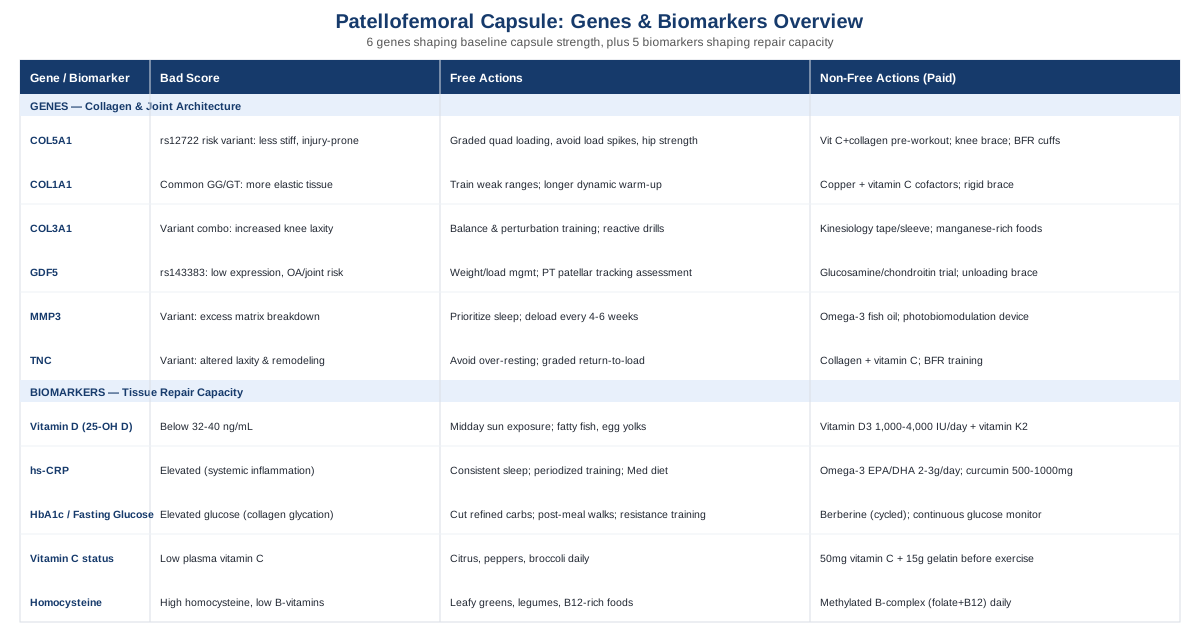
Before going deep into the science, here's the shape of what's coming. Six genes — COL5A1, COL1A1, COL3A1, GDF5, MMP3, and TNC — show up repeatedly in human studies of ligament rupture, joint laxity, and osteoarthritis risk, and each one has a plausible, specific mechanism tied to how a knee capsule handles mechanical stress. Some of these variants make tissue less stiff and more prone to spraining under load; others affect how aggressively the body remodels or breaks down capsule tissue during healing. For every gene, there's a version of "if this is working against you" and a concrete two-track plan — one relying on training and behavior alone, another adding supplements or simple equipment.
Genetics is only half the story, though, because DNA doesn't tell you what's happening in your tissue right now. That's where five trackable biomarkers come in — vitamin D, hs-CRP, fasting glucose or HbA1c, vitamin C status, and homocysteine/B-vitamin status — each one a live readout of whether your body currently has the raw materials and the inflammatory environment needed for capsule repair. Beyond genes and blood work, a widely discussed book reframes joint durability as a set of daily physical habits rather than a post-injury fix, and a short set of complementary approaches — from biofeedback to guided imagery — round out what actually has clinical evidence behind it for knee capsule recovery. Read on to see exactly where you might stand, and what to do about it.
What Your Genes May Be Telling You About Capsule And Ligament Strength
The genetics of soft tissue injury is a small but active research field, largely built on candidate-gene studies in athletes — skiers, soccer players, and runners who tore an ACL, ruptured an Achilles tendon, or dislocated a shoulder. The patellofemoral capsule is made of the same collagen-rich connective tissue as ligaments elsewhere in the knee, so variants associated with ACL and general ligament injury risk are reasonably assumed to apply to capsular tissue as well, even though most studies were not designed around the patellofemoral joint specifically.
Researchers like Ali Torkamani, a genomics scientist known for his work on polygenic risk interpretation, and Gary Brecka, who has popularized turning raw genetic data into actionable lifestyle protocols, have pushed the idea that consumer-accessible DNA data shouldn't just sit in a report — it should change what you actually do. Applied to a capsule sprain, that means treating each gene below as a probability shift, not a diagnosis, and pairing it with a concrete plan.
A caution worth stating up front: effect sizes for most of these variants are modest, several associations have been replicated inconsistently across populations, and none of them override the basics of good rehab. Where the evidence is mixed, it's noted explicitly.
COL5A1: The Fibril-Diameter Regulator
COL5A1 encodes part of type V collagen, a minor but structurally critical collagen that controls how thick and how densely packed type I collagen fibrils become as they assemble into ligament and capsule tissue. Variants here don't change how much collagen you have — they change its architecture, which affects tissue stiffness and elasticity.
The rs12722 (BstUI) polymorphism has been studied directly in ligament injury. A study of skiers found that a specific COL5A1 haplotype was associated with reduced risk of anterior cruciate ligament injury, suggesting the CC/CT genotype pattern produces comparatively less compliant, injury-resistant tissue in some populations (COL5A1 variants and reduced ACL injury risk in skiers). That said, larger meta-analyses across different athlete populations have found inconsistent results for this same variant, so treat this as a real but not universally reproduced signal.
If The Gene Variant Is Unfavorable: The Plan Without Supplements
If your genotype leans toward the higher-injury-risk variant, the priority is graded mechanical loading rather than avoidance. Isometric and slow eccentric quadriceps work (think tempo step-downs and wall sits progressing in duration) conditions the capsule and surrounding tendon tissue over weeks, not days. Avoid abrupt spikes in training volume — the "10% rule" for weekly load increases is a reasonable, evidence-informed default. Add hip external rotator and abductor strengthening, since poor hip control increases dynamic valgus load transmitted straight into the patellofemoral capsule. Do this three to four times per week, with no need to cycle — this is a standing practice, not a course of treatment.
If The Gene Variant Is Unfavorable: The Plan With Supplements Or Equipment
Fifteen grams of vitamin C-enriched collagen or gelatin taken about 60 minutes before rehab exercise or sport has been shown in a controlled human trial to measurably increase markers of collagen synthesis compared to placebo (vitamin C-enriched gelatin supplementation augments collagen synthesis). Take it on training days only, not continuously — there's no established benefit to dosing on rest days, and no meaningful side-effect profile at this dose beyond mild GI upset in sensitive individuals. A hinged knee brace or kinesiology taping during high-pivot sports can offload the capsule mechanically while tissue remodels; blood flow restriction (BFR) cuffs allow lower-load strength training during early rehab when full loading isn't tolerated yet.
COL1A1: The Stiffness Dial
COL1A1 encodes the alpha-1 chain of type I collagen, the dominant structural collagen in ligament, capsule, and bone. A regulatory variant at an Sp1 transcription factor binding site (rs1800012) changes how much of this collagen chain gets produced, which shifts overall tissue stiffness.
The rarer TT genotype has been associated with a protective effect against cruciate ligament rupture and shoulder dislocation, likely because it produces stiffer, less compliant tissue that resists sudden overstretch (COL1A1 Sp1 polymorphism and risk of cruciate ligament rupture or shoulder dislocation). A meta-analysis of sports-related tendon and ligament injuries found a similar pattern under a recessive genetic model. Most people carry the more common GG or GT genotype, which trends toward more elastic — and statistically more injury-prone — capsular tissue.
If The Gene Variant Is Unfavorable: The Plan Without Supplements
Because more elastic tissue tends to tolerate sudden multi-directional stress worse, the practical countermeasure is building strength through the ranges the capsule is weakest in — terminal knee extension, deep flexion under control, and rotational loading (lateral lunges, cutting drills at controlled speed before progressing to full intensity). Warm-up quality matters more here than for stiffer-tissue phenotypes: a genuine 8-10 minute dynamic warm-up before high-intensity movement measurably reduces peak tissue strain. This is a permanent training habit, done every session, not a cycled intervention.
If The Gene Variant Is Unfavorable: The Plan With Supplements Or Equipment
Copper and vitamin C both act as cofactors for lysyl oxidase, the enzyme responsible for cross-linking collagen fibers into stronger networks. A reasonable, low-risk approach is 1-2mg copper (from diet or a bisglycinate supplement) alongside daily vitamin C from food, without exceeding 2-3g/day of vitamin C, where GI tolerance drops off. There's no strong evidence for cycling this — it supports an ongoing biological process rather than a spike-and-recover mechanism. A rigid or semi-rigid patellofemoral brace during high-risk activities (cutting sports, skiing) gives mechanical stability while tissue quality is being built.
COL3A1: The Laxity Variable
Type III collagen, encoded by COL3A1, is more extensible than type I and is present in higher relative amounts in more mobile, less rigid connective tissue. A study examining COL1A1 and COL3A1 variants together found associations with knee joint laxity measurements, meaning certain genotype combinations correlate with objectively looser knee joints (COL1A1 and COL3A1 gene variants and knee joint laxity). Looser capsular tissue isn't inherently bad — it can support range of motion — but it also means the knee relies more heavily on active muscular control to stay stable, since passive restraint from the capsule is doing comparatively less of the job.
If The Gene Variant Is Unfavorable: The Plan Without Supplements
The single most effective lever here is neuromuscular control training: single-leg balance progressions, perturbation training (someone gently pushing you off balance on one leg), and reactive strength drills. These retrain the nervous system to stabilize a joint that has less passive restraint available. Three sessions a week for at least 8-12 weeks is a realistic minimum to see durable improvement in joint position sense, and this should be maintained indefinitely for anyone with a known hypermobility pattern, not just during rehab.
If The Gene Variant Is Unfavorable: The Plan With Supplements Or Equipment
Kinesiology tape or a light elastic knee sleeve provides proprioceptive feedback — it doesn't mechanically restrain the joint much, but it improves the brain's awareness of joint position, which is exactly what lax-capsule phenotypes benefit from. Manganese, a cofactor for enzymes involved in proteoglycan and collagen assembly, is present in most multivitamins at appropriate doses; there's no evidence supporting high-dose manganese supplementation, and doing so risks neurotoxicity, so food-level intake (whole grains, nuts, leafy greens) is the safer route here rather than isolated high-dose supplements.
GDF5: The Joint Architecture Gene
Growth differentiation factor 5 (GDF5) is involved in cartilage, ligament, and joint shape formation during development. The rs143383 variant, sitting in the gene's regulatory region, reduces GDF5 expression and has one of the most consistently replicated associations with knee osteoarthritis in the genetics literature, reaching genome-wide significance in some cohorts (GDF5 rs143383 polymorphism and knee osteoarthritis with genome-wide significance). For a capsule sprain specifically, the relevance is indirect but real: altered joint shape and cartilage quality change how load is distributed through the patellofemoral joint, which can put chronic extra strain on the capsule even after the original sprain has technically healed.
If The Gene Variant Is Unfavorable: The Plan Without Supplements
Since this variant affects joint architecture rather than tissue elasticity, the actionable lever is load management: maintaining a body weight that doesn't overload the patellofemoral joint, correcting movement patterns that increase patellar maltracking (commonly, weak hip external rotators letting the knee cave inward), and avoiding high-frequency deep knee flexion under load (deep squats, kneeling work) if symptoms flare. A physical therapist's patellar tracking assessment is worth the appointment here — this is one case where professional gait/movement analysis outperforms generic advice.
If The Gene Variant Is Unfavorable: The Plan With Supplements Or Equipment
Glucosamine sulfate and chondroitin (1500mg/1200mg daily) have mixed but not negligible trial evidence for cartilage-related knee symptoms; a reasonable approach is a 3-month trial with symptom tracking rather than indefinite use, since response is individual and not guaranteed. An unloading or patellar-stabilizing brace during aggravating activities reduces direct patellofemoral compression. There's no meaningful side-effect concern with glucosamine/chondroitin beyond mild GI effects, though people with shellfish allergies should check the source.
MMP3: The Remodeling Balance
Matrix metalloproteinase-3 (MMP-3, also called stromelysin-1) breaks down collagen, fibronectin, and proteoglycans as part of normal tissue turnover — a necessary process, since healing requires clearing damaged matrix before rebuilding. Certain MMP3 promoter and coding variants shift this process toward more aggressive breakdown relative to repair, and these variants have been associated with increased risk across multiple tendon and ligament injury types in a soft-tissue injury risk-profiling study spanning independent sample groups (the MMP3 gene in musculoskeletal soft tissue injury risk profiling).
If The Gene Variant Is Unfavorable: The Plan Without Supplements
Because this variant tilts the balance toward matrix breakdown, the practical priority is avoiding the conditions that spike MMP activity further: chronic sleep deprivation, unmanaged systemic inflammation, and overtraining without adequate recovery windows. A structured deload every 4-6 weeks of training (roughly 40-50% volume reduction for a week) gives remodeling processes time to catch up rather than staying in a constant breakdown-dominant state. This is a permanent periodization habit rather than a temporary fix.
If The Gene Variant Is Unfavorable: The Plan With Supplements Or Equipment
Omega-3 fatty acids (2-3g combined EPA/DHA daily) have some evidence for modulating inflammatory and metalloproteinase activity, though the direct data on injured ligament tissue in humans is still limited — treat this as a plausible, low-risk addition rather than a proven fix. Continuous daily use is appropriate; watch for increased bleeding risk if combined with anticoagulant medication. A photobiomodulation (low-level laser) device, discussed further below, has more direct evidence for supporting collagen remodeling in injured tissue and is a reasonable equipment-based addition during active rehab phases.
TNC: The Mechanical Signal
Tenascin-C is an extracellular matrix glycoprotein that gets upregulated specifically in response to mechanical stress and tissue remodeling — it's part of how healthy ligament and capsule tissue adapts to load. Variants in the TNC gene have been linked to knee joint laxity measurements in a study examining type XII collagen and tenascin-C genes together (genetic variants within type XII collagen and tenascin-C genes and knee joint laxity), though its association with ACL rupture risk specifically has been inconsistent across populations, with some studies finding no significant link. Separately, tenascin-C levels rise in synovial fluid after joint injury, tracking with markers of inflammation and matrix degradation, which makes it a biologically plausible marker of active capsule remodeling after a sprain.
If The Gene Variant Is Unfavorable: The Plan Without Supplements
Since tenascin-C expression is itself driven by mechanical loading, the single most relevant behavior is not over-resting the joint. Prolonged immobilization beyond what's medically necessary can blunt the very remodeling signal this gene product represents. A graded return-to-load protocol — moving from isometrics to controlled dynamic movement to sport-specific drills over roughly 6-8 weeks, guided by symptom response rather than a fixed calendar — keeps this adaptive signaling active without overloading a healing capsule.
If The Gene Variant Is Unfavorable: The Plan With Supplements Or Equipment
The same collagen-vitamin C-protein foundation described for COL5A1 applies here, since it supports the raw materials tenascin-C-driven remodeling depends on. Blood flow restriction training equipment is particularly useful for this variant profile, since it allows meaningful mechanical loading signal at low absolute joint stress — appropriate during the early-to-mid rehab window, used 2-3 times weekly, not daily, to avoid excessive cuff-related discomfort or bruising.
Turning Genetic Data Into An Actual Plan
If you already have raw data from a consumer DNA test, third-party interpretation tools can pull out the specific rsIDs mentioned above (rs12722, rs1800012, rs143383, and the relevant MMP3 and TNC variants) without needing a new clinical test. Where a gene shows a less favorable variant, the priority order is consistent across all six: get the loading progression right first, since mechanical stimulus does more for capsule resilience than any supplement; use nutrition and cofactor support as a genuine second layer, not a replacement; and reserve bracing or BFR equipment for periods of elevated risk or active rehab rather than permanent use. Retesting genes obviously isn't necessary — DNA doesn't change — but reassessing your functional response (joint laxity tests, strength benchmarks, symptom logs) every 8-12 weeks tells you whether the plan built around your genetic profile is actually working.
Genes describe your starting material. What's happening in your blood right now describes whether that material is being given what it needs to repair — which is where biomarkers come in.
Five Blood And Function Markers Worth Tracking Alongside Your Genes
Unlike genetic variants, these markers change month to month based on behavior, which makes them useful feedback loops rather than fixed risk labels. Clinicians like Peter Attia, Thomas Dayspring, and Allan Sniderman have popularized the idea of tracking a small set of high-yield biomarkers rather than a sprawling panel — the same philosophy applies well to soft tissue recovery, where a handful of markers capture most of the relevant biology.
25-Hydroxyvitamin D
Vitamin D regulates bone and connective tissue remodeling, supports muscle function that offloads the capsule during movement, and modulates the inflammatory response involved in healing. A review focused on athletes found vitamin D status connected to both performance and musculoskeletal injury risk, with researchers recommending serum levels at or above 32-40 ng/mL for those actively training or rehabbing (vitamin D in athletes: physical performance and musculoskeletal injuries).
How To Measure It
A standard 25(OH)D blood test costs roughly $30-$100 out of pocket where not covered by insurance; home fingerstick kits run about $50-70 and are a reasonable option for repeat testing.
If The Score Is Low: The Plan Without Supplements
Regular midday sun exposure on bare skin (15-20 minutes, several times weekly, adjusted for skin tone and latitude) and dietary sources like fatty fish and egg yolks are the non-supplement route, though they're often insufficient in winter months or northern latitudes.
If The Score Is Low: The Plan With Supplements Or Equipment
Vitamin D3, typically 1,000-4,000 IU daily depending on how low the baseline is, taken continuously rather than cycled, with retesting at 3 months to confirm the dose is working. Pairing with vitamin K2 (around 100mcg) is a common practice to support appropriate calcium utilization. Side effects are rare at these doses but sustained intake above 10,000 IU/day risks hypercalcemia, so periodic serum calcium checks are worthwhile for anyone supplementing long-term at the higher end.
High-Sensitivity C-Reactive Protein (hs-CRP)
hs-CRP is a general marker of systemic low-grade inflammation. It isn't specific to knee tissue, but persistently elevated levels suggest a body-wide inflammatory tone that can slow tissue repair and blunt training adaptation (C-reactive protein as a marker of tissue injury and inflammation).
How To Measure It
A standard blood draw, usually $15-40 standalone or bundled into broader panels ($100-300) of the kind Attia-style longevity clinics run.
If The Score Is High: The Plan Without Supplements
Prioritizing sleep consistency, periodizing training load so recovery windows aren't chronically skipped, maintaining a healthy body composition, and shifting toward a Mediterranean-style diet pattern are the highest-leverage, no-cost interventions for lowering hs-CRP.
If The Score Is High: The Plan With Supplements Or Equipment
Omega-3 fish oil (2-3g EPA/DHA daily, continuous use, watch for bleeding risk with blood thinners) and curcumin (500-1000mg standardized extract, typically cycled 8-12 weeks with a break, occasional GI upset) are the two most commonly used options, both with reasonable but not definitive human evidence for lowering inflammatory markers.
Fasting Glucose Or HbA1c
Chronic elevated blood sugar promotes non-enzymatic glycation of collagen, forming cross-links that make tendon and ligament tissue stiffer and more brittle rather than resiliently elastic — a mechanism demonstrated directly in tendon collagen studies (glycation cross-linking increases matrix stiffness in tendon collagen). For capsule tissue undergoing repair, this is a meaningful and underappreciated variable.
How To Measure It
Fasting glucose costs $10-30 as a standalone test; HbA1c runs $20-50. A continuous glucose monitor (CGM), often used experimentally the way Attia has discussed even in non-diabetic individuals, costs roughly $50-100/month.
If The Score Is High: The Plan Without Supplements
Reducing refined carbohydrate and added sugar intake, taking short walks after meals, and adding resistance training sessions (which improve insulin sensitivity independent of weight loss) are the foundational levers here.
If The Score Is High: The Plan With Supplements Or Equipment
Berberine (500mg, 2-3 times daily, cycled 8-12 weeks on with a 4-week break) has meaningful evidence for improving glycemic markers, though it can cause GI upset and interacts with several medications, so it warrants a conversation with a physician before starting. A CGM used for 2-4 weeks at a time provides direct, personalized feedback on which foods and habits move your glucose the most.
Vitamin C Status
Vitamin C is a required cofactor for the enzymes that hydroxylate proline and lysine during collagen synthesis — without adequate vitamin C, the body simply cannot build strong, properly cross-linked collagen, which is directly relevant to healing a sprained capsule (vitamin C-enriched gelatin supplementation and collagen synthesis).
How To Measure It
Plasma vitamin C isn't part of routine panels; specialty or functional medicine labs offer it for roughly $40-80.
If The Score Is Low: The Plan Without Supplements
Regular intake of citrus fruit, bell peppers, and broccoli covers most people's needs, though vitamin C has a short half-life in the body, so consistency matters more than occasional large servings.
If The Score Is Low: The Plan With Supplements Or Equipment
The specific protocol with human trial support is 50mg vitamin C paired with 15g gelatin or collagen peptides, taken about an hour before rehab exercise or sport, on training days rather than daily. Side effects are minimal at this dose; above roughly 2g/day, GI upset becomes more common, and very high sustained doses carry a small kidney stone risk in predisposed individuals.
Homocysteine And B-Vitamin Status
Homocysteine metabolism intersects with methylation pathways that support connective tissue maintenance, and adequate B12 and folate status are part of keeping that system running smoothly. This marker is less directly studied in capsule injury specifically, but it's a low-cost, high-information addition to a broader nutritional picture.
How To Measure It
A standard blood test costs $30-60 and is frequently bundled into cardiovascular or longevity-focused panels.
If The Score Is High: The Plan Without Supplements
Leafy greens, legumes, and adequate B12 intake through animal products, or fortified foods for those eating plant-based, generally cover requirements.
If The Score Is High: The Plan With Supplements Or Equipment
A methylated B-complex (methylfolate plus B12) taken daily, retested at 3 months, has a low side-effect profile and doesn't require cycling since it's supporting an ongoing metabolic process rather than a short-term spike.
Genes and blood markers describe the biology. The next piece is behavioral — how you actually move and load your joints day to day, which is where one particular book has reframed the conversation.
The Book That Reframes Joint Health As A Daily Practice, Not A Fix
Built to Move, by Kelly Starrett and Juliet Starrett, challenges a habit baked into most orthopedic care: waiting until something breaks, then rehabbing it back to a baseline that was never actually tested or tracked beforehand. The book's central idea — that a handful of simple physical tests function as vital signs, just like blood pressure or resting heart rate — is directly useful for anyone recovering from a capsule sprain and trying to prevent the next one. Below are the ten most useful ideas from that framework, applied specifically to knee and capsule health.
1. Getting Up Off The Floor Without Using Your Hands Is A Real Vital Sign
The sitting-rising test — going from standing to sitting on the floor and back up, losing a point for every hand or knee used as support — was validated in a cohort study as a predictor of all-cause mortality independent of cardiorespiratory fitness (ability to sit and rise from the floor as a predictor of all-cause mortality). For a patellofemoral capsule specifically, this test also directly stresses deep knee flexion under control — exactly the range many people avoid after a sprain, which lets it stay weak indefinitely.
2. Hip Rotation Range Predicts Knee Compensation Patterns
Limited hip internal or external rotation forces the knee to absorb rotational forces it wasn't built to handle, which transmits directly into the patellofemoral capsule during pivoting movements. Testing hip rotation seated, comparing side to side, is a five-minute screen worth doing before assuming the knee itself is the only problem.
3. A Deep, Heel-Down Squat Reveals Daily Capsule Loading Patterns
The ability to squat to full depth with heels flat isn't about performance — it's a proxy for whether the joint moves through its full range regularly in daily life. Joints that spend most of the day in a narrow mid-range (chairs, cars, couches) lose capsule tissue quality over time simply from disuse.
4. Ankle Stiffness Quietly Transfers Stress To The Knee Capsule
Restricted ankle dorsiflexion — commonly from tight calves or old ankle sprains — changes knee mechanics during walking, running, and squatting, often increasing patellofemoral load without the person ever suspecting their ankle is the upstream cause.
5. Walking Volume Is Non-Negotiable Tissue Medicine
Low-intensity, high-volume walking (the book suggests thinking in daily step totals rather than workout minutes) provides the kind of repetitive, low-load mechanical stimulus that keeps capsule and cartilage tissue nourished, since much of the knee's cartilage relies on movement-driven nutrient exchange rather than direct blood supply.
6. Grip Strength Is A Proxy For Whole-Body Tissue Resilience
Grip strength correlates with general connective tissue and muscular robustness across the body, making it a quick, equipment-light way to track whether your overall physical capacity — which supports joint-specific rehab — is trending up or down over time.
7. Protein Intake Is The Most Underrated "Supplement" For Capsule Repair
Adequate daily protein (the book advocates roughly 0.7-1g per pound of bodyweight for people actively training or rehabbing) supplies the amino acid pool collagen synthesis draws from — arguably a more foundational lever than any single supplement discussed elsewhere in this article.
8. Sleep Is The Repair Window Your Ligaments Depend On
Most collagen synthesis and tissue remodeling activity is concentrated during deep sleep phases, which makes consistent, adequate sleep duration a direct rehab intervention rather than a vague wellness suggestion.
9. Nasal Breathing And Movement Snacks Reduce Compensatory Strain
Short, frequent movement breaks throughout a sedentary day — a few minutes of mobility work every hour — prevent the joint stiffness that builds up during prolonged static positions and that later shows up as compensatory strain during actual training sessions.
10. Vital Signs Should Be Retested, Not Just Measured Once
The book's most practical challenge to conventional medical thinking is treating these physical tests as an ongoing dashboard, checked every few months, rather than a one-time assessment done only after an injury already happened.
These daily physical habits work alongside — not instead of — the more targeted, evidence-based therapies that follow, several of which have direct clinical evidence in knee and patellofemoral conditions specifically.
Complementary Approaches With Real Evidence For Knee Recovery
The approaches below were selected because they have actual human clinical evidence tied to knee or patellofemoral conditions, not because they're broadly popular in wellness circles. Evidence strength varies between them, and that's noted honestly in each case.
Biofeedback
Electromyographic (EMG) biofeedback gives real-time visual or auditory feedback on quadriceps muscle activation, helping retrain the specific timing and strength of muscle firing that stabilizes the patellofemoral joint — directly relevant when a capsule sprain has left proprioception and muscle activation patterns disrupted.
A randomized controlled trial in young adult athletes with patellofemoral pain syndrome combined patellar taping with EMG-biofeedback-guided isometric quadriceps training at multiple knee angles and found meaningful improvements in quadriceps strength and functional performance compared to sham treatment (patellar taping and EMG biofeedback training in patellofemoral pain syndrome: a randomized controlled trial), and a systematic review of biofeedback in patellofemoral pain treatment across a dozen RCTs supports its use as part of a broader conservative treatment plan (the role of biofeedback in patellofemoral pain conservative treatment: a systematic review).
Realistically, this requires access to a physical therapist with EMG-biofeedback equipment, typically used during supervised sessions two to three times weekly for 6-8 weeks alongside a standard strengthening program — it's a rehab addition, not a standalone replacement for structured quadriceps and hip strengthening.
Guided Imagery
Guided or motor imagery involves mentally rehearsing movement without physically performing it, which keeps neuromuscular pathways active during periods when full movement isn't possible or advisable — particularly relevant during the early, more protected phase after a capsule sprain.
A randomized controlled pilot trial found that guided imagery improved functional outcomes following anterior cruciate ligament repair compared to standard care alone (guided imagery to improve functional outcomes post-ACL repair: a randomized controlled pilot trial), and separate work has shown increased muscle activation following motor imagery practice during ACL rehabilitation, supporting a genuine neuromuscular mechanism rather than a purely psychological effect.
In practice, this means spending 10-15 minutes several times a week vividly visualizing controlled knee movement — walking, stair negotiation, or sport-specific motions — particularly useful in the first weeks after injury when physical loading is limited; evidence for its effect on pain and fear of re-injury specifically remains mixed, so it's best treated as a low-cost adjunct rather than a primary strategy.
Photobiomodulation (Low-Level Laser Therapy)
Photobiomodulation uses low-level laser light to stimulate cellular activity in soft tissue, with proposed effects on collagen remodeling, local circulation, and inflammation — mechanistically relevant to a healing capsule, though most direct ligament-healing evidence still comes from animal models rather than human capsule-specific trials.
A systematic review and meta-analysis of randomized placebo-controlled trials found low-level laser therapy reduced pain and disability in knee osteoarthritis (efficacy of low-level laser therapy on pain and disability in knee osteoarthritis: a systematic review and meta-analysis), and an animal study specifically found it improved early healing of medial collateral ligament injuries at the tissue level (low-level laser therapy and healing of medial collateral ligament injuries in rats) — useful mechanistic support, but a reminder that direct human capsule-specific evidence is still limited.
A reasonable, cautious approach is using a clinical-grade device under a physical therapist's guidance, 2-3 times weekly for several weeks during active rehab, since consumer-grade devices vary widely in power output and haven't been validated to the same standard as the devices used in these trials.
Tai Chi
Tai chi combines slow, controlled weight-shifting movements with sustained single-leg loading phases, directly training the kind of joint position sense and proprioceptive control that a capsule sprain often disrupts.
A pilot randomized controlled trial found tai chi practice improved knee-joint proprioception in people with knee osteoarthritis compared to a control condition (the influence of tai chi exercise on proprioception in patients with knee osteoarthritis: a pilot randomized controlled trial), and while this evidence base centers on osteoarthritis rather than acute capsule sprains, the proprioceptive mechanism plausibly transfers to post-sprain rehabilitation as well.
A practical starting point is a beginner class or instructor-led online program, twice weekly for at least 8-12 weeks, ideally once acute pain has settled enough to tolerate sustained single-leg weight-bearing — it pairs well as a lower-intensity complement to more direct strength-based rehab rather than a replacement for it.
Conclusion
A patellofemoral capsule sprain isn't just a generic injury — it happens inside tissue shaped by collagen genetics that influence stiffness and laxity, remodeling genes that determine how aggressively your body breaks down and rebuilds damaged matrix, and a current biochemical environment reflected in markers like vitamin D, inflammation, and blood sugar. None of these factors guarantee an outcome, but together they explain more about why your recovery looks the way it does than "rest and strengthen" ever could on its own.
The most useful next step isn't necessarily a genetic test — it's an honest inventory of what you already have access to: recent bloodwork you could revisit with these five markers in mind, a physical therapist who can properly assess hip, ankle, and patellar tracking mechanics, and a training log that tracks load progression instead of just pain levels. If swelling, instability, locking, or giving-way persists, that's a signal to see an orthopedic specialist or sports medicine physician directly rather than working through this list alone. Start with one change you can act on this week — a blood draw, a movement screen, or a loading adjustment — and build from there.
Musculoskeletal: Tendon & Ligament Conditions
Endocrine & Metabolic: Diabetes & Blood Sugar
Autoimmune: Inflammatory Conditions