This article contains AI generated content.
· UpdatedPatellofemoral Instability — 5 Genes and 6 Biomarkers to Track
Introduction
If your kneecap has ever slipped, caught, or felt unreliable during ordinary movements — walking down stairs, landing from a jump, or even sitting for too long — you already know how disorienting patellofemoral instability can be. The patella is supposed to glide smoothly in the trochlear groove of the femur. When the structures holding it there are compromised — whether through lax ligaments, poor trochlear morphology, muscle imbalances, or cartilage degradation — that guidance system breaks down. Pain, grinding, buckling, and recurrent dislocations are not just inconveniences. They erode confidence in your own body.
Most standard advice for this condition focuses on quadriceps strengthening and activity modification. These are not wrong, but they are incomplete. Two people with nearly identical anatomy can respond very differently to the same rehabilitation program. One recovers well; the other keeps re-dislocating. Generic protocols tend to ignore the underlying biological terrain — what is happening at the level of collagen structure, cartilage turnover, inflammation, and even the DNA that shaped the tissue in the first place.
That is the gap this article tries to fill. There are measurable biological signals — circulating biomarkers — that can tell you whether your cartilage is under active stress, whether inflammation is silently accelerating tissue breakdown, or whether key micronutrients critical to ligament and tendon integrity are deficient. There are also specific genetic variants that influence connective tissue quality, joint morphology, and matrix remodeling, which can help explain why some individuals are structurally more vulnerable from the outset.
Better information does not replace a qualified physiotherapist or orthopedic surgeon. But it can sharpen the conversation, guide more targeted interventions, and help you move beyond generic advice. This article covers six biomarkers you can realistically track, five genetic factors worth understanding, a deep dive into connective tissue regeneration science, and several evidence-supported complementary approaches — so you have more than one angle of attack.
6 Biomarkers That May Reveal What Is Driving Your Patellar Instability
Biomarkers are measurable biological signals in blood, urine, or tissue. They do not diagnose patellofemoral instability — imaging and clinical assessment do that. But they reveal the quality of the biological environment your joint is living in: how fast cartilage is breaking down, how inflamed the tissue is, whether nutritional deficiencies are limiting repair. Each marker below has a practical implication and a response plan.
1. COMP — Cartilage Oligomeric Matrix Protein
Why it matters
COMP is a non-collagenous protein concentrated in cartilage, tendons, and ligaments. When mechanical load damages these tissues — or when chronic inflammation accelerates matrix breakdown — COMP leaks into the bloodstream. Elevated serum COMP levels are one of the earliest detectable signals of cartilage stress, often appearing before structural changes show up on MRI. In the context of patellofemoral instability, abnormal patellar tracking generates repeated focal overload on the retropatellar cartilage, which increases COMP release. Tracking this marker over time can indicate whether your current training load or rehabilitation program is protecting the cartilage or quietly degrading it.
How to measure it
COMP is measured from a fasting blood draw and is available through specialized labs (standard panels at most clinics do not include it). Cost ranges from approximately $100 to $350 depending on the laboratory. Reference ranges vary by lab but elevated values are generally considered above 12–15 U/L in adults. Repeat testing every 3–6 months during rehabilitation is reasonable to track trends.
If the score is elevated: the plan without supplements
The first priority is load management. Elevated COMP is a biological signal that mechanical demand is exceeding tissue capacity. Temporarily shifting to low-impact aerobic activity — cycling, swimming, water running — reduces compressive patellofemoral forces while maintaining conditioning. Alongside this, begin a structured hip abductor and external rotator strengthening program; research consistently shows that hip muscle deficits increase lateral patellar tracking deviation and focal cartilage overload. The goal is not rest but redistribution of mechanical stress so the tissue can recover. Proprioception training on unstable surfaces also reduces aberrant joint mechanics.
If the score is elevated: the plan with supplements or equipment
Collagen peptides at 10–15 grams per day, taken alongside 500 mg of vitamin C approximately 30–60 minutes before exercise, have been shown in research by Keith Baar and colleagues to increase collagen synthesis in periarticular connective tissues. This timing is important — the vitamin C and amino acid surge appears to amplify the anabolic response during subsequent loading. Combine this with a patellar tracking brace or McConnell taping during activity to mechanically unload the lateral cartilage while healing occurs. Omega-3 fatty acids at 2–3 g EPA+DHA daily have anti-inflammatory effects that may slow COMP release by reducing cytokine-driven cartilage catabolism. Recheck COMP at 12 weeks.
2. uCTX-II — Urinary Type II Collagen Crosslinks
Why it matters
Type II collagen is the primary structural protein of articular cartilage. When it degrades, its crosslinked fragments — specifically C-terminal crosslinking telopeptides — are released into the bloodstream and excreted in urine. The urinary form, uCTX-II, is a sensitive and specific marker of the rate at which articular cartilage is breaking down. Unlike imaging, which captures structural damage after it has already occurred, uCTX-II tracks an active process in real time. In patellofemoral instability, repeated subluxation episodes and aberrant mechanics accelerate retropatellar cartilage matrix catabolism. A high uCTX-II level suggests that degradation is currently outpacing synthesis.
How to measure it
uCTX-II is measured from a spot urine sample, corrected for creatinine concentration to account for hydration variability. It is typically available through specialized labs or functional medicine panels. Cost is approximately $80–$200. Values are usually expressed in ng/mmol creatinine. Elevated values vary by age and sex, so reference ranges from the specific lab should be used. Testing every 3–6 months during active management is informative.
If the score is elevated: the plan without supplements
The most evidence-supported non-pharmacological intervention for reducing cartilage catabolism is neuromuscular control training — specifically exercises that improve dynamic valgus control and patellar alignment. VMO (vastus medialis oblique) activation training in closed-chain patterns and terminal knee extensions reduces lateral patellar tilt and redistributes compressive forces. Swimming and cycling preserve muscle without aggravating the marker further. Anti-inflammatory dietary patterns — Mediterranean-style, emphasizing whole foods, olive oil, fatty fish, and reducing ultra-processed foods — lower the systemic cytokine environment that drives cartilage catabolism.
If the score is elevated: the plan with supplements or equipment
Avocado-soybean unsaponifiables (ASU) at 300 mg per day have the strongest evidence among natural supplements for reducing cartilage degradation markers, with multiple randomized controlled trials showing benefit in knee OA populations. Undenatured type II collagen (UC-II) at 40 mg daily — a lower dose than hydrolyzed collagen — works through oral tolerance mechanisms to reduce collagen-specific autoimmune activity. Curcumin (BCM-95 form, 500 mg twice daily for bioavailability) has shown meaningful reductions in cartilage degradation markers in several trials. Avoid NSAIDs as a long-term solution, as they inhibit proteoglycan synthesis and can paradoxically worsen cartilage matrix over time.
3. High-Sensitivity CRP (hs-CRP) — Systemic Inflammation Index
Why it matters
C-reactive protein produced by the liver in response to inflammatory cytokines — particularly IL-6 — is one of the most accessible and broadly informative biomarkers in medicine. In the context of patellofemoral instability, chronic low-grade inflammation, even at levels well below those typically associated with disease, significantly impairs connective tissue repair, reduces ligament tensile strength, and accelerates cartilage breakdown. An hs-CRP above 1 mg/L suggests an inflammatory burden that may be silently working against your rehabilitation. Above 3 mg/L, the inflammatory environment becomes a meaningful biological obstacle to tissue recovery.
How to measure it
hs-CRP is one of the most widely available and affordable biomarkers in this list. A standard blood draw at any lab will return the result; cost ranges from $15 to $50. Optimal target is below 0.5 mg/L. Values between 1–3 mg/L suggest moderate, clinically relevant inflammation. Values above 3 mg/L warrant investigation of root causes (sleep, body composition, diet, dental health, gut health).
If the score is elevated: the plan without supplements
Sleep is the most potent non-pharmacological anti-inflammatory lever available. A consistent 7–9 hours of quality sleep reduces IL-6 and TNF-alpha, the upstream drivers of CRP. Addressing visceral adiposity through dietary quality and daily movement (10,000 steps baseline) has been shown in multiple trials to meaningfully lower CRP. Eliminate ultra-processed foods, refined seed oils, and high-glycemic carbohydrates. Regular, moderate aerobic exercise — 150 minutes per week — has a consistent CRP-lowering effect; however, high-volume endurance training in the presence of already-elevated CRP can paradoxically raise it further, so context matters.
If the score is elevated: the plan with supplements or equipment
Omega-3 fatty acids (EPA + DHA, 3–4 grams per day in triglyceride form) are one of the most evidence-backed anti-inflammatory supplements and consistently lower hs-CRP in meta-analyses. Vitamin D3 supplementation, when levels are deficient, also reduces inflammatory markers. Curcumin (500–1000 mg of a bioavailable form) has meaningful CRP-lowering effects. Magnesium deficiency independently elevates CRP, so addressing magnesium status (see below) is often a hidden variable. Continuous glucose monitors (CGMs), now widely available at $50–$100 per sensor, can reveal post-meal glucose spikes that chronically elevate CRP — a surprisingly actionable insight for many people.
4. 25-OH Vitamin D — The Musculoskeletal Baseline
Why it matters
Vitamin D is a steroid hormone with receptors in muscle cells, chondrocytes, osteoblasts, and immune cells. Its relevance to patellofemoral instability is multi-layered: deficiency impairs skeletal muscle strength (including the VMO, the primary dynamic patellar stabilizer), reduces bone mineral density at the patella and femur, increases systemic inflammation, and impairs proprioception by affecting muscle spindle function. Holick's landmark 2007 review in the New England Journal of Medicine documented the extraordinary prevalence of vitamin D insufficiency and its broad musculoskeletal consequences. For anyone with recurrent patellar instability, unchecked vitamin D deficiency is a remediable biological obstacle.
How to measure it
25-hydroxyvitamin D (25-OH-D3) is measured from a blood draw and is available at virtually every lab, typically covered by insurance or available for $30–$80 out of pocket. Optimal range for musculoskeletal function is generally considered 40–60 ng/mL (100–150 nmol/L), which is above the conventional "sufficient" threshold of 30 ng/mL. Testing twice per year — once in late summer and once in late winter — captures seasonal variability.
If the score is low: the plan without supplements
Midday sun exposure — 15 to 30 minutes of direct skin exposure on arms and legs around solar noon — can meaningfully raise vitamin D levels in fair-skinned individuals during summer months. Weight-bearing exercise itself modestly stimulates vitamin D receptor expression. Dietary sources (fatty fish, egg yolks, liver) provide modest amounts but are rarely sufficient alone to resolve deficiency. Move toward a nutrient-dense whole-food dietary pattern that includes these foods regularly.
If the score is low: the plan with supplements or equipment
Vitamin D3 (cholecalciferol) — not D2 — is the preferred supplementation form. For levels between 20–30 ng/mL, 2000–4000 IU daily is a common starting dose; for levels below 20 ng/mL, 5000 IU daily for 8–12 weeks followed by retesting is often appropriate. Always pair vitamin D3 with vitamin K2 (MK-7 form, 100–200 mcg daily), as K2 directs calcium to bone and away from soft tissues. Magnesium is required for vitamin D hydroxylation — without adequate magnesium, vitamin D supplementation has a blunted effect. Retest at 12 weeks. Side effects at normal supplementation doses are rare but toxicity is possible above 10,000 IU per day without medical supervision.
5. RBC Magnesium — The Overlooked Neuromuscular Marker
Why it matters
Serum magnesium — the standard test — is a poor indicator of total body magnesium status because the body tightly defends serum levels by drawing from intracellular stores. Red blood cell (RBC) magnesium reflects tissue stores more accurately. Magnesium is a cofactor in over 300 enzymatic reactions, including those governing muscle contraction, protein synthesis, and collagen cross-linking. In the context of patellofemoral instability, low RBC magnesium impairs VMO activation timing, reduces neuromuscular coordination, and limits the body's capacity to produce and maintain connective tissue. Chronic magnesium deficiency is also independently associated with elevated hs-CRP, creating a compounding inflammatory burden.
How to measure it
RBC magnesium is ordered specifically — not the standard serum magnesium test. Most functional medicine labs and some hospital labs offer it. Cost is approximately $50–$80. Target range is 5.5–7.0 mg/dL for RBC magnesium. Many individuals eating a typical Western diet test below 5.5 mg/dL, even with normal serum levels. Retest 8–12 weeks after initiating supplementation.
If the score is low: the plan without supplements
Dietary magnesium can be meaningfully increased through regular consumption of leafy greens (spinach, Swiss chard), pumpkin seeds, hemp seeds, dark chocolate (85%+), black beans, and almonds. Reducing alcohol and excessive caffeine — both of which increase renal magnesium excretion — is equally important. Epsom salt baths (magnesium sulfate) provide modest transdermal absorption and can be a useful adjunct, particularly for muscle relaxation after training.
If the score is low: the plan with supplements or equipment
Magnesium glycinate (300–400 mg elemental magnesium per evening) is the most bioavailable and best-tolerated form for most people, with minimal laxative effect. Magnesium malate is preferable if fatigue is also a concern. Avoid magnesium oxide — it has poor absorption and is largely wasted. Timing matters: evening supplementation improves sleep quality alongside replenishing stores. Cycling is not typically necessary; magnesium can be taken continuously. Diarrhea at higher doses signals the need to reduce the dose. Pair with vitamin B6 (P5P form, 25–50 mg) which enhances intracellular magnesium uptake.
6. Omega-3 Index — The Membrane Inflammation Gauge
Why it matters
The omega-3 index measures the percentage of EPA (eicosapentaenoic acid) and DHA (docosahexaenoic acid) in red blood cell membranes. It reflects dietary fatty acid intake over the prior 3–4 months and is a highly stable, reproducible marker. An omega-3 index below 4% is associated with a markedly pro-inflammatory cellular environment — elevated prostaglandins, leukotrienes, and cytokines that promote synovial inflammation, accelerate cartilage degradation, and impair ligament healing. An index above 8% is associated with reduced inflammatory signaling throughout the body, including in joint tissue. For patellofemoral instability, this matters because synovial inflammation amplifies pain sensitivity, worsens cartilage catabolism, and creates a biological headwind against connective tissue repair.
How to measure it
The omega-3 index is available as a finger-prick dried blood spot test (several commercial labs offer home kits for $50–$100) or as a standard blood draw panel. It is not routinely ordered by most physicians but is widely available through labs such as OmegaQuant. Target: above 8%. Most people eating a typical Western diet test between 3–5%. Retest every 4–6 months when actively supplementing.
If the index is low: the plan without supplements
Two to three servings per week of fatty cold-water fish — wild-caught salmon, sardines, mackerel, anchovies, or herring — can meaningfully raise the omega-3 index over 2–3 months. Sardines and mackerel are the most cost-effective sources and the lowest in environmental contaminants. This dietary shift also reduces omega-6 polyunsaturated fat intake (from processed vegetable oils), which is as important as increasing omega-3s, since the ratio between them determines net inflammatory signaling.
If the index is low: the plan with supplements or equipment
High-quality fish oil at 2–4 grams EPA+DHA per day in triglyceride (re-esterified) form has superior bioavailability compared to ethyl ester forms. Algae-based omega-3 (providing EPA + DHA) is an effective alternative for those avoiding fish products. Take with the largest meal of the day for optimal absorption. Avoid flaxseed oil as a sole omega-3 source — ALA has very poor conversion to EPA and DHA in most adults. No serious cycling is required; continuous daily dosing is appropriate. High doses above 5 grams daily may modestly increase bleeding time — relevant if surgical intervention is being considered.
The Genetic Side of the Story: 5 Variants That Influence Patellar Stability
Genetics do not determine destiny, but they can explain why identical training programs produce very different outcomes in different people. Several well-characterized genetic variants affect the quality of connective tissue, the architecture of the patellofemoral joint, and the rate at which the extracellular matrix is remodeled. Understanding your genetic profile — through direct-to-consumer testing or more comprehensive panels — can help you make better-informed decisions about training load, supplementation, and injury prevention.
COL5A1 — The Ligament Laxity Gene
What this gene does
COL5A1 encodes the alpha-1 chain of type V collagen, a critical structural component of tendons, ligaments, and the medial patellofemoral ligament (MPFL) — the primary passive restraint against lateral patellar dislocation. Type V collagen regulates the diameter and spacing of type I collagen fibrils; without adequate COL5A1 function, collagen fibers become thicker but less mechanically organized, reducing tensile strength. The rs12722 C/T polymorphism has been studied in ligament and tendon injury populations. Individuals with the TT genotype are consistently overrepresented in populations with ligament laxity, tendon injuries, and recurrent joint instability. September et al. (2009, British Journal of Sports Medicine) identified COL5A1 variants as associated with Achilles tendinopathy and related connective tissue injuries, with relevance extending to other ligamentous structures.
If the gene is bad: the plan without supplements
Individuals with the TT genotype should prioritize gradual load progression over rapid intensity increases — a principle sometimes called "tissue conditioning." Tendons and ligaments adapt slowly; they require weeks of consistent loading at sub-maximal intensity before they can safely tolerate higher demands. Prioritize eccentric loading exercises (single-leg lowering movements, Nordic-style exercises for hamstrings, step-down movements) which generate greater collagen remodeling stimulus per unit of joint stress. Hip abductor strengthening — particularly gluteus medius — reduces the lateral pull on the patella, which decreases MPFL stress. Avoid hypermobility provocation: end-range passive knee extension under load, deep twisting movements without control, and high-impact landing without adequate deceleration strength.
If the gene is bad: the plan with supplements or equipment
The collagen synthesis protocol is especially relevant here: 10–15 grams of hydrolyzed collagen peptides with 500 mg vitamin C, taken 30–60 minutes before a connective-tissue-focused exercise session (not just any workout). This timing window, identified in Keith Baar's research, aligns peak amino acid availability with the anabolic signaling triggered by exercise. Frequency: daily or at least 5 days per week. Copper (2–3 mg daily) and zinc (15–25 mg daily) are cofactors in lysyl oxidase, the enzyme responsible for collagen cross-linking — deficiencies directly impair ligament tensile strength. Patellar tracking orthoses or custom knee bracing can mechanically protect the MPFL while tissue adaptation occurs. Cycling for COL5A1 support is not required; continuous supplementation is appropriate.
COL1A1 — The Structural Integrity Blueprint
What this gene does
COL1A1 encodes alpha-1 type I collagen, the most abundant protein in the body and the dominant structural material in tendons, ligaments, and bone. The Sp1 binding site polymorphism (rs1800012) alters transcription factor binding efficiency, affecting how much COL1A1 is produced. The SS genotype (wild-type) produces more collagen; the Ss and ss genotypes are associated with reduced collagen output, lower ligament tensile strength, and higher rates of musculoskeletal injury across multiple joint systems. For patellofemoral instability, a reduced-output COL1A1 genotype means the patellar retinaculum, MPFL, and patellar tendon may be structurally less robust from the outset — not due to injury, but due to inherent tissue quality.
If the gene is bad: the plan without supplements
The therapeutic priority shifts from developing strength through the tissue to developing strength around it. Isometric quadriceps exercises (particularly at 60–90° of knee flexion) place less shear on the patellofemoral joint while building VMO hypertrophy and motor control. Slow eccentric step-downs on a decline board (15–25° decline) specifically load the patellar tendon in a manner that stimulates collagen synthesis while avoiding ballistic stress. Proprioception retraining is essential: individuals with structurally compromised ligaments rely more heavily on neuromuscular control to compensate — single-leg balance drills, wobble board training, and perturbation training all build this secondary stabilization system.
If the gene is bad: the plan with supplements or equipment
Glycine supplementation (3–5 grams per day) directly provides the dominant amino acid in collagen synthesis; human data shows that dietary glycine intake is consistently below the threshold required for optimal collagen production. Proline (1–3 grams) and lysine (1–2 grams) round out the collagen-building amino acid profile. Vitamin C remains essential for prolyl and lysyl hydroxylation reactions in collagen maturation. Silicon — found in orthosilicic acid form (BioSil, 10 mg daily) — has shown in small human trials to increase collagen synthesis markers. A patellar sleeve brace worn during high-demand activities provides passive lateral restraint for individuals whose ligamentous tissue cannot fully compensate independently.
GDF5 — The Joint Architecture Gene
What this gene does
Growth differentiation factor 5 (GDF5) is a bone morphogenetic protein involved in the embryological development of joints, including the formation of the trochlear groove and the patellar articular surface. The rs143384 A allele of GDF5 has been associated with altered joint morphology and increased risk of knee osteoarthritis and joint instability in multiple large-scale genome-wide association studies. Individuals with unfavorable GDF5 variants may have a shallower trochlear groove (trochlear dysplasia being a well-established anatomical risk factor for patellar instability), smaller patella, or altered patellar height — all morphological factors that increase the risk of lateral dislocation even with otherwise normal soft tissue mechanics. Evidence in this area is growing but comes primarily from association studies rather than mechanistic clinical trials.
If the gene is bad: the plan without supplements
For individuals with GDF5 risk alleles and confirmed shallow trochlear anatomy (on MRI), soft tissue and neuromuscular compensation becomes even more critical because structural anatomy cannot be altered without surgery. VMO activation training, hip external rotator strengthening (glute medius, piriformis, obturator), and foot arch management (reducing dynamic valgus through arch support or foot strengthening) all address the lateral force vectors that the trochlear groove cannot adequately resist. Blood flow restriction (BFR) training at low loads (20–30% of 1RM) provides high neuromuscular stimulus with minimal patellofemoral compression — a particularly relevant tool here.
If the gene is bad: the plan with supplements or equipment
While no supplement alters trochlear anatomy, protecting the cartilage within the shallow groove is critical. Glucosamine sulfate (1500 mg per day) and chondroitin sulfate (1200 mg per day) have the strongest long-term evidence among structural supplements for joint tissue support, with particular benefit in populations with morphological joint compromise. Custom patellar-tracking orthotics and dynamic valgus-correcting knee braces provide biomechanical compensation during loading. For contact sports or high-risk activities, prophylactic patellar stabilizing braces significantly reduce dislocation rates in individuals with shallow trochlear anatomy.
MMP3 — The Matrix Degradation Regulator
What this gene does
Matrix metalloproteinase 3 (MMP3), also called stromelysin-1, is an enzyme that degrades type II and III collagen, aggrecan, fibronectin, and other structural matrix proteins. The promoter polymorphism rs3025058 creates either a 5A (high-expression) or 6A (low-expression) variant. Individuals with the 5A/5A genotype have significantly higher MMP3 activity, which accelerates breakdown of connective tissue matrix under inflammatory conditions. This becomes particularly relevant after a patellar subluxation event — the inflammatory cascade that follows injury massively upregulates MMP3 in the synovium, and individuals with the 5A/5A genotype may experience a longer and more destructive post-injury inflammatory window. Over time, this accelerated matrix catabolism contributes to early cartilage deterioration and reduced ligament structural integrity.
If the gene is bad: the plan without supplements
Cortisol — the stress hormone — is one of the most potent endogenous upregulators of MMP3. Chronic psychological stress, sleep deprivation, and overtraining all elevate cortisol and thereby amplify MMP3-driven matrix degradation. This creates a non-obvious but actionable connection: stress management and sleep quality are literally tissue-preservation strategies for MMP3 high-expressers. A Mediterranean dietary pattern — with high polyphenol content from olive oil, berries, and dark vegetables — reduces the inflammatory cytokine milieu that activates MMP3. Post-injury, aggressive cryotherapy and compression in the acute phase (first 48–72 hours) limits the synovial cytokine surge that drives MMP3 upregulation.
If the gene is bad: the plan with supplements or equipment
EGCG (epigallocatechin gallate), the primary polyphenol in green tea extract, is one of the best-studied natural inhibitors of MMP3 activity. Doses of 400–600 mg standardized green tea extract daily have shown MMP-inhibiting effects in cartilage and connective tissue cell studies. Curcumin (bioavailable form, 500 mg twice daily) inhibits NF-κB, the primary transcription factor driving inflammatory MMP3 upregulation. Resveratrol (100–200 mg trans-resveratrol daily) has shown in preclinical and some human studies to inhibit MMP expression in joint tissue. Cycling for these polyphenols is not strictly necessary; 3-month continuous periods with reassessment are a reasonable approach.
TNXB — The Connective Tissue Integrity Gene
What this gene does
Tenascin-X, encoded by TNXB, is an extracellular matrix glycoprotein that regulates collagen fibril spacing, assembly, and mechanical behavior in ligaments, tendons, and skin. TNXB haploinsufficiency — caused by partial loss of function in one copy of the gene — produces a recognizable clinical syndrome: joint hypermobility, skin hyperextensibility, and chronic musculoskeletal pain that overlaps significantly with hypermobile Ehlers-Danlos syndrome (hEDS). Individuals with unrecognized TNXB haploinsufficiency frequently present with patellofemoral instability as part of a broader pattern of ligamentous laxity — recurrent ankle sprains, shoulder subluxations, and sacroiliac joint dysfunction often accompany it. Complete TNXB deficiency is rare; partial variants are more common and frequently undiagnosed.
If the gene is bad: the plan without supplements
The fundamental rehabilitation principle for TNXB-related hypermobility is to prioritize strength and neuromuscular control over flexibility. Stretching an already-hypermobile joint is counterproductive — it further reduces the passive restraint that these individuals are short on. The program should emphasize isometric holds, closed-chain strengthening in the mid-range (avoiding end-range joint positions), and progressive resistance training with an emphasis on co-contraction of antagonist muscle pairs. Proprioception training — balance board work, perturbation training, functional stability exercises — is especially important because the passive restraint deficit must be compensated almost entirely by active neuromuscular control.
If the gene is bad: the plan with supplements or equipment
Comprehensive collagen-supportive nutrition is foundational: vitamin C (500–1000 mg daily), copper (2 mg), zinc (25 mg), manganese (5–10 mg), and lysine (1–2 g daily) collectively support the enzymatic pathways required for collagen cross-linking and fibril maturation. Vitamin B6 (P5P form, 25–50 mg) enhances connective tissue metabolism. Magnesium is often depleted in hypermobile individuals and should be supplemented as above. For equipment: rigid patellar stabilizing braces (not just sleeves) provide significant external lateral restraint and are particularly valuable during high-demand activities for individuals with TNXB-related instability. Proprioceptive-enhancing footwear or custom orthotics address the foundation — ankle and foot hypermobility contribute significantly to proximal knee mechanics.
What Andrew Huberman's Connective Tissue Science Can Teach You About Healing Smarter
One of the most practically impactful areas of health science communication in recent years has been the synthesis of connective tissue research into actionable protocols — a topic Andrew Huberman and collaborating researchers like Dr. Keith Baar have discussed at length. The core insight challenges what many physiotherapists and sports medicine physicians still commonly recommend: rest, passive rehabilitation, and avoidance of load. The biology of tendons and ligaments tells a fundamentally different story.
Here are the ten most impactful principles from this body of work:
1. Connective Tissue Does Not Respond to Rest — It Responds to Load
Unlike skeletal muscle, tendons and ligaments have low vascularity and a slow cellular turnover rate. The primary stimulus for collagen synthesis in these structures is mechanical loading — specifically, cyclic tensile stress applied at appropriate frequencies and magnitudes. Rest deprives connective tissue of this signal and leads to matrix disorganization over time.
2. The Vitamin C and Gelatin Protocol Is Timing-Dependent
Research from Keith Baar's lab at UC Davis demonstrated that consuming collagen or gelatin (15 g) with 500 mg vitamin C, 30–60 minutes before a short bout of targeted loading exercise, significantly increases collagen synthesis in tendons and ligaments. The vitamin C is required for prolyl and lysyl hydroxylation — without it, procollagen strands cannot be properly cross-linked. Shaw et al. (2017, American Journal of Clinical Nutrition) confirmed this in human subjects using a jump-rope protocol.
3. Low-Force, High-Frequency Loading Is Optimal for Tendons
Tendons are most stimulated by loading frequencies around 0.25–1 Hz with tension held for brief periods. Short, frequent sessions (10–15 minutes of targeted loading, 3–5 times per week) are more effective for tendon collagen synthesis than longer sessions performed less frequently. This suggests that brief daily VMO and patellar tendon loading sessions may outperform standard physiotherapy scheduling.
4. Eccentric Loading Is a Disproportionately Powerful Stimulus
Eccentric contractions — where muscle generates force while lengthening — create a greater collagen synthesis stimulus per unit of exercise than concentric movements. For patellofemoral instability, slow eccentric step-downs and Spanish squats (isometric wall sits with eccentric transitions) provide targeted connective tissue loading with manageable patellofemoral compression.
5. Blood Flow Restriction Training Can Load Connective Tissue at Safe Pressures
BFR training at 20–30% of one-rep maximum, combined with venous occlusion cuffs at 40–60% of limb occlusion pressure, produces substantial mechanical stimulus on tendons with minimal joint loading. This is especially relevant for patellofemoral instability, where full compressive loads can be painful or damaging during early rehabilitation. BFR cuffs are available for $30–$100 at home.
6. Sleep Is When Connective Tissue Repairs
Growth hormone — the primary anabolic driver of collagen synthesis — is secreted almost exclusively during slow-wave sleep. Individuals averaging fewer than 7 hours of sleep show significantly impaired connective tissue repair rates. Prioritizing sleep is not a soft recommendation; it is a biological prerequisite for ligament and cartilage healing.
7. Chronic Inflammation Actively Degrades Connective Tissue
Elevated prostaglandins and cytokines (particularly IL-1β and TNF-α) upregulate MMPs — the collagenases that degrade the very matrix you are trying to build. This creates a biochemical paradox where inflammation from one cause (poor diet, poor sleep, excess stress) directly sabotages connective tissue repair elsewhere in the body. Anti-inflammatory lifestyle factors are load-bearing elements of any connective tissue recovery strategy.
8. Proprioception Can Be Trained and Is Critical for Stability
Dynamic joint stability is approximately 80% neuromuscular and 20% passive (ligaments, capsule) during functional movement. Targeted proprioception training — single-leg stance with perturbation, balance board work, reactive stepping — rebuilds the sensorimotor loop that is disrupted after patellar subluxation or in the presence of ligamentous laxity. Neuroplasticity of motor patterns is a trainable, not fixed, variable.
9. Hip and Foot Function Determine Knee Mechanics
The patellofemoral joint does not exist in isolation. Tibial internal rotation (from weak hip external rotators) and dynamic valgus (from hip abductor weakness or flat foot mechanics) dramatically increase lateral patellar tracking deviation. Addressing the hip-knee-foot chain — not just the knee — is essential for durable stability, and this insight is increasingly recognized in sports science but remains underrepresented in standard physiotherapy for knee complaints.
10. Nagging, Chronic Instability Often Signals Nutritional Deficits, Not Just Structural Damage
Chronically poor connective tissue quality — soft, lax ligaments that never seem to fully recover — is often a symptom of long-standing micronutrient deficiencies (vitamin C, magnesium, zinc, copper) combined with a pro-inflammatory diet, inadequate sleep, and insufficient loading stimulus. Addressing these upstream biological inputs often produces more meaningful improvement than continued passive therapy alone.
Complementary Approaches With Meaningful Evidence for Patellofemoral Instability
Biofeedback
Electromyographic (EMG) biofeedback involves placing sensors over the VMO and lateral vastus lateralis (VL), with real-time audio or visual feedback allowing the patient to consciously modify muscle activation patterns during exercise. The theoretical basis is well-grounded: in patellofemoral instability, there is a consistent pattern of VMO activation delay relative to VL — the lateral stabilizer fires before the medial stabilizer, creating a net lateral patellar pull. Standard strengthening alone does not always correct this timing deficit because the brain has already learned to use the lateral pathway preferentially. Biofeedback introduces a conscious corrective loop.
Multiple randomized controlled trials have demonstrated that EMG biofeedback training produces significantly greater VMO:VL activation ratios and greater pain reduction than exercise alone in patellofemoral pain patients. A systematic review by Crossley and colleagues in the British Journal of Sports Medicine identified biofeedback-augmented rehabilitation as a superior approach for correcting muscle imbalances around the patella compared to exercise in isolation. The effect appears durable when biofeedback-trained patterns are practiced consistently.
Practically, EMG biofeedback sessions with a physiotherapist are available in most sports medicine clinics at $80–$150 per session; a 6–8 week program of 2 sessions per week is a reasonable trial. Consumer-grade EMG sensors are also available for home use at $150–$400, enabling daily practice. The best exercises to pair with biofeedback are terminal knee extensions, short arc quads, and mini squats at 30–60° — exercises where VMO recruitment is most correctable without high patellofemoral compressive load.
Low-Level Laser Therapy (Photobiomodulation)
Low-level laser therapy (LLLT) — also called photobiomodulation — uses red and near-infrared light (wavelengths of 630–1000 nm) at low power densities to stimulate cellular mitochondrial function, reduce local inflammation, and accelerate tissue repair. In joint tissues, photobiomodulation has been shown to reduce synovial inflammation, modulate macrophage polarization toward repair phenotypes, and stimulate chondrocyte and tenocyte collagen synthesis. Its relevance to patellofemoral instability lies primarily in managing the synovial and periarticular inflammation that perpetuates the pain-inhibition cycle and impairs neuromuscular rehabilitation.
A meta-analysis by Rayegani et al. (2017) covering randomized trials of LLLT in knee osteoarthritis found statistically significant improvements in pain and functional scores versus sham. While most trials focus on OA rather than instability specifically, the synovial anti-inflammatory mechanisms are shared. Wavelengths of 780–860 nm at fluences of 4–10 J/cm² appear to be in the most effective therapeutic range. Clinical treatment protocols typically involve 10–15 sessions over 3–5 weeks.
For practical application, clinical LLLT is available through many physiotherapy clinics at $40–$100 per session. Home devices — class 2 and class 3R laser and LED-based photobiomodulation panels — are available at $300–$1500 and provide ongoing access. Target the medial and suprapatellar retinaculum, the infrapatellar fat pad, and the patellar tendon insertion. Avoid applying heat simultaneously. LLLT has an excellent safety profile and no serious side effects at therapeutic doses; however, it should not be applied directly over any active infection or over the eye.
Yoga
Yoga's relevance to patellofemoral instability is not primarily about flexibility — which is already excessive in many individuals with this condition — but about controlled neuromuscular loading of the hip and lower limb muscles in functional ranges of motion. Specific poses that strengthen hip external rotators (Warrior II, Goddess Pose, Chair Pose with external rotation cues), hip abductors (Side Plank with leg lifts, Warrior III), and the VMO (single-leg squat variations, Crescent Lunge) directly address the muscle imbalances that contribute to lateral patellar tracking. Yoga also trains proprioception through balance-demanding single-leg postures, addressing the neuromuscular deficit common in unstable joints.
A 2021 randomized trial (Swain et al.) published in the Journal of Orthopaedic & Sports Physical Therapy demonstrated that a structured 8-week yoga program focused on hip and knee alignment significantly improved patellofemoral pain scores and functional outcomes compared to standard advice. The yoga program specifically avoided deep knee flexion beyond 90° in loaded positions and focused on alignment cues to prevent dynamic valgus. This kind of condition-specific yoga is more effective than generic yoga classes.
The practical protocol: 3–4 sessions per week of 30–45 minutes, focusing on alignment-conscious lower limb strengthening rather than flexibility. Avoid Lotus and deep pigeon-style hip openers that may stress the medial retinaculum. Modify poses that create retropatellar pain. Work with an instructor who has experience with knee pathologies or who can adapt cues for individuals with hypermobility. An 8–12 week program is a reasonable minimum trial for assessing benefit.
Tai Chi
Tai chi is a low-impact mind-body practice that develops balance, proprioceptive sensitivity, coordinated lower limb strength, and single-leg stability through slow, continuous, weight-shifting movements. Its relevance to patellofemoral instability is built on the consistent evidence base for its effectiveness in knee joint function, proprioception, and pain reduction in older adults and those with knee OA — conditions that share neuromuscular control deficits with patellofemoral instability. The slow, controlled weight-shifting characteristic of tai chi provides a sustained, non-threatening proprioceptive stimulus that reinforces neuromuscular stability pathways without exposing the joint to high loads.
A well-cited randomized trial by Wang et al. (2009, published in Arthritis & Rheumatism) demonstrated that 12 weeks of tai chi practice produced significant improvements in knee pain, physical function, and self-efficacy compared to attention-control in individuals with knee OA. Proprioceptive improvements were specifically documented. While direct tai chi trials in patellofemoral instability populations are lacking, the overlapping mechanisms — neuromuscular control, single-leg stability, coordination — are directly applicable.
For practical use, beginners should seek a Yang-style tai chi class or online program, 3 sessions per week of 30–45 minutes. The "Brush Knee" and "White Crane" sequences involve significant lateral weight shifting that specifically challenges patellofemoral joint stability. Avoid styles that incorporate very deep knee flexion (some competitive wushu forms). A 12-week commitment is typically required to see meaningful functional benefit. Tai chi requires no equipment, can be performed indoors, and carries essentially no injury risk.
Conclusion
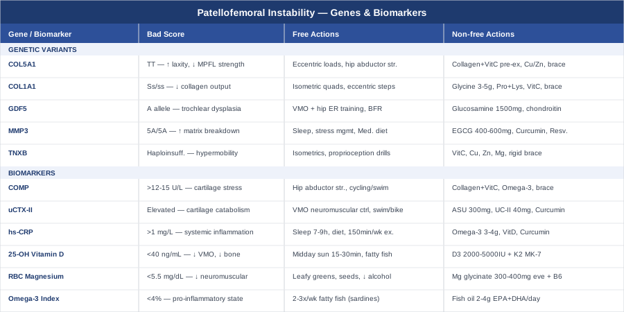
Patellofemoral instability is not a simple mechanical problem. It is the product of tissue quality, joint architecture, neuromuscular control, nutritional status, and — increasingly clearly — a genetic blueprint that varies considerably from person to person. The six biomarkers covered here — COMP, uCTX-II, hs-CRP, vitamin D, RBC magnesium, and the omega-3 index — give you a measurable starting point for understanding the biological terrain your joint is working within. The five genetic variants outlined offer a lens for understanding why some individuals are more vulnerable and what compensatory strategies make the most sense for them specifically.
None of this replaces clinical care. But it changes the quality of the conversation you can have with your physiotherapist, sports medicine physician, or orthopedic surgeon. The next smart step is practical: pick one or two of the most accessible biomarkers from this article, arrange the blood work, and bring the results to your next clinical appointment. Better data, more targeted interventions, and a clearer biological picture are the foundation of outcomes that actually stick.
Musculoskeletal: Joint Conditions Tendon & Ligament Conditions Sports Injuries
Autoimmune: Inflammatory Conditions Connective Tissue Conditions