This article contains AI generated content.
Pellegrini-Stieda Syndrome: 3 Genes and 6 Biomarkers to Track
Introduction
If you've been told you have Pellegrini-Stieda syndrome — that patch of calcification sitting at the inner side of your knee, just above the joint line — you've probably already noticed that most of what's written about it is short, generic, and unsatisfying. A paragraph on what it is, a mention of rest and anti-inflammatories, maybe a note that it "usually resolves." That's not wrong, but it isn't much to work with when you're the one dealing with stiffness, a tender lump, or a knee that never quite feels the same since the original injury.
Part of the problem is that Pellegrini-Stieda syndrome sits in an odd spot: it's not a disease you're born with, not an autoimmune process, and not something with a dedicated research pipeline the way arthritis or osteoporosis have. It's a mechanical aftermath — bone forming where it shouldn't, at the site of an old ligament injury. That means generic knee advice misses the specific biology at play, and generic "genetics and biomarkers" content risks inventing connections that simply aren't there.
This article takes a more careful approach. Rather than pretending Pellegrini-Stieda syndrome has a tidy genetic fingerprint, it starts from what is actually documented — the biology of how trauma turns into calcification — and builds outward to the lab values, imaging patterns, and biological pathways worth understanding if you're trying to make sense of your own case. Where the evidence is thin or borrowed from closely related conditions, that's stated plainly rather than dressed up.
None of this promises a cure or a shortcut. What it offers is a clearer map: which biomarkers are worth tracking and why, what the underlying gene pathways suggest (and don't), what broader recovery science adds to the picture, and which complementary approaches have at least some human evidence behind them. Better information doesn't guarantee a faster recovery, but it does let you ask sharper questions — of your own body, and of the clinicians managing your care.
Summary
Pellegrini-Stieda syndrome is calcification or ossification at the femoral attachment of the knee's medial collateral ligament, almost always following a prior injury such as an MCL sprain. It is not classically inherited — the NIH's own genetic reference database lists no associated genes for it. So instead of manufacturing a genetics story, this article leads with the six biomarkers most worth tracking: vitamin D, calcium and parathyroid hormone, bone-specific alkaline phosphatase, inflammatory markers (CRP/ESR and IL-6), and imaging-based staging of the calcification itself. Each section explains what the marker reveals, roughly what it costs to check, and two levels of response — a no-supplement plan and a supplement-or-equipment plan, both with realistic dosing, cycling, and side-effect notes.
After that, a shorter bonus section looks at the three biological pathways — ACVR1/BMP signaling, RUNX2, and the IL-6/TNF-alpha inflammatory axis — that drive ectopic bone formation in general, what happens when direct-to-consumer genetic reports flag "unfavorable" variants in these pathways, and how that risk might realistically be offset. A further section pulls out ten evidence-grounded ideas from pain-recovery neuroscience — the kind of material Andrew Huberman's podcast often summarizes — and a final section reviews which complementary therapies (photobiomodulation, massage, biofeedback, mindfulness) have any real human evidence behind them for calcific and post-traumatic knee conditions, and which don't.
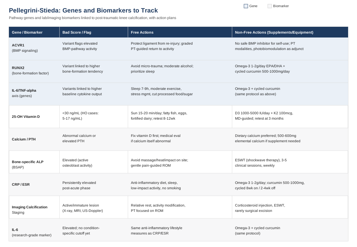
6 Biomarkers Worth Tracking for Pellegrini-Stieda Syndrome
Before getting into individual markers, it's worth being explicit about what these biomarkers can and can't do. None of them diagnoses Pellegrini-Stieda syndrome — that's done with an X-ray or MRI showing the characteristic calcification at the MCL's femoral origin, as described in the core clinical reference on the condition. What these markers can do is help you and your clinician understand the biological environment your knee is healing in — whether it's tilted toward more inflammation and more calcification, or toward quieter, more organized tissue repair.
1. 25-hydroxyvitamin D (25-OH-D)
Vitamin D status is one of the more concrete, modifiable pieces of this puzzle. In patients who develop heterotopic ossification (ectopic bone formation, the broader category Pellegrini-Stieda belongs to) after conditions like spinal cord injury, low 25-OH-D levels are strikingly common — one study found 11 of 12 patients with heterotopic ossification had levels between 5 and 17 ng/mL, well below the normal range, as reported in this analysis of vitamin D and secondary hyperparathyroidism in heterotopic ossification. That's not proof vitamin D deficiency causes ectopic calcification in the knee specifically, but it's a strong enough association in the broader ossification literature to be worth checking.
How to measure it
A standard 25-OH-D blood test, available through a primary care visit or direct-to-consumer lab service. Typical cost is $40–$80 out of pocket if not covered by insurance; many routine physicals include it at no extra charge. Optimal range for most adults is generally considered 30–50 ng/mL, though labs vary slightly.If the score is bad, the plan without supplements
Fifteen to twenty minutes of midday sun exposure on bare skin several times a week (more in winter or at higher latitudes, less if you have fair skin or a personal/family history of skin cancer), combined with regular intake of fatty fish, egg yolks, and fortified dairy. Retest after 8–12 weeks of consistent sun and dietary changes before assuming this alone will normalize a significant deficiency — for many people it won't, and that's a normal outcome, not a failure.If the score is bad, the plan with supplements or equipment
Vitamin D3, typically 1,000–2,000 IU per day for mild deficiency, higher (2,000–5,000 IU/day) short-term under physician guidance for more significant deficiency, paired with vitamin K2 (MK-7, ~100 mcg/day) to support appropriate calcium routing. Retest at 3 months, then adjust. One caution specific to this condition: a case report of heterotopic ossification following stroke explicitly noted that calcium and vitamin D repletion can, in theory, aggravate an already-active, immature ossification site — see this case discussion. Practical takeaway: correcting a genuine deficiency is reasonable, but megadosing "just in case" while a calcification is still actively forming is not — this is a conversation to have with your physician, not a self-directed high-dose protocol. Side effects of excess vitamin D include hypercalcemia, nausea, and kidney strain; stay within tested, monitored ranges.2. Serum calcium and parathyroid hormone (PTH)
Calcium and PTH sit upstream of any calcification process — they govern how much calcium is circulating and available to be deposited in soft tissue. Abnormal PTH, often secondary to low vitamin D, shows up alongside heterotopic ossification in the same population referenced above. These aren't usually abnormal in an isolated, otherwise-healthy person with Pellegrini-Stieda syndrome, but checking them rules out an underlying metabolic driver, particularly if calcification has been unusually extensive or recurrent.
How to measure it
A basic metabolic panel (includes calcium) plus a separate intact PTH test. Combined cost is usually $30–$100, often bundled into a standard blood panel your doctor already orders.If the score is bad, the plan without supplements
If PTH is elevated secondary to low vitamin D, correcting the vitamin D deficiency (as above) is the primary lever — PTH usually normalizes as 25-OH-D improves. If calcium itself is abnormal, this warrants direct medical evaluation rather than a self-directed plan, since both high and low calcium have causes well beyond a knee condition (parathyroid gland issues, kidney function, medication effects).If the score is bad, the plan with supplements or equipment
This is one of the few markers on this list where supplementation should follow, not precede, a clear diagnosis. If your physician confirms low calcium intake as a contributing factor, dietary calcium (dairy, leafy greens, fortified foods) is generally preferred over supplements, since high-dose calcium supplementation has been associated with vascular calcification risk in some populations. If a supplement is recommended, it's typically 500–600 mg elemental calcium per dose (better absorbed than a single large dose), taken with food. Side effects include constipation and, at high doses, kidney stone risk.3. Bone-specific alkaline phosphatase (BSAP)
This enzyme is produced by osteoblasts — the cells responsible for laying down new bone — and rises when bone-forming activity increases, including in ectopic sites. Research on calcific rotator cuff disease, a well-studied analog to Pellegrini-Stieda, shows that tendon cells can transform into alkaline-phosphatase-producing, calcium-depositing chondrocyte-like cells during the active calcification phase, as detailed in this study of rotator cuff tenocyte differentiation. A rising BSAP can be a rough signal that a calcification is still in an active, forming phase rather than a quiet, resorbing one.
How to measure it
A dedicated bone-specific ALP test (distinct from the ALP included in a standard liver panel). Cost ranges $50–$150 depending on the lab, and it's not always ordered by default — you may need to specifically request it.If the score is bad, the plan without supplements
There's no dietary "fix" for elevated BSAP — it's a downstream readout of ongoing osteoblast activity, not a target to suppress directly. The most useful non-supplement response is behavioral: avoid aggressive massage, heat, or high-impact loading directly over an actively calcifying site, since mechanical irritation during the active phase may prolong the inflammatory-osteogenic cycle. Gentle, pain-guided range-of-motion work is generally preferred over rest or aggressive stretching during this phase.If the score is bad, the plan with supplements or equipment
There's no supplement shown to lower BSAP directly, and attempting to suppress it isn't necessarily desirable — bone formation is also part of normal healing. Where equipment has a role is in guided treatment: extracorporeal shockwave therapy (ESWT) has reasonable trial evidence for calcific tendinopathy, shown in this network meta-analysis and a more recent 2024 update, typically delivered as 3–5 sessions spaced a week apart in a clinical setting. It's not something to self-administer, and mild bruising or transient pain flare after sessions is common and usually resolves within days.4. C-reactive protein (CRP) and erythrocyte sedimentation rate (ESR)
These are general inflammation markers, not specific to this condition, but they're useful for context: heterotopic ossification is driven in part by an inflammatory cascade involving cytokines like TNF-alpha and IL-1beta acting on osteogenic precursor cells, as reviewed in this study of cytokine effects on osteogenic differentiation. Persistently elevated CRP or ESR after the acute injury phase has passed can suggest ongoing low-grade inflammation worth investigating, rather than a knee healing quietly on its own timeline.
How to measure it
Both are inexpensive, widely available blood tests, usually $20–$50 combined, and often included in a standard workup if a clinician suspects an inflammatory component.If the score is bad, the plan without supplements
An anti-inflammatory eating pattern (more vegetables, fatty fish, olive oil; less ultra-processed food and added sugar), consistent sleep timing, and moderate low-impact activity such as walking or swimming, which tends to lower systemic inflammatory markers over weeks to months. Smoking cessation matters here too — smoking measurably impairs soft tissue healing and is worth addressing directly if relevant.If the score is bad, the plan with supplements or equipment
Omega-3 fatty acids (1–2 g/day combined EPA/DHA) have reasonably consistent evidence for modestly lowering CRP over 8–12 weeks; mild fish-burp or GI upset is the main side effect, and doses above 3 g/day can increase bleeding risk, particularly alongside blood thinners. Curcumin (500–1,000 mg/day with piperine for absorption) is a reasonable adjunct, cycled 8 weeks on and 2–4 weeks off rather than taken indefinitely, with GI upset as the most common side effect and a genuine interaction risk with anticoagulants — flag this to your doctor if you're on one. Neither of these is a substitute for medical evaluation if CRP or ESR is significantly elevated.5. Imaging-based calcification staging
This isn't a blood test, but it functions as a biomarker in the sense that matters most here: it tells you where the calcification is in its life cycle. Plain X-ray typically shows the calcification as a linear or curvilinear density becoming visible around three weeks after the original injury, while MRI can additionally show surrounding marrow edema and ligament thickening in the acute phase, per the StatPearls clinical overview. Ultrasound with Doppler can add information about local vascularity — more blood flow generally corresponds to a more active, still-forming lesion, and less to a mature, stable one. A case report using ultrasound-guided percutaneous lavage to treat this condition, drawing an explicit parallel to calcific rotator cuff disease, is described here.
How to measure it
X-ray costs roughly $100–$300 depending on region and insurance; MRI runs considerably higher, typically $500–$2,000 without insurance; ultrasound is often the most affordable follow-up option at $150–$400 and can be repeated more frequently without radiation exposure.If the score is bad, the plan without supplements
If imaging shows an actively forming, still-inflamed lesion, the standard non-supplement approach is relative rest from aggravating movements, activity modification rather than complete immobilization, and physical therapy focused on maintaining range of motion without direct mechanical irritation of the calcified area. Most cases resolve within roughly five to six months with this conservative approach, per the same clinical reference above.If the score is bad, the plan with supplements or equipment
For a mature, symptomatic, non-resolving lesion, options escalate to corticosteroid injection or ESWT under medical supervision, and rarely surgical excision if the mass remains painful and mechanically limiting long after the expected healing window. These are procedural decisions for an orthopedic clinician, not self-directed choices — the value of tracking imaging over time is knowing when to have that conversation rather than waiting indefinitely on "it'll probably go away."6. Interleukin-6 (IL-6) — a research-grade marker
This one is more advanced and less routinely available, but worth knowing about if you want to go deeper. IL-6 is a cytokine implicated in ligament and soft-tissue ossification processes, shown for example in research on ossification of the spinal ligamentum flavum, a mechanistically related process, in this study. It isn't a standard clinical test for Pellegrini-Stieda syndrome specifically, and there's no established "normal versus concerning" cutoff for this exact condition — it's included here as an example of where the mechanistic research is heading, not as something to routinely order.
How to measure it
Specialty or research-oriented labs offer IL-6 testing, typically $80–$200, and it's not something most primary care practices order by default. For most readers, this is more useful as a concept to understand than a test to pursue right now.If the score is bad, the plan without supplements
The same general anti-inflammatory lifestyle measures described under CRP/ESR apply — sleep, diet, moderate activity, and avoiding smoking all influence systemic IL-6 levels over time.If the score is bad, the plan with supplements or equipment
Given the lack of condition-specific cutoffs, an aggressive supplement protocol aimed narrowly at lowering IL-6 isn't well justified here. The same omega-3 and curcumin approach outlined above has the best general evidence for modulating this cytokine family, at the same doses and cycling pattern already described.What the Genetics Research Actually Suggests
It's worth stating this plainly: Pellegrini-Stieda syndrome is not currently listed with any associated genes in the NIH's own genetics reference database, MedGen. This is a post-traumatic condition — it happens after a specific injury, not because of an inherited predisposition in the way that, say, familial hypercholesterolemia or hereditary hemochromatosis do. So rather than inventing a gene panel for a condition that doesn't have one, this section looks at the biological pathways known to drive ectopic bone formation in general, and what direct-to-consumer genetic reports touching on these pathways might realistically mean for you.
Researchers and public figures like Ali Torkamani, whose work at Scripps Research focuses heavily on polygenic risk scoring, and Gary Brecka, known for popularizing biomarker- and genetics-informed wellness protocols, have helped mainstream the idea that raw genetic data can flag directional tendencies — not diagnoses, but leanings worth knowing about. Neither has published research specifically on Pellegrini-Stieda syndrome, and no such research exists yet. What follows applies that same directional-tendency framing to the three pathways most implicated in ectopic ossification generally.
1. ACVR1 (the BMP signaling gene)
ACVR1 encodes a receptor in the bone morphogenetic protein (BMP) pathway, the master signaling system that tells cells to become bone. A specific, rare mutation in this gene (R206H) causes fibrodysplasia ossificans progressiva, a severe hereditary ossification disorder — see the mechanism described in this review. That specific mutation has nothing to do with Pellegrini-Stieda syndrome. What's relevant is the broader pathway: BMP signaling activity, influenced by common (non-pathogenic) variation across many genes, plausibly affects how readily any individual forms ectopic bone after trauma, and animal research shows that inhibiting this pathway with a BMP type I receptor blocker measurably reduces heterotopic ossification, per this Nature Medicine study.
If the gene is flagged, the plan without supplements
If a consumer genetic report flags elevated BMP-pathway activity variants, the most sensible response is behavioral, not pharmacological: protect any healing ligament from repeated re-injury, since each new trauma is a fresh trigger for the ossification cascade, and follow a graded, physical-therapy-guided return to activity rather than pushing through pain.If the gene is flagged, the plan with supplements or equipment
There is no supplement that directly and safely inhibits BMP signaling in humans outside of investigational drug trials — this isn't a pathway to self-medicate. The realistic equipment-based lever is early, appropriately timed physical therapy modalities (not aggressive massage) during the acute phase, and photobiomodulation devices, discussed further below, which have some evidence in analogous calcific conditions.2. RUNX2 (the master bone-formation transcription factor)
RUNX2 is the key transcription factor that pushes precursor cells toward becoming bone-forming osteoblasts, and it's directly activated by the same inflammatory cytokines (TNF-alpha, IL-1beta) present at an injury site, as shown in the osteogenic differentiation study referenced earlier. Common genetic variation in RUNX2 has been linked in broader bone-density research to individual differences in bone formation tendency — again, a directional signal, not a diagnosis.
If the gene is flagged, the plan without supplements
Since RUNX2 activity is amplified by local inflammation, minimizing unnecessary inflammatory flare-ups around the injury site is the main non-supplement lever — this means avoiding repeated micro-trauma, moderating alcohol intake during the healing window (it independently impairs tissue repair), and prioritizing sleep, which regulates several of the same inflammatory pathways.If the gene is flagged, the plan with supplements or equipment
The same anti-inflammatory supplement approach described under the CRP/ESR biomarker above (omega-3s, cycled curcumin) is the most defensible option here, since it targets the inflammatory drivers of RUNX2 activation rather than attempting to alter gene expression directly. Frequency and side-effect notes are identical to those given earlier — this is not a case for a separate, more aggressive protocol.3. The IL-6/TNF-alpha inflammatory axis
This isn't a single gene but a cluster of genes governing cytokine production, and it's the pathway most consistently implicated across the heterotopic ossification literature, summarized comprehensively in this review. People with genetic variants associated with higher baseline inflammatory cytokine production may, in theory, be more prone to an exaggerated healing response after ligament trauma — though this remains an extrapolation from general HO biology rather than a finding specific to Pellegrini-Stieda syndrome.
If the gene is flagged, the plan without supplements
Behavioral inflammation management is the first-line response regardless of genetic background: consistent sleep (7–9 hours), regular moderate exercise once cleared by a clinician, stress management, and reducing ultra-processed food and added sugar intake, all of which have measurable effects on circulating cytokine levels over weeks to months.If the gene is flagged, the plan with supplements or equipment
Again, omega-3 fatty acids and cycled curcumin are the best-supported general options, at the doses and cycling schedule already described under the biomarker section. There's no justification for a more aggressive or novel supplement stack based on a genetic flag alone — the evidence doesn't support it, and unnecessary supplementation carries real (if modest) cost and side-effect burden for no proven benefit in this specific condition.What Pain-and-Recovery Science Adds to the Picture
No Andrew Huberman episode, Peter Attia interview, or dedicated book focuses specifically on Pellegrini-Stieda syndrome or heterotopic ossification — that should be said plainly rather than papered over. What does exist, and what's genuinely useful, is a body of general pain-neuroscience and recovery science that Huberman's podcast has repeatedly summarized for a mainstream audience, drawing on decades of peer-reviewed research. Applied thoughtfully — not as condition-specific treatment, but as general context for anyone recovering from a soft-tissue injury — these ideas are worth knowing.
1. Inflammation is the first phase of repair, not simply the problem
The inflammatory cascade that can drive ectopic calcification is the same cascade that clears debris and recruits repair cells after any injury, as the heterotopic ossification review makes clear. The goal isn't to eliminate inflammation entirely — it's to avoid letting it run unchecked for longer than the healing process requires.2. Timing matters more than total suppression
Blunting inflammation too early and too aggressively (for instance, high-dose anti-inflammatory use in the first 48–72 hours) may interfere with the initial repair signaling your knee needs. This is a conversation for your physician, not a DIY decision, but it's a useful frame for understanding why "just take anti-inflammatories" isn't universally the right first move.3. Pain perception is modulated by the brain, not just the injury site
Gate control theory — the well-established idea that non-painful sensory input can reduce the perception of pain signals reaching the brain — explains part of why techniques like massage, heat, or even distraction can meaningfully change how a knee feels, independent of the tissue itself changing.4. Fear of movement can slow functional recovery
Kinesiophobia — avoiding movement out of fear of re-injury — is a well-documented factor that can prolong disability beyond what the tissue damage alone would predict. Gradual, guided movement, even when imperfect, generally outperforms rigid protective avoidance.5. Deep rest states support recovery physiology
Structured relaxation practices that shift the body toward parasympathetic dominance are associated with better recovery markers across multiple injury contexts, likely by reducing sustained sympathetic-driven inflammation.6. Breath pattern influences pain and stress in real time
Slow, exhale-emphasized breathing shifts autonomic balance measurably within minutes, and is a low-cost, zero-side-effect tool worth using during flare-ups of knee pain or stiffness.7. Sleep quality directly affects tissue repair
Growth hormone release and much of connective tissue remodeling happen preferentially during deep sleep stages. Poor sleep doesn't just make pain feel worse subjectively — it plausibly slows the biological repair process itself.8. Mental rehearsal can preserve motor patterns during forced rest
If movement is restricted during an active calcification phase, motor imagery — mentally rehearsing normal gait and knee mechanics — has some evidence for helping preserve neuromuscular coordination that would otherwise degrade during prolonged inactivity.9. Controlled loading beats both total rest and premature high-impact activity
Mechanotransduction — the process by which mechanical load signals cells to remodel tissue in an organized way — favors graded, progressive loading over both extremes. This aligns with why physical therapy protocols for this condition emphasize gradual return to motion rather than prolonged immobilization.10. Expectation shapes outcomes
How a condition is framed to a patient — as a dangerous, permanent mass versus a well-understood, usually self-limiting post-traumatic process — measurably influences reported pain and functional recovery in the broader placebo/nocebo literature. Understanding that Pellegrini-Stieda syndrome typically resolves within months, as the clinical literature indicates, is itself a useful piece of information to hold onto.Complementary Approaches With Real (Though Often Indirect) Evidence
No complementary therapy has been studied specifically in Pellegrini-Stieda syndrome — the condition is simply too uncommon to have generated dedicated trials. The approaches below are the ones with the strongest human evidence in closely related contexts: calcific tendinopathy (a close biological cousin) and post-traumatic knee rehabilitation. That distinction matters, and it's called out honestly below rather than implied away.
Photobiomodulation (low-level laser therapy)
Photobiomodulation uses low-level laser or LED light to influence cellular activity in tendon and soft tissue, with proposed effects on mitochondrial function and local inflammation. It's relevant here because the closest condition-specific evidence comes from calcific tendinopathy, which shares the calcium-deposition biology seen in Pellegrini-Stieda syndrome.
A 2025 randomized controlled trial specifically tested photobiomodulation in calcific rotator cuff tendinopathy and found a positive short-term effect, described in this study, and broader tendinopathy meta-analyses have found dose-dependent benefits, summarized in this review.
Realistically, this means: if you have access to a clinical-grade photobiomodulation device through a physical therapist, a course of 2–3 sessions weekly for 4–6 weeks is a reasonable, low-risk adjunct to standard care — not a replacement for it, and not something to expect dramatic results from given the evidence is extrapolated from a related condition rather than this one directly.
Massage therapy
Massage is relevant here mainly through its role in general knee-injury rehabilitation and pain modulation via the gate-control mechanism described earlier, rather than through any direct effect on calcification itself.
A 2025 randomized trial found percussion massage improved outcomes during ACL-reconstruction rehabilitation, detailed here, and a knee osteoarthritis dose-finding trial found measurable benefit from regular massage sessions, described here — both knee-adjacent, not condition-specific.
Applied cautiously, gentle massage of the surrounding musculature (not direct, deep pressure over an actively calcifying lesion, which could aggravate an inflamed area) once or twice weekly is a reasonable comfort-and-mobility measure during the subacute and later phases of recovery.
Biofeedback
Biofeedback, typically EMG-based, trains conscious control over muscle activation patterns and has a reasonable track record in post-operative knee rehabilitation, though the evidence for pain outcomes specifically is mixed.
An EMG biofeedback trial in ACL reconstruction rehab showed functional benefit, found here, while a broader systematic review found heterogeneous results without a clear pain advantage, noted here — worth knowing before expecting too much from this modality alone.
Where a physical therapist has EMG biofeedback equipment available, it can reasonably be incorporated into quadriceps and hamstring strengthening work during the later stages of recovery, once the acute calcification phase has settled.
Mindfulness-based stress reduction (MBSR)
Mindfulness practices are relevant less for the tissue itself and more for managing the frustration and low-grade anxiety that can accompany a slow-resolving, visible knee mass.
A systematic review and meta-analysis of MBSR for chronic low back pain found short-term benefit compared to usual care, though not clearly superior to other active treatments, described here — again, a related musculoskeletal pain context rather than this specific condition.
A realistic application is a standard 8-week MBSR course, or a shorter daily 10–15 minute guided practice, used as a parallel support alongside physical therapy rather than a primary treatment — genuinely useful for coping and pain perception, not for resolving the calcification itself.
Conclusion
Pellegrini-Stieda syndrome is a post-traumatic condition, not a hereditary one — the honest starting point is that there's no gene panel that predicts or explains it. What's useful instead is tracking the biology actually in play: vitamin D and calcium status, inflammatory markers, bone-formation activity, and the calcification's own stage on imaging. Together, these give you and your clinician a clearer sense of whether the knee is still in an active, forming phase or settling toward the resolution most cases eventually reach.
The genetic pathways behind ectopic bone formation (BMP signaling, RUNX2, inflammatory cytokines), the broader recovery-science principles popularized by pain researchers, and the complementary approaches with the best (if indirect) evidence all point in the same direction: manage inflammation sensibly, protect the healing ligament from repeat trauma, load it gradually rather than avoiding it entirely, and address the modifiable biomarkers — especially vitamin D — with your physician rather than alone.
The next practical step is simple: ask your doctor for a baseline panel covering 25-OH-D, calcium, and inflammatory markers, get a clear read on your imaging stage, and use that information to set realistic expectations for the months ahead rather than guessing.
Musculoskeletal: Bone Conditions Joint Conditions