This article contains AI generated content.
· UpdatedPemphigus Vulgaris: 6 Genes and 7 Biomarkers to Track
Introduction
Pemphigus vulgaris is one of the more disorienting autoimmune diagnoses to receive. The blisters appear on skin and mucous membranes, treatment is aggressive, and the disease can feel entirely beyond your control. If you have been through cycles of flares, high-dose corticosteroids, and cautious tapering, you know that "management" often feels more like guesswork than science. That frustration is legitimate.
What most people are not told is that pemphigus vulgaris is not a monolithic disease. It sits at the intersection of specific genetic vulnerabilities, measurable immune dysregulation, and environmental triggers that interact differently in each person. Generic advice — eat less sugar, reduce stress — is not wrong, but it is far too coarse to help someone navigating a condition driven by anti-desmoglein autoantibodies and dysfunctional B-cell regulation.
This article takes a different approach. Rather than repeating standard guidance, it focuses on what you can actually measure and what the evidence says about those measurements. That includes the specific genes that increase susceptibility and the biomarkers that track disease activity and treatment response in real time.
Better information does not guarantee better outcomes, but it does narrow the gap between what your doctors can monitor and what you can monitor. The sections below cover first the most actionable biomarkers — what to test, what the numbers mean, and what to do if they are off — and then a deeper look at the genetic variants most consistently linked to pemphigus vulgaris, including what current evidence suggests about compensating for them.
Summary
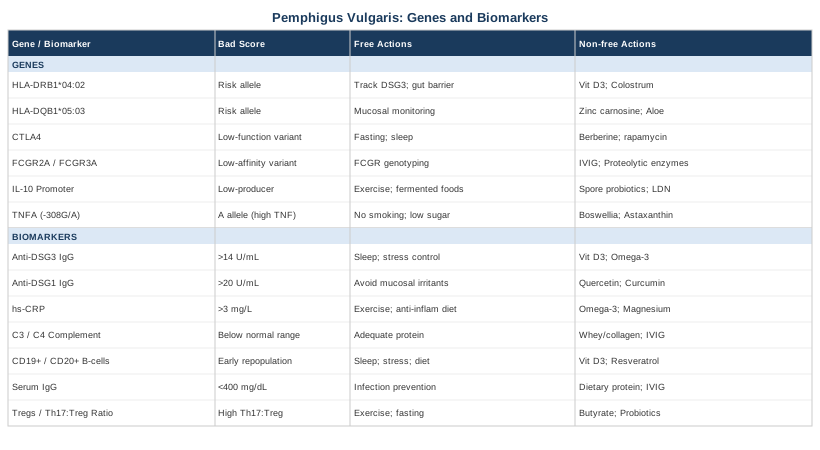
This article covers 7 biomarkers and 6 genes directly relevant to pemphigus vulgaris, with practical plans for each abnormal result — both with and without supplements. You will find:
- Anti-DSG3 and anti-DSG1 antibodies: the two markers that track disease activity most precisely, including how to interpret titer changes and what to do when they rise - hs-CRP, complement levels, and B-cell counts: the inflammatory and immune markers that give early warning before a flare becomes visible - HLA-DRB1 and HLA-DQB1: the two most powerful genetic risk factors for pemphigus vulgaris, and why your immune system may have been set up for this long before symptoms appeared - A practical section on the Autoimmune Protocol (AIP) as a structured dietary intervention with clinical backing for autoimmune blistering conditions - A candid look at mindfulness, microbiome therapy, and other complementary approaches with condition-specific evidence
---
7 Biomarkers to Track in Pemphigus Vulgaris
Tracking biomarkers in pemphigus vulgaris is not about collecting numbers — it is about building a picture of where your immune system is at any given moment. The seven markers below range from the disease-specific antibodies that are essentially diagnostic to the general inflammatory and immunological markers that tell you how your body is handling the condition and its treatment.
1. Anti-Desmoglein 3 (DSG3) IgG Antibodies
Why it matters: Anti-DSG3 IgG antibodies target desmoglein 3, the adhesion protein that holds keratinocytes together in mucous membranes and deep skin layers. These are the defining autoantibodies in pemphigus vulgaris and correlate strongly with disease activity. In mucosal-dominant disease, this is often the only elevated desmoglein antibody. According to research published in the Journal of Investigative Dermatology, anti-DSG3 levels track clinical severity and can predict relapse when monitored longitudinally.
What it reveals: Elevated titers during clinical remission signal a high risk of relapse — sometimes months before visible blistering returns. Falling titers during treatment indicate the therapy is working at the immunological level.
How to measure it: ELISA test ordered by a dermatologist or immunologist. Cost: $80–$200 depending on the lab and insurance coverage. Units are typically U/mL, with values above 14 U/mL generally considered positive (lab cutoffs vary).
If the score is high — the plan without supplements
- Work with your specialist to assess whether the elevation is rising, stable, or falling. A single high value matters less than the trend. - Identify and eliminate known immune triggers: consider food antigen testing, rule out concurrent infection (Helicobacter pylori has been associated with PV flares in several case series), and reduce UV exposure and physical trauma to skin. - Practice sleep optimization — 7–9 hours, consistent schedule. Immune dysregulation is profoundly sensitive to sleep debt; CD4+ T-cell activity and regulatory T-cell function both decline with poor sleep. - Stress reduction is not filler here: cortisol suppresses regulatory T-cells and can amplify B-cell autoantibody production. Implement at minimum 10–20 minutes daily of structured breathwork or meditation (box breathing, 4-7-8 technique).
If the score is high — the plan with supplements or equipment
- Vitamin D3 (2,000–5,000 IU/day) with K2 (100–200 mcg): low vitamin D is consistently associated with autoimmune severity. Aim for serum 25-OH-D between 50–80 ng/mL. Do not exceed 10,000 IU without medical supervision. - Omega-3 fatty acids (2–4 g EPA+DHA/day) from fish oil or algae: EPA in particular modulates B-cell activation and leukotriene synthesis. Cycle: 3 months on, reassess antibody levels. Side effects: fishy breath, mild GI loosening at high doses. - Low-level laser therapy (LLLT) / photobiomodulation: preliminary evidence in mucosal wound healing suggests benefit for active lesions. Frequency: 3×/week on active mucosal lesions; avoid during active systemic flare without physician guidance. - Discuss N-acetylcysteine (600 mg 2–3×/day) with your physician: it supports glutathione, which modulates oxidative-driven B-cell hyperactivation. Cycling: 8 weeks on, 2 weeks off. Monitor for GI side effects.
---
2. Anti-Desmoglein 1 (DSG1) IgG Antibodies
Why it matters: DSG1 is the primary adhesion protein in the superficial epidermis. Anti-DSG1 antibodies are elevated in patients with mucocutaneous pemphigus vulgaris — where blistering affects both skin and mucosa — but are typically absent in purely mucosal disease. Their presence shifts the clinical picture and is associated with more extensive skin involvement.
What it reveals: Rising anti-DSG1 in a patient previously showing only anti-DSG3 may indicate disease progression toward mucocutaneous involvement. This marker is particularly useful for monitoring patients after rituximab, where B-cell depletion should reduce both antibody subtypes.
How to measure it: Same ELISA panel as DSG3, often run together. Cost: bundled with DSG3 testing, typically $120–$250 for both. Reference range: values above 20 U/mL are generally considered positive.
If the score is high — the plan without supplements
- Track both DSG1 and DSG3 simultaneously to understand the clinical phenotype. A rising DSG1 with stable DSG3 warrants closer dermatological review. - Implement a strict elimination of known mucosal irritants: spicy foods, hard-textured foods, alcohol, and tobacco — all of which can worsen mucosal barrier dysfunction and provoke local inflammation that amplifies autoantibody-driven damage. - Review any medications recently started: drugs such as penicillamine, ACE inhibitors, and certain NSAIDs have been associated with drug-induced pemphigus in susceptible individuals.
If the score is high — the plan with supplements or equipment
- Quercetin (500–1,000 mg/day with food): flavonoid with demonstrated inhibitory effects on mast cell degranulation and IgE-mediated B-cell activity. Not specific to pemphigus, but relevant in autoantibody-driven conditions. Cycle: 8–12 weeks, then reassess. Side effects: mild GI at high doses. - Curcumin phytosome (500–1,000 mg/day): anti-NF-κB activity may reduce downstream B-cell stimulation. Use phospholipid-bound form for absorption. Cycling: 12 weeks on, 4 weeks off. Do not combine with blood thinners without guidance.
---
3. High-Sensitivity C-Reactive Protein (hs-CRP)
Why it matters: CRP is a systemic inflammation marker produced by the liver in response to IL-6 signaling. In pemphigus vulgaris, elevated hs-CRP reflects the inflammatory backdrop that sustains autoantibody production and may signal concurrent infection — a critical distinction, since infections in immunosuppressed pemphigus patients can trigger or mimic flares.
What it reveals: Persistently elevated hs-CRP (above 3 mg/L) in a patient in apparent clinical remission may suggest ongoing subclinical inflammation, a secondary infection, or inadequate immunosuppression of the underlying autoimmune process. Very high CRP (above 10 mg/L) should prompt evaluation for infection.
How to measure it: Standard blood draw, available at any laboratory. Cost: $10–$40. Target: below 1 mg/L is optimal; 1–3 mg/L is borderline; above 3 mg/L warrants investigation.
If the score is high — the plan without supplements
- Rule out concurrent infection first — this is non-negotiable in immunosuppressed patients. - Evaluate sleep, exercise tolerance, and dietary pattern. A diet high in refined carbohydrates and vegetable oils consistently elevates CRP through prostaglandin pathways. - Introduce resistance exercise 2–3×/week: moderate-intensity weight training lowers IL-6 and CRP over 8–12 weeks in most populations. During active skin disease, avoid exercises that cause skin friction or pressure on affected areas. - Consider an anti-inflammatory dietary shift: Mediterranean-pattern or Autoimmune Protocol (AIP) eating — detailed in the complementary approaches section.
If the score is high — the plan with supplements or equipment
- Omega-3s (3–4 g EPA+DHA/day): one of the most evidence-backed supplements for lowering hs-CRP. Effect is dose-dependent and takes 6–8 weeks to appear. Side effects: bleeding risk at doses above 4 g/day; inform your physician if on anticoagulants. - Magnesium glycinate (300–400 mg before bed): low magnesium is independently associated with elevated CRP. Cycling: continuous supplementation is generally safe for most people; monitor bowel tolerance.
---
4. Complement Levels (C3 and C4)
Why it matters: The complement system is activated by antigen-antibody complexes, including those formed by anti-desmoglein autoantibodies. In active pemphigus, C3 and C4 may be consumed faster than they are produced, leading to low serum levels. Tracking complement helps distinguish active immune complex-mediated disease from other causes of inflammation and helps monitor treatment response.
What it reveals: Low C3 or C4 suggests active complement consumption — a sign that the autoimmune process is producing significant immune complex activity. Normal complement in a patient with elevated anti-DSG3 may indicate the antibodies are present but not yet actively driving tissue destruction. This nuance matters for treatment decisions.
How to measure it: Standard blood draw, often included in complement panels. Cost: $40–$100. Normal ranges: C3: 90–180 mg/dL; C4: 16–47 mg/dL.
If the score is low — the plan without supplements
- Low complement in pemphigus vulgaris generally reflects active disease rather than a complement deficiency — the priority is optimizing immunosuppressive therapy under physician guidance. - Ensure adequate protein intake (1.2–1.6 g/kg body weight/day): complement proteins are synthesized by the liver and their production is protein-dependent. Malnourished or hypoalbuminemic patients may have impaired complement synthesis.
If the score is low — the plan with supplements or equipment
- Whey or collagen protein supplementation (if tolerated) to support hepatic synthesis of complement proteins. 20–30 g/day in patients with documented low protein intake. Collagen peptides may additionally support dermal integrity. - Discuss intravenous immunoglobulin (IVIG) with your physician: in refractory pemphigus, IVIG can modulate complement activation and accelerate autoantibody clearance. This is a medical intervention, not a supplement, but relevant for patients with persistently low complement and active disease.
---
5. CD19+ and CD20+ B-Cell Counts
Why it matters: Pemphigus vulgaris is fundamentally a B-cell disease — autoreactive B-cells produce the anti-desmoglein antibodies that destroy epidermal adhesion. Rituximab, now a first-line therapy for moderate-to-severe pemphigus, works by depleting CD20+ B-cells. Monitoring B-cell counts is therefore essential for patients on rituximab and provides direct insight into the disease mechanism for all patients.
What it reveals: In patients on rituximab, CD19/CD20 depletion below 1% of lymphocytes signals effective B-cell suppression. B-cell repopulation — return to above 1–2% — often precedes antibody titer rise and clinical relapse by several months, making this a valuable early warning signal. According to data supporting rituximab's use in pemphigus, B-cell depletion correlates with durable remission in a significant proportion of patients.
How to measure it: Flow cytometry panel (lymphocyte subset analysis). Cost: $150–$400. Often ordered quarterly in rituximab-treated patients.
If the score is abnormal — the plan without supplements
- If B-cells are repopulating ahead of schedule, this should be communicated to your dermatologist — it may trigger earlier re-dosing of rituximab or closer monitoring of anti-DSG titers. - For patients not on rituximab, elevated B-cell counts in the context of active disease are expected. Focus on lifestyle factors that modulate B-cell activity: sleep, stress, and dietary antigen load.
If the score is abnormal — the plan with supplements or equipment
- Vitamin D3 (optimize to 60–80 ng/mL serum 25-OH-D): vitamin D receptors are expressed on B-cells, and D3 signaling reduces B-cell proliferation and promotes regulatory B-cell activity. Frequency: ongoing. Monitor serum calcium if using doses above 5,000 IU/day. - Resveratrol (250–500 mg/day): preliminary evidence suggests resveratrol modulates B-cell differentiation through SIRT1 and NF-κB pathways. Not specific to pemphigus, but mechanistically relevant. Cycling: 8 weeks on, 4 weeks off. Side effects: mild estrogenic effects at high doses; caution in hormone-sensitive conditions.
---
6. Serum IgG (Total and Subclasses)
Why it matters: Anti-desmoglein autoantibodies are IgG antibodies. Total serum IgG and IgG subclasses (particularly IgG1 and IgG4) provide context around the autoimmune response and flag hypogammaglobulinemia — a serious complication of long-term rituximab use that increases infection risk.
What it reveals: IgG4 anti-DSG3 is the pathogenic subclass in pemphigus vulgaris. Tracking IgG subclasses can reveal whether the immune response is shifting during treatment. Total IgG below 400 mg/dL in a rituximab-treated patient signals significant immunodeficiency requiring intervention.
How to measure it: Blood draw with quantitative immunoglobulin panel. Cost: $80–$200. Frequency: every 3–6 months in rituximab-treated patients.
If the score is low — the plan without supplements
- Low total IgG warrants immediate physician attention — IVIG replacement may be necessary. - Prioritize infection prevention: avoid crowds during active hypogammaglobulinemia, practice rigorous hand hygiene, stay current on applicable vaccines (discuss timing around rituximab cycles with your physician).
If the score is low — the plan with supplements or equipment
- Adequate dietary protein (1.4–1.8 g/kg/day): immunoglobulins are proteins; sustained protein deficiency impairs their production. For patients with oral pemphigus limiting food intake, consider protein-enriched smoothies or enteral supplementation. - IVIG infusion under medical supervision for IgG below 400 mg/dL — this is not optional.
---
7. Regulatory T-Cells (Tregs) and the Th17/Treg Ratio
Why it matters: Pemphigus vulgaris involves a fundamental imbalance between pro-inflammatory Th17 cells and suppressive regulatory T-cells (Tregs). Studies in pemphigus patients have documented reduced Treg frequency and function, allowing autoreactive B-cells and Th2 cells to drive anti-desmoglein production unchecked. The Th17/Treg ratio is an emerging marker of autoimmune activity that several researchers consider more mechanistically informative than general inflammation markers.
What it reveals: A high Th17/Treg ratio indicates the immune system is tilted toward attack rather than tolerance — the exact pattern underlying pemphigus pathology. Normalization of this ratio under treatment may predict durable remission more reliably than antibody titers alone.
How to measure it: Advanced flow cytometry panel, available at university hospitals and specialized immunology labs. Cost: $200–$600. Not standard in routine care but valuable in research settings or treatment-refractory patients. Research published in Clinical Immunology has documented the Treg deficit in active pemphigus vulgaris.
If the score is unfavorable — the plan without supplements
- Timed exercise: aerobic exercise (30–45 minutes, 70% max heart rate) acutely expands Treg populations and suppresses Th17 signaling. 3–4 sessions per week. Avoid skin-contact sports during active lesions. - Short-term fasting or time-restricted eating (14:10 or 16:8): caloric restriction and fasting states promote FOXP3+ Treg expansion in animal models and early human data. Discuss with physician before implementing during active disease. - Cold exposure (cold showers, 2–3 minutes): stimulates norepinephrine release, which downregulates IL-17 production. Begin with 30 seconds and build tolerance. Avoid directly over active skin lesions.
If the score is unfavorable — the plan with supplements or equipment
- Short-chain butyrate supplementation (600–1,200 mg/day tributyrin or sodium butyrate): butyrate is produced by gut bacteria fermenting dietary fiber and is a potent inducer of FOXP3+ Tregs in the gut-associated immune tissue. Cycling: 12 weeks on, 4 weeks off. Mild GI adjustment in first 1–2 weeks. - Probiotics with Lactobacillus rhamnosus and Bifidobacterium longum: these strains are associated with Treg induction via gut immune modulation. Frequency: daily. Choose enteric-coated formulations. Side effects: initial bloating; starts at 1 capsule/day and increase.
---
Moving from what you can measure today to the underlying architecture of susceptibility, the genetics of pemphigus vulgaris offer a different kind of insight — one that explains why some people develop this disease at all, and what that might mean for long-term immune management.
The Genetic Foundation of Pemphigus Vulgaris: 6 Key Genes
Pemphigus vulgaris is not random. The most consistent genetic finding across populations — from Ashkenazi Jewish to South Asian to East Asian cohorts — is a tight clustering around specific HLA alleles. But the HLA region is not the whole story. Several non-HLA genes modulate antibody clearance, cytokine balance, and regulatory immune cell function in ways that can either amplify or dampen the HLA-driven risk.
1. HLA-DRB1*04:02 — The Dominant Risk Allele
What it is: HLA-DRB1 encodes part of the MHC class II complex, the molecular "presentation table" where immune cells display peptide fragments for T-cell inspection. The *04:02 variant is by far the strongest genetic risk factor for pemphigus vulgaris in Ashkenazi Jewish and several European populations, conferring an odds ratio for disease above 20 in some studies.
What it may affect: HLA-DRB1*04:02 creates a peptide-binding groove with a specific shape that accommodates desmoglein-derived peptides efficiently. This means the immune system is more likely to "see" desmoglein fragments as foreign during immune activation — a setup for autoimmunity in the presence of environmental triggers. Research published in the Journal of Clinical Investigation documented this antigen-presentation mechanism in detail.
If the gene is unfavorable — the plan without supplements
- HLA alleles cannot be changed, but their expression of disease can be modified by reducing the inflammatory milieu that allows autoreactive T-cells to become activated. - Minimize molecular mimicry triggers: certain bacterial and viral proteins share structural homology with desmoglein. Maintaining gut barrier integrity — through dietary choices, sleep, and stress management — reduces the antigen load reaching systemic immune circuits. - Track anti-DSG3 titers 1–2×/year even during apparent remission if you carry this allele.
If the gene is unfavorable — the plan with supplements or equipment
- Vitamin D3 optimization (target 60–80 ng/mL serum 25-OH-D): vitamin D modulates MHC class II expression and T-cell responsiveness to self-antigens. This is one of the few interventions with mechanistic relevance at the HLA level. - Colostrum supplementation (2–4 g/day): contains proline-rich peptides (PRPs) that modulate T-cell-mediated immune hyperactivity. Human data are limited, but mechanism is consistent with HLA-related autoimmunity. Cycling: 8 weeks on, 4 weeks off.
---
2. HLA-DQB1*05:03 — The Mucosal Disease Modifier
What it is: HLA-DQB1*05:03 is frequently found in linkage disequilibrium with HLA-DRB1*14:01 and is particularly associated with pemphigus vulgaris in Mediterranean, South Asian, and East Asian populations. It is thought to contribute to the mucosal-dominant disease phenotype — where oral and mucosal blistering precedes or dominates over skin disease.
What it may affect: Like DRB1*04:02, this allele creates a permissive antigen-presentation environment for anti-DSG3 T-cell activation, particularly in mucosal tissue where DSG3 is more abundantly expressed.
If the gene is unfavorable — the plan without supplements
- Patients with this allele should prioritize oral mucosal monitoring: self-examine monthly for early erosions, and report new oral discomfort or altered eating patterns to a physician promptly — earlier detection means earlier treatment, which generally correlates with better outcomes. - Maintain a low-abrasion diet during remission: avoid hard, sharp, or acidic foods that stress the mucosal barrier.
If the gene is unfavorable — the plan with supplements or equipment
- Zinc carnosine (75 mg/day): has documented mucosal-protective effects in the oral cavity and upper GI tract through upregulation of mucosal defense proteins. Not specific to pemphigus, but mechanistically sound for mucosal protection. Cycling: continuous; assess at 12 weeks. - Aloe vera (inner leaf juice, preservative-free, 50–100 mL/day): preliminary evidence for mucosal healing and anti-inflammatory effects in oral mucositis. Use cold-processed inner gel only. Side effects: laxative effect at high doses.
---
3. CTLA4 — The Immune Checkpoint Gene
What it is: CTLA4 (cytotoxic T-lymphocyte antigen 4) encodes an inhibitory receptor that brakes T-cell activation after an immune response. Certain CTLA4 polymorphisms reduce this braking mechanism, allowing T-cells — including autoreactive ones — to remain activated longer than they should.
What it may affect: In pemphigus vulgaris, reduced CTLA4 function means autoreactive T-helper cells that should be shut down after encountering desmoglein peptides instead continue to provide help to anti-desmoglein-producing B-cells. This amplifies autoantibody titers and prolongs disease activity. Several studies have found CTLA4 polymorphisms overrepresented in pemphigus cohorts, though the effect size is smaller than HLA associations.
If the gene is unfavorable — the plan without supplements
- Caloric restriction and intermittent fasting: CTLA4 expression on activated T-cells is modulated by mTOR signaling. Intermittent fasting (16:8 protocol) reduces mTOR activity and may improve CTLA4-mediated suppression. Discuss with physician before starting during active disease. - Prioritize adequate sleep (7–9 hours): CTLA4 expression and T-cell homeostasis are highly sleep-dependent.
If the gene is unfavorable — the plan with supplements or equipment
- Berberine (500 mg 2–3×/day with meals): AMPK activator that downregulates mTOR, creating an environment more favorable to CTLA4-mediated T-cell suppression. Cycling: 8 weeks on, 4 weeks off. Side effects: GI discomfort, particularly in the first week; avoid with CYP3A4-sensitive drugs. - Rapamycin (low-dose, off-label): direct mTOR inhibitor; increasingly studied in autoimmune conditions for its Treg-promoting, T-cell suppression-normalizing effects. Not a supplement — requires prescription and monitoring. Mention to your physician as an area of active research.
---
4. FCGR2A and FCGR3A — Antibody Clearance Efficiency
What it is: FCGR2A and FCGR3A encode Fc gamma receptors on innate immune cells. These receptors bind the tail (Fc region) of IgG antibodies, including anti-desmoglein IgG, and facilitate their clearance from circulation. Polymorphisms in these genes affect how efficiently antibody-antigen complexes are eliminated.
What it may affect: Low-affinity FCGR variants result in slower clearance of anti-desmoglein antibodies, allowing higher titers to persist longer. This may partly explain why some patients have disproportionately elevated antibody titers relative to their clinical severity and why some respond more slowly to B-cell depletion therapies. It also affects how well the immune system clears rituximab-coated B-cells, potentially influencing rituximab efficacy.
If the gene is unfavorable — the plan without supplements
- Discuss FCGR genotyping with your physician before rituximab initiation, if available at your center — some data suggest FCGR3A polymorphisms influence rituximab response in autoimmune conditions. - Plasmapheresis (plasma exchange) is an option in severe, refractory cases to mechanically clear circulating antibodies — particularly relevant if antibody titers remain high despite B-cell depletion.
If the gene is unfavorable — the plan with supplements or equipment
- IVIG (intravenous immunoglobulin): beyond replacing low immunoglobulin, IVIG competitively saturates Fc receptors and accelerates autoantibody catabolism via FcRn saturation — a mechanism that may partially compensate for FCGR inefficiency. Medical intervention, not supplement. - Proteolytic enzyme supplements (bromelain, serrapeptase): very preliminary evidence for reducing circulating immune complex burden. Not pemphigus-specific. Cycling: 4 weeks on, assess, then continue or stop. Side effects: bleeding risk; avoid with anticoagulants.
---
5. IL-10 Promoter Polymorphisms — Anti-Inflammatory Capacity
What it is: IL-10 is a potent anti-inflammatory cytokine produced by Tregs and tolerogenic dendritic cells. Several promoter polymorphisms at positions -1082, -819, and -592 create low-producer haplotypes that reduce endogenous IL-10 output, tilting the cytokine environment toward inflammation.
What it may affect: Low IL-10 production in pemphigus vulgaris patients has been associated with more severe disease and slower response to treatment in some cohort studies. IL-10 normally suppresses B-cell autoantibody production and promotes Treg differentiation — both deficits that characterize active pemphigus.
If the gene is unfavorable — the plan without supplements
- Regular aerobic exercise is one of the most consistently documented stimulators of systemic IL-10 production. 30–45 minutes of moderate-intensity cardio 4–5×/week raises post-exercise IL-10 levels acutely and blunts pro-inflammatory cytokine responses chronically. - Fermented foods daily (kefir, kimchi, sauerkraut, miso): emerging evidence from human trials suggests fermented food consumption increases IL-10-producing immune cells. The 2021 Sonnenburg lab trial published in Cell demonstrated that high-fermented food intake increased immune regulation markers including IL-10.
If the gene is unfavorable — the plan with supplements or equipment
- Spore-based probiotics (Bacillus subtilis, B. coagulans): these strains colonize the gut transiently and stimulate IL-10 production by colonic immune cells. Dose: 1–2 billion CFU/day. Cycling: continuous. - Low-dose naltrexone (LDN, 1.5–4.5 mg/night): off-label use increasingly supported in autoimmune conditions; proposed mechanism includes upregulation of endogenous opioid signaling, which indirectly increases IL-10 through immune modulation. Requires prescription. Best discussed with a physician familiar with LDN in autoimmune disease.
---
6. TNFA — The Inflammatory Amplifier
What it is: The TNFA gene encodes tumor necrosis factor alpha, a central pro-inflammatory cytokine. The -308G/A promoter polymorphism (rs1800629) creates a high-producer allele (A variant) associated with enhanced TNF-alpha transcription.
What it may affect: TNF-alpha drives NF-κB activation, which promotes B-cell survival and immunoglobulin class switching — directly relevant to autoantibody production. Patients carrying the high-producer TNFA variant may have a more inflammatory disease course and greater susceptibility to triggers such as infections and UV exposure.
If the gene is unfavorable — the plan without supplements
- Minimize known TNF-alpha triggers: smoking is a powerful inducer of TNF-alpha; cessation is non-negotiable. Sun exposure on unprotected skin stimulates keratinocyte TNF-alpha release — daily broad-spectrum SPF 50+ is relevant both for UV protection and local inflammatory control. - Reduce refined sugar and trans-fat intake: both independently activate NF-κB signaling, amplifying TNF-alpha transcription in high-producer genotypes.
If the gene is unfavorable — the plan with supplements or equipment
- Boswellia serrata extract (200–400 mg AKBA, 3×/day): clinically studied inhibitor of 5-LOX and NF-κB pathways, reducing downstream TNF-alpha activity. Cycling: 12 weeks on, 4 weeks off. Side effects: mild GI; use with food. - Astaxanthin (8–12 mg/day): potent carotenoid antioxidant with documented suppression of NF-κB activity and TNF-alpha production in human trials. Cycling: continuous. Safe profile; slight skin/urine discoloration at high doses. - Red light therapy / photobiomodulation (660–850 nm): modulates NF-κB at the cellular level through mitochondrial signaling; 10–20 minutes, 3–4×/week. Avoid direct irradiation of active blistered areas.
---
The Autoimmune Protocol and Other Evidence-Based Approaches
Biomarkers and genes define the terrain. What you do within that terrain — through diet, lifestyle, and targeted complementary approaches — shapes how that terrain expresses itself day to day. The following modalities have meaningful human clinical evidence applicable to autoimmune blistering conditions.
The Autoimmune Protocol (AIP) — Sarah Ballantyne's Framework
What it is and why it matters: The Autoimmune Protocol, developed by Dr. Sarah Ballantyne (author of The Paleo Approach), is a structured elimination-and-reintroduction dietary framework specifically designed for autoimmune conditions. It removes foods associated with gut permeability, molecular mimicry, and immune hyperactivation — including grains, legumes, dairy, eggs, nightshades, nuts, seeds, alcohol, and refined oils — while emphasizing nutrient-dense whole foods that support immune regulation, gut integrity, and Treg function.
The protocol and the evidence: AIP is not based on a single trial for pemphigus specifically, but it has accumulated meaningful human clinical trial data in related autoimmune conditions. A 2017 pilot trial published in Inflammatory Bowel Diseases showed significant symptom improvement in Crohn's and colitis patients following AIP for 6 weeks. A 2019 study in Journal of the Academy of Nutrition and Dietetics found improvements in thyroid autoantibodies and quality of life in Hashimoto's thyroiditis patients following AIP for 10 weeks. The shared mechanisms — gut barrier restoration, Treg promotion, reduction of antigen load — are relevant to any IgG-mediated autoimmune condition, including pemphigus vulgaris. Ballantyne's approach is particularly notable for its systematic citation of peer-reviewed research on each eliminated food group's immune effects.
How to apply it for pemphigus vulgaris: Begin with a strict 30–60 day elimination phase. Given that oral pemphigus causes eating difficulty, adapt food textures as needed — smoothies, soups, and soft cooked foods are compatible with AIP. Focus on: organ meats (for dense nutrient delivery), wild-caught fish, leafy greens, sweet potato, and fermented vegetables. Reintroduce foods methodically one at a time, monitoring anti-DSG3 titers and symptom response. Do not attempt AIP without medical supervision if actively immunosuppressed, as dietary changes can interact with medication absorption and energy requirements.
---
Mindfulness Meditation and MBSR
What it is and why it matters: Mindfulness-Based Stress Reduction (MBSR) is an 8-week structured program developed by Jon Kabat-Zinn combining meditation, body scanning, and yoga-based movement. In autoimmune conditions, psychological stress is not merely a quality-of-life issue — it is a documented immunological trigger. Cortisol and catecholamines released during chronic stress suppress Treg function, elevate IL-17, and dysregulate B-cell antibody class switching. Pemphigus flares are frequently preceded by stressful life events, and this connection is mechanistically grounded.
The protocol and the evidence: A randomized controlled trial published in Psychoneuroendocrinology demonstrated that MBSR reduced IL-6 and CRP in stressed adults. Multiple meta-analyses have documented reductions in cortisol and inflammatory markers across MBSR participants. Though no randomized trial exists specifically for pemphigus vulgaris, the immune mechanisms modulated by MBSR overlap directly with those driving pemphigus pathology.
How to apply it realistically: Enroll in a local or online MBSR course (typically 8 weeks, 2.5 hours/week plus daily home practice of 30–45 minutes). Apps such as Insight Timer, Waking Up, or Ten Percent Happier can bridge the gap before a formal program is available. Aim for consistency over intensity — 20 minutes daily of mindful breathing has measurable effects on cortisol within 4–6 weeks.
---
Microbiome-Directed Therapies
What it is and why it matters: The gut microbiome is increasingly recognized as a regulator of systemic immune tolerance. Dysbiosis — an imbalance in gut microbial populations — is associated with reduced Treg induction, increased intestinal permeability, and elevated systemic inflammatory load. In pemphigus vulgaris, altered gut microbiome composition has been documented in at least one Chinese cohort study, with reductions in short-chain fatty acid-producing bacteria and increases in pro-inflammatory species.
The protocol and the evidence: A 2021 trial published in Cell (Wastyk et al.) demonstrated that a high-fermented food diet robustly increased microbiome diversity and reduced 19 inflammatory markers including IL-6 and IL-12p70 — cytokines relevant to autoimmune B-cell stimulation. This is arguably the strongest dietary intervention data for immune modulation currently available.
How to apply it for pemphigus vulgaris: Introduce 3–6 servings of fermented foods daily over 4–6 weeks (plain kefir, sauerkraut, kimchi, kombucha, unsweetened yogurt with live cultures). For oral pemphigus, cold preparations are easier to tolerate. Supplement with a multi-strain probiotic including Lactobacillus rhamnosus GG and Bifidobacterium infantis. Track stool consistency and any GI changes — adjustment is common in the first 2 weeks.
---
Breathing-Based Therapies
What it is and why it matters: Conscious breath control — particularly slow diaphragmatic breathing (4–6 breaths/minute) and techniques like box breathing and resonance frequency breathing — directly modulates the autonomic nervous system, increasing vagal tone and reducing sympathetic drive. Vagal tone is inversely correlated with IL-6 and TNF-alpha production through the cholinergic anti-inflammatory pathway. For pemphigus patients managing chronic disease-related stress and the HPA axis dysregulation that accompanies corticosteroid treatment, this is particularly relevant.
The protocol and the evidence: A randomized controlled trial in Frontiers in Human Neuroscience demonstrated that slow breathing at 6 cycles/minute significantly increased heart rate variability (HRV) — a surrogate of vagal tone — and reduced self-reported stress. Elevated HRV is associated with better immune regulation and lower CRP.
How to apply it for pemphigus vulgaris: Practice box breathing (4 seconds in, 4 hold, 4 out, 4 hold) for 5–10 minutes before sleep and during any high-stress period. Use a free HRV-tracking app (Welltory, HRV4Training) to observe your baseline and track progress. A measurable shift in HRV typically appears within 3–4 weeks of consistent daily practice.
---
What the Research on Autoimmune Disease Immune Regulation Keeps Showing
One resource worth knowing is the body of work synthesized by Dr. Terry Wahls — particularly her work applying a nutrient-dense, mitochondria-focused dietary protocol to autoimmune neurological disease. While her primary research context is multiple sclerosis (she published a pilot trial in Journal of Alternative and Complementary Medicine in 2014), the underlying framework maps directly to any autoimmune condition driven by T-cell dysfunction, mitochondrial stress, and antigen presentation abnormalities.
10 Things the Wahls-Informed Framework Says About Autoimmunity and Pemphigus
1. Mitochondrial function drives immune cell differentiation. Tregs and effector T-cells have different metabolic profiles — Tregs prefer fatty acid oxidation, Th17 cells use glycolysis. A diet that supports mitochondrial efficiency (adequate B vitamins, CoQ10, sulfur-containing vegetables) shifts the balance toward tolerance.
2. Sulfur-containing vegetables matter specifically. Kale, broccoli, onions, garlic, and leeks support glutathione synthesis and mitochondrial Complex II function. Three cups of these per day is a recurring recommendation in the Wahls Protocol. Glutathione modulates NF-κB and TNF-alpha activity directly.
3. Three cups of deeply pigmented vegetables daily. Anthocyanins, carotenoids, and flavonoids in deeply colored vegetables suppress oxidative stress in immune cells and reduce the inflammatory cytokine output that drives B-cell autoantibody production.
4. Eliminating gluten and dairy has a structural rationale. Gliadin peptides and casein-derived peptides have been shown to increase intestinal permeability in genetically susceptible individuals, allowing antigen translocation that could amplify ongoing autoimmune reactivity through molecular mimicry.
5. The gut-immune axis is the central battleground. Wahls frames gut health not as a side issue but as the primary driver of systemic immune tone. This aligns with the microbiome data in pemphigus and the evidence for AIP described above.
6. Omega-3 to omega-6 ratio matters, not just omega-3 intake. The ratio of EPA+DHA to arachidonic acid in cell membranes determines whether inflammatory cascades are amplified or suppressed. Reducing corn, soy, and sunflower oils while increasing fatty fish is the practical implementation.
7. Detoxification pathways need support. Glucuronidation and sulfation — liver detox pathways dependent on glycine, taurine, and sulfur amino acids — help clear inflammatory mediators and immune complexes. Bone broth, organ meats, and cruciferous vegetables support these.
8. Sleep is not recovery time — it is immune recalibration. Cytokine balance, Treg/Th17 ratios, and glymphatic clearance of neuro-immune signaling molecules all occur primarily during deep sleep. Wahls consistently identifies poor sleep as a primary driver of autoimmune flare.
9. Chronic stress overrides dietary intervention. The HPA axis — cortisol specifically — counteracts the Treg-promoting effects of diet and exercise. Wahls frames stress management as a non-negotiable, not an optional add-on. Cortisol chronically suppresses FOXP3 expression in Tregs.
10. Nutrients from food outperform equivalent supplements. The synergistic matrix of cofactors, enzymes, and phytonutrients in whole foods delivers effects that isolated supplements cannot replicate. The Wahls Protocol prioritizes food density over supplementation stacking.
---
Conclusion
Pemphigus vulgaris is not a condition that yields to generic health advice. It is driven by specific autoantibodies, enabled by particular genetic variants, and monitored through biomarkers that most patients are never taught to track. The seven biomarkers covered in this article — from anti-DSG3 titers to Treg ratios — give you a concrete, measurable picture of where your immune system stands at any given time. The six genes described in the bonus section explain why your immune system arrived at this place to begin with, and what that architecture might mean for long-term management.
None of what is covered here replaces the judgment of a qualified dermatologist or immunologist. But it does put you in a position to ask better questions, interpret your lab results with more context, and make more informed decisions about lifestyle, diet, and adjunctive approaches. The next smart step is to bring this framework to your next medical appointment, ask which of these markers are already being tracked, and identify which ones might be worth adding. Better monitoring leads to earlier intervention, and earlier intervention leads to better control. That is not a promise — it is the straightforward logic of how chronic autoimmune disease management works when it works well.