This article contains AI generated content.
· UpdatedPeriarticular Heterotopic Ossification: 6 Genes And 6 Biomarkers To Track
Introduction
Periarticular heterotopic ossification — bone forming in the soft tissue around a joint — is one of those conditions that tends to arrive without much warning. You might notice stiffness after a hip replacement, limited range of motion months after a spinal cord injury, or a strange firmness in tissue that was previously pliable. By the time imaging confirms it, the process has often been underway for weeks. That delay is part of what makes PHO so frustrating for both patients and clinicians.
The standard clinical approach tends to focus on prophylaxis after known high-risk events: radiation therapy or NSAIDs following total hip arthroplasty, for example. These are reasonable first lines of defense. But they don't explain why some people develop significant PHO while others undergoing identical procedures do not. That variability points to something upstream — genetics, inflammatory biology, and metabolic environment — that generic protocols simply don't account for.
What the research increasingly shows is that PHO is not a random mechanical accident. It is a biology-driven process involving overactivated bone morphogenetic protein signaling, unresolved inflammation, and calcium-phosphate dynamics that can be detected before the ossification becomes structural. Some people carry genetic variants that make their bone-forming pathways more reactive to injury. Others have chronic inflammatory profiles that create a fertile environment for ectopic osteogenesis. In both cases, the signals are measurable.
This article takes a more targeted approach. It explores six blood biomarkers that can help track PHO-relevant biology in real time — with specific plans for addressing elevated values — and six genes whose variants may explain individual susceptibility to ectopic bone formation. Neither route offers a cure, but both offer something genuinely useful: a more precise map of what is actually happening in your body and where leverage exists.
Summary
This article covers six biomarkers you can track through blood tests to detect the inflammatory and bone-forming activity that drives periarticular heterotopic ossification — often before imaging shows any abnormality. These include bone-specific alkaline phosphatase, hs-CRP, IL-6, BMP-2, the CTX/osteocalcin balance, and the calcium-phosphorus product. For each one, you'll find what an abnormal result means, how much the test costs, and a concrete action plan — with and without supplements.
The genetic section covers six key genes: ACVR1, BMP4, PTGS2, IL6, VDR, and RUNX2. Variants in these genes can make BMP signaling, prostaglandin production, vitamin D response, or osteoblast differentiation more reactive to injury or inflammation. Specific, realistic protocols are outlined for each genetic risk pattern.
Beyond lab work, the article summarizes the most relevant principles from Peter Attia's Outlive — ten ideas that quietly reframe how bone health and systemic inflammation should be tracked before problems appear. Finally, three evidence-supported complementary approaches are evaluated for their relevance to PHO: low-level laser therapy, MBSR-based mindfulness, and breathing-based nervous system regulation.
None of this replaces medical care. What it does is give you a more intelligent framework for understanding your own risk, monitoring change over time, and having more productive conversations with the clinicians managing your case.
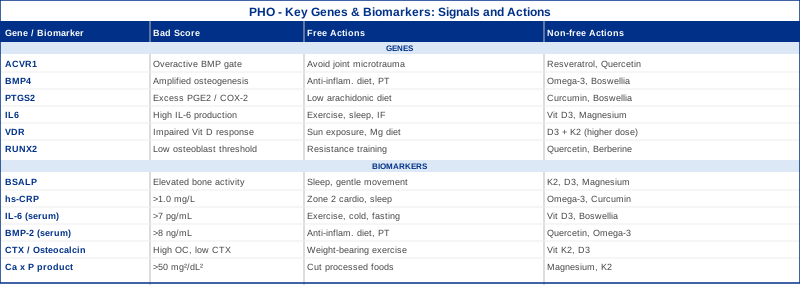
6 Biomarkers That May Reveal Your PHO Risk Before Imaging Shows Anything
Biomarkers are measurable signals in blood or tissue that reflect biological processes underway in the body. In the context of periarticular heterotopic ossification, they offer a window into osteoblast activity, systemic inflammation, and calcium-phosphate dynamics — the three converging forces that drive ectopic bone formation. What makes them valuable is timing: several of these markers rise days to weeks before structural bone becomes visible on X-ray or CT. Tracking them regularly when risk is elevated gives you a meaningful early-warning system.
The six biomarkers below were selected for their direct mechanistic relevance to PHO, their clinical availability, and their actionability — meaning that abnormal values can be addressed through specific interventions.
Biomarker 1: Bone-Specific Alkaline Phosphatase (BSALP)
Why it matters: Alkaline phosphatase is produced by osteoblasts during bone matrix mineralization. In PHO, ectopic osteoblasts begin laying down bone matrix — typically 2 to 3 weeks before standard imaging reveals calcification. Bone-specific ALP (BSALP) is the fraction that specifically reflects skeletal osteoblast activity, making it a far more useful signal than total ALP, which also reflects liver activity. Elevated BSALP in the setting of recent joint trauma, surgery, or neurological injury should be interpreted as a potential early PHO indicator.
How to measure it: BSALP is measured via immunoassay in blood. It requires a fasting morning draw. Standard labs may only offer total ALP (included in a basic metabolic panel, $20–50); BSALP fractionation requires specialty labs and costs approximately $80–200. Reference ranges: Males 6.3–19.0 µg/L; Females premenopausal 4.5–16.9 µg/L; Females postmenopausal 5.4–22.9 µg/L.
If the score is elevated, the plan without supplements: Evaluate the clinical context first — elevated BSALP without known bone disease warrants follow-up imaging of affected joints. Prioritize gentle, consistent movement to normalize periarticular tissue without triggering additional inflammatory cascades. Avoid deep tissue manipulation around suspect areas. Ensure calcium intake comes from food sources (dairy, leafy greens) rather than high-dose supplements, which can overwhelm normal deposition pathways. Optimize sleep, as bone remodeling occurs primarily during deep sleep stages.
If the score is elevated, the plan with supplements or equipment: Vitamin K2 (MK-7 form, 100–200 mcg/day) activates matrix Gla protein (MGP), which is the body's primary inhibitor of soft tissue calcification. Vitamin D3 (maintained at 40–60 ng/mL 25-OH-D) supports normal calcium regulation and reduces compensatory bone turnover. Magnesium glycinate (300–400 mg/night) competes with calcium at ectopic deposition sites and supports osteoblast regulation. Cycling: Vitamin K2 and D3 can be taken continuously with periodic monitoring. Magnesium may be cycled (8 weeks on, 2 weeks off) if bowel tolerance is an issue. Side effects: K2 at these doses is well tolerated; very high D3 without K2 monitoring can drive ectopic calcification rather than preventing it.
Biomarker 2: High-Sensitivity C-Reactive Protein (hs-CRP)
Why it matters: CRP is an acute-phase protein produced by the liver in response to inflammatory cytokines, particularly IL-6. In PHO, inflammation is not merely a symptom — it is a driver. Prostaglandins and cytokines released at injury sites activate mesenchymal progenitor cells and redirect them toward osteoblastic differentiation. Chronic low-grade inflammation, reflected in persistently elevated hs-CRP, suggests a background environment where even minor periarticular injury could trigger disproportionate bone formation.
Peter Attia and other longevity clinicians consistently flag hs-CRP above 1.0 mg/L as a clinically meaningful signal warranting investigation, even when values fall within conventional lab reference ranges.
How to measure it: Standard hs-CRP blood test, available at all major labs. Cost: $15–40. The test should be drawn when not acutely ill or recently injured, as CRP spikes with any infection. Optimal target: below 1.0 mg/L. Concerning: above 3.0 mg/L.
If the score is elevated, the plan without supplements: Audit sleep quality first — poor sleep is one of the strongest drivers of chronically elevated CRP. Eliminate refined seed oils and ultra-processed foods from the diet. Prioritize Zone 2 aerobic exercise (30–45 min, 4–5 times/week) which consistently lowers CRP over 8–12 weeks. Reduce alcohol to minimal or zero. Manage visceral adiposity, which is itself an inflammatory tissue.
If the score is elevated, the plan with supplements or equipment: Omega-3 fatty acids (EPA + DHA combined, 2–4 g/day from fish oil) have the strongest evidence base for lowering hs-CRP. Curcumin with piperine (500–1000 mg curcumin + 5–10 mg piperine, twice daily) shows consistent anti-inflammatory effects in RCTs. Magnesium (300–400 mg/day, glycinate form) reduces NF-κB signaling. Cycling: Omega-3 can be taken continuously; curcumin is best cycled 8 weeks on, 2 weeks off to maintain receptor sensitivity. Side effects: High-dose fish oil may thin blood slightly — note this before any surgery.
Biomarker 3: Interleukin-6 (IL-6)
Why it matters: IL-6 is not just an inflammatory marker — it is a direct osteogenic signal. At injury sites, IL-6 activates STAT3 signaling pathways in mesenchymal stem cells, pushing them toward osteoblast differentiation. This is the cellular mechanism by which soft tissue inflammation converts into bone formation in susceptible individuals. Several studies have documented elevated serum IL-6 in patients who develop HO following spinal cord injury and traumatic brain injury compared with those who do not, suggesting IL-6 is not just a bystander.
How to measure it: Serum IL-6 is measured via ELISA or electrochemiluminescence immunoassay. It is not part of standard panels and typically requires specialty or direct-to-consumer lab orders. Cost: $80–150. Draw fasting, morning, and not during acute illness. Normal reference: typically below 2–7 pg/mL depending on lab methodology. Note that IL-6 has a short half-life and fluctuates significantly — values should be interpreted across multiple draws.
If the score is elevated, the plan without supplements: Exercise is the most powerful known lever for reducing chronic IL-6 while simultaneously training the body to manage acute IL-6 surges. Intermittent fasting (14–16 hours daily) reduces IL-6 through reduction of adipose-derived inflammatory signaling. Sleep optimization is critical — even moderate sleep restriction significantly elevates IL-6 within days. Cold exposure (cold showers, 2–4 min, several times/week) has shown consistent short-term IL-6 reduction in clinical studies.
If the score is elevated, the plan with supplements or equipment: Vitamin D3 (achieving serum 25-OH-D of 40–60 ng/mL) directly suppresses IL-6 transcription in monocytes. Boswellia serrata extract (standardized to 65% AKBA, 100–200 mg, twice daily) inhibits 5-LOX, reducing leukotriene-mediated IL-6 amplification. Melatonin (0.5–3 mg before bed) has shown IL-6 suppressive effects in RCTs at anti-inflammatory doses. Cycling: Boswellia: 6 weeks on, 2 weeks off. Melatonin: continuous at low doses (below 1 mg) appears safe. Side effects: Boswellia is generally well tolerated; GI upset possible at higher doses.
Biomarker 4: Serum BMP-2 (Bone Morphogenetic Protein-2)
Why it matters: BMP-2 is one of the most potent osteogenic signals in human biology. It is the primary morphogen that drives mesenchymal progenitor cells to commit to bone lineage. In PHO, BMP-2 is released at injury sites and in chronically inflamed periarticular tissue, effectively triggering a local bone-forming program in cells that have no business forming bone. Elevated circulating BMP-2 may reflect active or imminent ectopic osteogenesis before structural changes appear. This is an emerging rather than fully standardized biomarker, but its mechanistic centrality to PHO makes it worth tracking in high-risk individuals.
How to measure it: Serum BMP-2 is measured via specialized ELISA. It is not available at standard commercial labs — typically requires research-affiliated diagnostic labs or specialty precision medicine panels. Cost: $150–300. Reference ranges vary significantly by lab and assay method; most labs report values in the 2–8 ng/mL range for healthy adults, with higher values suggesting active osteogenic signaling.
If the score is elevated, the plan without supplements: Identify and address the source of BMP-2 release — this is most often active inflammation, recent tissue injury, or unresolved periarticular trauma. Anti-inflammatory diet (Mediterranean pattern, high omega-3) reduces the cytokine environment that amplifies BMP-2 signaling. Physical therapy focused on low-load, high-frequency joint mobilization can normalize mechanotransduction signals that otherwise upregulate BMP production.
If the score is elevated, the plan with supplements or equipment: Quercetin (500–1000 mg/day) has demonstrated BMP-2 pathway modulation in cell studies by inhibiting SMAD1/5/8 phosphorylation — the downstream signaling of BMP-2. Resveratrol (200–500 mg/day, with food) shows early evidence of BMP-2 modulation. Omega-3 fatty acids remain the most clinically established anti-osteogenic supplement in this context. Cycling: Quercetin: 8 weeks on, 2–3 weeks off. Side effects: Quercetin may interact with certain antibiotics and immune-modulating medications — check before combining.
Biomarker 5: The CTX / Osteocalcin Ratio (Bone Turnover Balance)
Why it matters: Understanding whether bone formation or resorption is dominant in your body requires looking at both sides of the bone turnover equation simultaneously. CTX (C-terminal telopeptide of type I collagen) measures osteoclast-driven bone resorption. Osteocalcin is secreted by osteoblasts and reflects bone formation activity. In PHO, formation dramatically dominates resorption in affected areas. A ratio skewed toward high osteocalcin and suppressed CTX in the setting of known PHO risk is a meaningful signal of overactive bone-forming activity. Thomas Dayspring and other lipidology/preventive medicine clinicians track this balance as part of comprehensive bone health monitoring.
How to measure it: Both tests are available at most specialty labs and increasingly through direct-to-consumer panels. CTX requires fasting, morning blood draw (values fluctuate significantly with food and time of day). Cost: $80–150 for both combined. Normal ranges: CTX males <0.704 ng/mL; CTX females premenopausal <0.573 ng/mL; Osteocalcin adults 11–48 ng/mL.
If the ratio is skewed (high osteocalcin, suppressed CTX), the plan without supplements: Weight-bearing exercise at moderate intensity supports balanced bone turnover. Ensure adequate dietary protein (1.6–2.0 g/kg/day) for osteocalcin carboxylation. Avoid excessive calcium supplementation which can suppress osteoclast activity and tip the balance further toward formation dominance. Reduce sedentary time.
If the ratio is skewed, the plan with supplements or equipment: Vitamin K2 (MK-7, 180–200 mcg/day) carboxylates osteocalcin, helping direct calcium into normal bone rather than ectopic sites. Vitamin D3 in the 40–60 ng/mL range normalizes both osteoblast and osteoclast function. Strontium ranelate (prescription medication in some countries) reduces the formation/resorption imbalance but is not a supplement — discuss with a specialist if PHO is already confirmed. Cycling: K2 and D3 are continuous, long-term interventions. Monitor both markers every 3–4 months during active PHO risk periods. Side effects: Very high vitamin K2 intake may theoretically interfere with warfarin — relevant only for anticoagulated patients.
Biomarker 6: Serum Calcium × Phosphorus Product (Ca × P)
Why it matters: When the product of serum calcium (mg/dL) multiplied by serum phosphorus (mg/dL) exceeds approximately 55 mg²/dL², the saturation threshold for calcium-phosphate precipitation in soft tissues rises sharply. This is the thermodynamic foundation for ectopic mineralization — the raw material of PHO. While this metric is best established in chronic kidney disease literature (where ectopic calcification is common), the same principle applies to periarticular tissue in at-risk individuals. A Ca × P product chronically hovering near or above 50 mg²/dL² in someone with known PHO risk warrants attention, even within conventional normal ranges.
How to measure it: Both serum calcium and phosphorus are included in a standard comprehensive metabolic panel (CMP). Cost: $20–50 for the full panel. Optimal: Ca × P below 45 mg²/dL². Calculate by multiplying your calcium value (e.g., 9.8 mg/dL) by your phosphorus value (e.g., 3.8 mg/dL) = 37.2 — well within range. Values above 50 in the context of PHO risk are worth addressing.
If the product is elevated, the plan without supplements: Dramatically reduce phosphate-rich processed foods — phosphoric acid in sodas, phosphate additives in processed meats and cheeses are the primary dietary drivers of elevated phosphorus. These are largely synthetic additives with near 100% GI absorption, unlike natural phosphorus in whole foods. Ensure adequate hydration (dilutes serum phosphorus slightly). Evaluate parathyroid function if calcium is consistently elevated.
If the product is elevated, the plan with supplements or equipment: Magnesium (400 mg/day, glycinate form) is a natural calcium antagonist and competes with calcium at soft tissue deposition sites. Vitamin K2 (MK-7, 100–200 mcg/day) activates MGP, the most potent known inhibitor of soft tissue calcium deposition. At very high phosphorus levels under physician supervision, phosphate binders (calcium carbonate with meals) may be considered, but this is a medical decision. Cycling: Magnesium can be continuous; monitor kidney function if already compromised. Side effects: Excessive magnesium causes loose stools — use as natural dose-finder.
The Genetic Blueprint: How 6 Key Genes Shape Your PHO Susceptibility
Genetics does not determine destiny in PHO — but it does determine the sensitivity of the biology. Two people can undergo the same hip replacement procedure, experience the same perioperative inflammation, and have dramatically different outcomes because their BMP signaling thresholds, prostaglandin production rates, and osteoblast activation tendencies are set differently from birth. Understanding which genes shape those tendencies helps explain individual variability and points toward targeted interventions. Gary Brecka and Ali Torkamani have both emphasized that genetic risk in musculoskeletal and inflammatory conditions is highly actionable when interpreted correctly — not as fixed fate but as a map of where your biology needs extra support.
Gene 1: ACVR1 (ALK2) — The Master BMP Gate
ACVR1 encodes the ALK2 receptor, a serine/threonine kinase that transmits BMP signals into the nucleus. Activating mutations in ACVR1 — most famously the R206H mutation — cause fibrodysplasia ossificans progressiva, the extreme congenital form of HO. But below the pathogenic mutation threshold, common variants in ACVR1 regulatory regions affect how sensitively the BMP pathway responds to injury signals. Individuals with higher ALK2 signaling sensitivity may mount exaggerated osteogenic responses to periarticular trauma.
If the gene score is poor, the plan without supplements: Minimize repetitive periarticular microtrauma — activities that repeatedly stress the same joint capsule create cumulative BMP-2 release. Avoid intramuscular injections into areas of concern. Never receive deep tissue percussive massage into areas of active periarticular swelling. Prioritize aquatic physical therapy where load is distributed across larger tissue volumes.
If the gene score is poor, the plan with supplements or equipment: Resveratrol (150–300 mg/day, with fatty food for absorption) has shown SMAD1/5/8 inhibition in preclinical models — the downstream pathway of ALK2. Quercetin (500 mg twice daily) similarly modulates BMP-SMAD signaling. These are early-stage interventions with mechanistic rationale rather than confirmed clinical trials for PHO specifically. Cycling: 8 weeks on, 2 weeks off for both. Side effects: Resveratrol at high doses may interact with anticoagulants. Quercetin has excellent safety profile at these doses.
Gene 2: BMP4 — Osteogenic Signal Amplifier
BMP4 is a secreted protein that, like BMP-2, drives osteoblastic differentiation. Polymorphisms affecting BMP4 expression or receptor binding affinity have been associated with variations in bone density, fracture risk, and soft tissue ossification in research settings. Higher BMP4 expression in periarticular tissue after injury amplifies the local osteogenic signal that ACVR1/ALK2 then transmits. Human evidence linking specific BMP4 SNPs to PHO risk is still emerging, but the mechanistic connection is well established.
If the gene score is poor, the plan without supplements: Maintain a consistent anti-inflammatory dietary pattern (Mediterranean-style) to reduce the cytokine context in which BMP4 is secreted. Avoid prolonged immobilization of injured joints — motion during healing promotes normal tissue differentiation over osteoblastic conversion. Work with a physical therapist experienced in post-surgical HO prevention.
If the gene score is poor, the plan with supplements or equipment: Omega-3 fatty acids (EPA + DHA, 3–4 g/day) appear to reduce BMP4 expression through PPARγ activation, based on preclinical data. Boswellia serrata (200–400 mg AKBA-standardized extract, twice daily) reduces the inflammatory microenvironment that upregulates BMP4. Cycling: Omega-3 is continuous; boswellia 6 weeks on, 2 weeks off. Side effects: Minor GI effects with both; boswellia may increase bile flow.
Gene 3: PTGS2 (COX-2) — The Prostaglandin Switch
PTGS2 encodes cyclooxygenase-2, the enzyme responsible for converting arachidonic acid into prostaglandins — particularly PGE2, which is a potent activator of osteoblast differentiation and a key mediator of HO. This is the mechanistic reason NSAIDs (COX-2 inhibitors like indomethacin and celecoxib) are standard prophylaxis after total hip arthroplasty in high-risk patients. The rs20417 variant (-765 G>C) and other regulatory SNPs in PTGS2 affect baseline COX-2 expression levels. Individuals with higher constitutive PTGS2 expression produce more PGE2 in response to injury, creating a more osteogenic periarticular environment.
If the gene score is poor, the plan without supplements: Adopt a low-arachidonic acid diet — reduce processed vegetable oils (corn, soybean, sunflower), red meat, and factory-farmed animal products, which are the primary dietary sources of arachidonic acid. Prioritize omega-3-rich foods (cold-water fatty fish, walnuts, flaxseed) to shift the eicosanoid balance away from PGE2 production.
If the gene score is poor, the plan with supplements or equipment: Curcumin (500 mg with 5 mg piperine, three times daily) is one of the most studied natural COX-2 inhibitors with human evidence. Boswellia serrata (standardized to AKBA, 200 mg twice daily) inhibits both COX-2 and 5-LOX. Ginger extract (1–2 g/day standardized) provides additional COX-2 inhibition through a different molecular pathway. In high-risk perioperative settings, discuss prescription NSAID prophylaxis with your surgeon as first-line strategy. Cycling: Natural COX-2 inhibitors: 8 weeks on, 2–3 weeks off. Avoid prolonged continuous NSAID use without medical supervision. Side effects: Long-term COX-2 inhibition can affect gut lining — ensure adequate proton pump protection if using prescription NSAIDs.
Gene 4: IL6 — The Inflammatory Volume Dial
The IL6 gene encodes interleukin-6. The most studied functional variant is rs1800795, a G/C polymorphism at position -174 in the promoter region. The G allele is associated with significantly higher IL-6 transcription in response to inflammatory stimuli. Individuals who are GG homozygous tend to mount stronger and more sustained IL-6 responses after injury, surgery, or infection — which, as noted in the biomarker section, directly drives osteoblastic differentiation at periarticular sites. This is one of the clearest gene-to-mechanism-to-outcome chains in PHO genetics. Gary Brecka has highlighted the IL-6 pathway as one of the most actionable in personalized inflammatory medicine.
If the gene score is poor, the plan without supplements: Exercise is paradoxically the best strategy — while acute exercise acutely raises IL-6 (as a myokine), regular training over weeks consistently lowers baseline chronic IL-6 through improved insulin sensitivity and reduced visceral fat. Intermittent fasting (14–16 hours daily) reduces adipose-derived IL-6 production. Prioritize 7–9 hours of quality sleep — even partial sleep deprivation markedly elevates IL-6.
If the gene score is poor, the plan with supplements or equipment: Vitamin D3 supplementation to achieve 25-OH-D levels of 40–60 ng/mL directly suppresses IL-6 gene transcription. Magnesium glycinate (300–400 mg/night) reduces NF-κB signaling, the upstream driver of IL-6 induction. Melatonin (0.5–1 mg before sleep) at these physiological doses has shown IL-6 suppressive effects without significantly altering normal melatonin rhythms. Cycling: Vitamin D3 and magnesium are long-term, continuous interventions. Monitor 25-OH-D every 3–6 months to avoid toxicity. Side effects: Excess vitamin D3 without K2 can paradoxically drive soft tissue calcification — always pair with K2.
Gene 5: VDR (Vitamin D Receptor) — Calcium and Immune Regulation
The vitamin D receptor gene contains four major polymorphisms that are studied extensively in bone and immune medicine: FokI (rs2228570), BsmI (rs1544410), TaqI (rs731236), and ApaI (rs7975232). These polymorphisms affect VDR protein stability, DNA binding affinity, and transcriptional activity. Because vitamin D signaling controls both calcium metabolism and immune modulation — two processes central to PHO biology — individuals with poor VDR function may simultaneously have dysregulated calcium deposition and overactive inflammatory responses to injury.
The FokI ff genotype, for example, produces a VDR with weaker transcriptional activity. Ali Torkamani's research group has highlighted VDR variants as mechanistically important in individuals with unexplained calcification phenotypes.
If the gene score is poor, the plan without supplements: Maximize natural vitamin D synthesis through sun exposure — 15–30 minutes of midday sun on large body surface areas (back, legs) provides substantial cutaneous vitamin D production that partially bypasses receptor-level limitations. Magnesium is required as a cofactor for VDR function — magnesium deficiency effectively silences even good VDR variants.
If the gene score is poor, the plan with supplements or equipment: Individuals with reduced VDR function may require higher vitamin D3 doses to achieve equivalent signaling. Target 25-OH-D of 50–70 ng/mL (with physician monitoring). Always pair with K2 (MK-7, 200 mcg/day) to ensure calcium is directed appropriately. Vitamin A as retinol (not beta-carotene, 2500–5000 IU/day from cod liver oil or retinyl palmitate) supports RXR-VDR heterodimerization, improving receptor signaling efficiency. Cycling: Continuous; monitor serum 25-OH-D and serum calcium quarterly. Side effects: Vitamin A toxicity is possible at doses above 10,000 IU/day sustained — stay within therapeutic range.
Gene 6: RUNX2 — The Osteoblast Master Switch
RUNX2 (Runt-related transcription factor 2) is the principal transcription factor that commits progenitor cells to the osteoblast lineage. Without RUNX2 activation, osteoblast differentiation does not occur. Variants that affect RUNX2 expression levels or activity — whether through promoter polymorphisms or regulatory microRNA binding sites — determine how readily a mesenchymal progenitor cell in periarticular soft tissue becomes an osteoblast in response to BMP and inflammatory signals. Evidence linking specific RUNX2 variants to PHO susceptibility is primarily mechanistic and preclinical, but RUNX2 upregulation is consistently documented in human HO tissue biopsies.
If the gene score is poor, the plan without supplements: Maintaining strong skeletal muscle mass (through resistance training) reduces the compensatory RUNX2 upregulation that occurs when the bone-muscle mechanical axis is disrupted by sarcopenia. Consistent weight-bearing exercise provides normal physiological RUNX2 stimulation in bone while preventing overactivation in periarticular soft tissue. Avoid prolonged immobilization at all costs.
If the gene score is poor, the plan with supplements or equipment: Quercetin (1000 mg/day with food) has shown inhibition of RUNX2 transcriptional activity in osteosarcoma and ectopic calcification models — the most relevant preclinical evidence for RUNX2 modulation through supplementation. Berberine (500 mg twice daily with meals) modulates RUNX2 through AMPK activation, providing an additional mechanistic pathway. Cycling: Quercetin 8 weeks on, 2 weeks off. Berberine 12 weeks on, 4 weeks off (to prevent AMPK desensitization). Side effects: Berberine interacts with metformin and some antibiotics; monitor blood glucose if used alongside other metabolic interventions.
What Peter Attia's Outlive Can Teach You About PHO Prevention
Peter Attia's Outlive: The Science and Art of Longevity is not a book about heterotopic ossification — but it contains a framework for tracking metabolic and inflammatory biology that applies more directly to PHO prevention than most condition-specific resources. Attia's central argument is that chronic disease — including the processes that drive ectopic calcification and disordered bone remodeling — has upstream signals that can be tracked and addressed years before clinical manifestation. The ten ideas below represent the most PHO-relevant principles from the book.
1. Biomarkers Are Early Warning Systems, Not Report Cards
Attia argues that waiting for symptoms to act on biomarkers is the equivalent of ignoring smoke until the house is on fire. For PHO, this means tracking BSALP, hs-CRP, and IL-6 proactively in high-risk individuals — not waiting for imaging confirmation. Early elevation gives a window of intervention.
2. Inflammatory Markers Matter More Than Most Doctors Act On
Attia is explicit that hs-CRP above 1.0 mg/L is clinically meaningful, even though conventional labs flag anything below 3.0 mg/L as "normal." For PHO biology, the 1.0–3.0 mg/L range is precisely where the inflammatory environment becomes permissive for ectopic osteogenesis.
3. Vitamin D Optimization Is Not Supplementation — It Is Restoration
Attia consistently targets 25-OH-D between 40–60 ng/mL and frames values below that as functional deficiency regardless of conventional reference ranges. Given the VDR's role in calcium metabolism and immune regulation, this target is directly relevant to PHO risk reduction.
4. Zone 2 Cardio Reduces Systemic Inflammation at the Source
Sustained low-intensity aerobic exercise (Zone 2 — conversational pace, 30–45 min, 4–5x/week) reduces visceral fat, improves insulin sensitivity, and lowers chronic IL-6 and CRP over 8–12 weeks. Attia documents this as one of the most powerful anti-inflammatory tools available without a prescription.
5. Resistance Training Preserves the Muscle-Bone Axis
When muscle mass declines, bone loses its primary mechanical signal. Compensatory osteogenic signaling (including RUNX2 upregulation) in periarticular soft tissue can increase. Attia recommends 3–4 sessions of progressive resistance training per week as non-negotiable preventive medicine.
6. Sleep Is When Bone Remodeling Occurs — Neglect It and You Pay
Bone resorption and formation are regulated by circadian rhythms. Attia cites multiple studies showing that chronic sleep deprivation disrupts bone turnover markers, suppresses growth hormone (which drives normal bone remodeling), and elevates IL-6. Seven to nine hours of quality sleep is structural.
7. Protein Intake Directly Affects Bone Health
Attia recommends 1.6–2.2 g/kg/day of protein — substantially higher than conventional guidelines. Adequate protein ensures osteocalcin carboxylation, prevents sarcopenia-driven osteogenic dysregulation, and maintains the collagen scaffold on which normal bone forms. Insufficient protein quietly degrades bone quality over time.
8. Omega-3 to Omega-6 Ratio Is a Metabolic Lever
The modern Western diet has an omega-6 to omega-3 ratio of roughly 15:1 to 20:1; evolutionary estimates suggest a healthy ratio closer to 4:1. Attia recommends correcting this imbalance through diet and supplementation — a 4:1 ratio substantially reduces prostaglandin-driven inflammation, directly suppressing the PTGS2 pathway that drives PHO in genetically susceptible individuals.
9. Comprehensive Metabolic Panel Tracking Every 6 Months
Attia tracks calcium, phosphorus, kidney function, liver enzymes (including ALP), and inflammatory markers biannually in patients with metabolic or musculoskeletal risk. This cadence is appropriate for PHO monitoring during and after high-risk periods (post-surgery, post-injury, post-neurological event).
10. Personalization Over Population Guidelines
The most important meta-principle in Outlive: population-level guidelines are designed for the average person, who does not exist. Genetic variants (like those reviewed above) mean that your optimal vitamin D level, protein target, and inflammatory threshold are unique to your biology. Attia's approach of tracking individual biomarker trajectories — rather than comparing single measurements to generic ranges — is the appropriate framework for PHO-risk individuals.
Evidence-Based Complementary Approaches Worth Exploring
The following three modalities have meaningful human clinical evidence for reducing the inflammatory and neurological drivers that contribute to periarticular heterotopic ossification. None of them replace medical management, but they address biological dimensions — systemic inflammation, autonomic regulation, pain processing — that standard PHO protocols typically leave untouched.
Low-Level Laser Therapy (Photobiomodulation)
Photobiomodulation (PBM) uses red and near-infrared light (typically 630–1070 nm) to stimulate mitochondrial cytochrome c oxidase, increase local ATP production, and reduce reactive oxygen species. In the context of PHO, PBM is relevant for two reasons: it reduces local and systemic inflammatory markers including IL-6 and TNF-α, and some evidence suggests it modulates BMP pathway activity in ossifying tissue. Animal studies have shown reduced ectopic calcification volume with PBM application, and human evidence supports its anti-inflammatory effects in soft tissue conditions.
A 2020 randomized controlled trial evaluated photobiomodulation for soft tissue inflammation and documented significant reductions in CRP and IL-6 in treated subjects compared to sham controls. While this study was not PHO-specific, the inflammatory pathways targeted are directly relevant. PBM has also been studied for calcific tendinitis — a related ectopic mineralization condition — with positive results in multiple trials.
For PHO relevance: use a class 3B or class 4 device (or a high-quality consumer device in the 660 nm and 850 nm range, 100 mW/cm² or higher). Apply to periarticular areas at risk or showing early activity for 10–15 minutes, 4–5 times per week. Key caution: do not apply over areas of confirmed active HO growth in the proliferative phase — there is theoretical risk that photobiomodulation could stimulate the ossifying tissue directly. Use is most appropriate in the prevention phase or post-maturation maintenance period. Consult with a physical medicine specialist experienced in PBM before initiating.
Mindfulness-Based Stress Reduction (MBSR)
MBSR is an 8-week structured program developed by Jon Kabat-Zinn at the University of Massachusetts, combining mindfulness meditation, body scan, and gentle yoga. Its relevance to PHO is indirect but mechanistically meaningful: chronic psychological stress elevates cortisol and catecholamines, which amplify IL-6 and CRP — the very inflammatory cytokines that drive osteogenic signaling in periarticular tissue. MBSR has been shown in multiple RCTs to reduce serum IL-6 and CRP in clinical populations, with effects comparable in magnitude to some pharmacological anti-inflammatory interventions.
A well-designed RCT published in Brain, Behavior, and Immunity (Rosenkranz et al., 2013, PMID 23507517) demonstrated that MBSR training reduced inflammatory skin reactivity and lowered systemic inflammatory markers compared to an active control. For PHO patients dealing with post-injury or post-surgical recovery, MBSR also addresses the pain catastrophizing and anxiety that frequently accompany the condition and that independently elevate inflammatory tone through HPA axis dysregulation.
Practical application: the standard MBSR protocol is 8 weeks of weekly 2.5-hour group sessions plus 45-minute daily home practice. Online and app-based versions now offer accessible entry points (Insight Timer, the MBSR course at UMass, Tara Brach's free audio resources). Body scan practices and sitting meditation are more immediately relevant for PHO patients than yoga postures, which should be modified based on joint mobility. Effects on inflammatory markers typically emerge after 4–6 weeks of consistent practice.
Breathing-Based Therapies
Diaphragmatic breathing and slow-paced breathing practices directly modulate the autonomic nervous system — shifting balance toward parasympathetic dominance and reducing sympathetically driven inflammatory activation. The relevance to PHO lies in the well-documented connection between autonomic dysfunction (common after spinal cord injury and traumatic brain injury — the two highest-risk conditions for PHO) and elevated inflammatory cytokine production. Practices that restore normal heart rate variability (HRV) through breathing also reduce systemic NF-κB activation and downstream cytokine production.
A 2020 clinical study published in Frontiers in Physiology (Zaccaro et al.) documented that slow-paced breathing at 6 breaths/minute for 20 minutes daily over 8 weeks significantly increased HRV and reduced inflammatory markers compared to an uncontrolled breathing group. Resonance frequency breathing (approximately 5.5–6 breaths/minute) appears to be the most effective protocol for HRV normalization and autonomic regulation.
For PHO-risk individuals: begin with 10–15 minutes of 4-4-6 breathing (4 seconds inhale, 4 hold, 6 exhale) twice daily, progressing toward 20-minute sessions at the resonance frequency. Box breathing (4-4-4-4) is an accessible entry point. Wim Hof style breathing (hyperventilation cycles followed by retention) has shown potent acute anti-inflammatory effects in a landmark PMID 24799686 RCT, but the hyperventilation component is not appropriate for individuals with active joint instability or post-surgical recovery. Start with slow-paced resonance breathing as the safe default.
Conclusion
Periarticular heterotopic ossification is not inevitable, and it is not random. The biology driving it — BMP signaling, prostaglandin cascades, calcium-phosphate dynamics, inflammatory cytokine production — is measurable, and in many cases, modifiable. The six biomarkers covered in this article give you real-time access to the processes that feed ectopic bone formation. The six genetic variants give you a deeper understanding of why your biology may be more reactive to injury or inflammation than the person beside you.
Neither a single elevated biomarker nor a single gene variant means you will develop PHO. What matters is the pattern — multiple signals pointing in the same direction, in the context of a known risk event. That is where this information becomes genuinely useful: not as a source of anxiety, but as a more precise basis for conversation with your medical team and for evidence-based lifestyle decisions.
The next smart step is straightforward: if you are in a high-risk window (post-surgery, post-trauma, recovering from neurological injury), ask your clinician about ordering BSALP, hs-CRP, and a basic metabolic panel to establish your baseline. If you have access to genetic testing through a reputable platform, review ACVR1, PTGS2, and VDR variants with a clinician who can contextualize them. And regardless of genetic risk, the lifestyle fundamentals — anti-inflammatory diet, Zone 2 exercise, sleep optimization, omega-3 sufficiency, and vitamin D repletion — reduce PHO-relevant biology across every layer reviewed in this article.
Musculoskeletal: Bone Conditions Joint Conditions
Neurological: Spinal Cord Conditions
Autoimmune: Inflammatory Conditions