This article contains AI generated content.
· UpdatedPeriosteal Chondroma: 7 Genes And 6 Biomarkers To Track
Introduction
If you or someone you care for has been diagnosed with a periosteal chondroma, you already know the experience of navigating a condition that most physicians encounter only rarely. The reassurance that it is benign is welcome, but it rarely answers the questions that follow: Why did it develop? Will it come back after excision? Is there any risk of it behaving differently over time? Generic advice about "monitoring" and "follow-up imaging" leaves a meaningful gap between what you have been told and what you actually need to know.
Periosteal chondroma, also called juxtacortical chondroma, is a benign cartilage-producing tumor that arises from the outer surface of bone rather than from bone marrow. It most commonly appears in the small bones of the hands and long bones of young adults. Surgical excision is the standard of care and recurrence is rare, but the molecular underpinnings of why these tumors form, how to distinguish them confidently from their malignant relatives, and how to support the biology that keeps them from progressing, deserve more attention than most clinical consultations allow.
This article takes a deeper approach. There is now a growing body of evidence linking specific gene mutations and epigenetic changes to cartilaginous tumor development, and a set of measurable biomarkers that can reveal how the body's bone and cartilage metabolism is functioning. Neither genetics nor biomarkers will replace your surgeon or radiologist, but they can meaningfully inform surveillance, lifestyle, and supplementation decisions. Understanding what is happening at the molecular level can turn a passive waiting posture into an informed one.
What follows is organized around two core strategies. The first and deeper one covers the key genes and epigenetic factors implicated in periosteal chondroma biology, what each one means, and what you can do in response. The second covers six measurable biomarkers that can help you track the underlying metabolic environment that influences cartilaginous tissue health. Together, they provide a framework for going beyond the X-ray and acting on the best available science.
The Genetics of Periosteal Chondroma: What the Evidence Actually Shows
Periosteal chondroma is rare enough that dedicated genetic studies are limited. Most of what is known is extrapolated from the broader biology of cartilaginous tumors, including enchondromas and chondrosarcomas, which share developmental origins and, in some cases, overlapping mutations. The picture that has emerged over the past fifteen years is coherent and increasingly actionable.
Two types of genetic findings matter in this context. The first is somatic mutations, changes that occur in the tumor cells themselves and are not inherited. These help explain why a specific tumor formed and are critical for diagnostic accuracy. The second is germline polymorphisms and epigenetic tendencies, constitutional factors that shape the cartilage environment over a lifetime and can be partially modified. Both are covered below.
Gene 1: IDH1 (Isocitrate Dehydrogenase 1)
IDH1 mutations are the single most important molecular finding in cartilaginous tumors. A landmark study published in Nature Genetics by Amary and colleagues (2011, PMID 21892188) demonstrated that IDH1 and IDH2 mutations are present in a substantial proportion of enchondromas, Ollier disease, and Maffucci syndrome lesions, and are also detectable in periosteal chondromas. The mutation — most commonly a single amino acid substitution at codon 132 (R132H) — causes the IDH1 enzyme to produce an abnormal metabolite called 2-hydroxyglutarate (2-HG) instead of its normal product, alpha-ketoglutarate (α-KG).
2-HG is an oncometabolite. It competitively inhibits a family of enzymes called alpha-KG-dependent dioxygenases, which includes the TET DNA demethylases and histone demethylases. The result is a hypermethylated epigenetic state — the so-called CpG Island Methylator Phenotype (CIMP) — that impairs normal chondrocyte differentiation. Chondrocytes in this state are more likely to proliferate and less likely to mature correctly, which provides the biological soil from which a tumor can grow.
If the tumor carries an IDH1 mutation: the plan without supplements
Because IDH1 mutations in chondromas are somatic (they live in the tumor, not in all your cells), you cannot modify this mutation systemically. What you can do is ensure consistent and appropriately timed imaging follow-up. An IDH1-mutant periosteal chondroma should be reviewed with your orthopedic oncologist every 12 to 24 months post-excision for the first five years, since IDH1 mutations, when present in incompletely excised or recurrent lesions, carry a small but real risk of transformation to low-grade chondrosarcoma. Aerobic exercise has been shown in multiple cancer biology contexts to reduce circulating 2-HG-like metabolic dysregulation indirectly through improved mitochondrial metabolism. Aim for 150 minutes per week of moderate-intensity aerobic activity. Avoid prolonged sedentary behavior, which dysregulates α-KG-dependent pathways through impaired oxidative phosphorylation.
If the tumor carries an IDH1 mutation: the plan with supplements or equipment
Alpha-ketoglutarate (AKG) supplementation is the most mechanistically relevant option. AKG is the normal product of IDH1 and a cofactor for TET enzymes. Exogenous AKG has been studied primarily in longevity and metabolic contexts, but its rationale here is the competitive restoration of enzyme activity inhibited by 2-HG. Human data in IDH-mutant cancers using AKG analogs is early but growing. A commonly cited dose is 1 to 3 grams of alpha-ketoglutaric acid daily, often as calcium AKG. Cycling: take for 8 to 12 weeks, then a 4-week break. Side effects are generally mild (mild GI discomfort at higher doses). Do not combine with high-dose antioxidants that might paradoxically reduce oxidative signaling needed for differentiation. Vitamin C (ascorbate) at 500–1000 mg per day supports TET enzyme activity and has been studied in IDH-mutant gliomas in human trials (not specifically in chondromas). Take in the morning with food. Cycling is not strictly necessary. Side effects at these doses are rare. Use buffered ascorbate if GI-sensitive.
Gene 2: IDH2 (Isocitrate Dehydrogenase 2)
IDH2 shares a near-identical functional role to IDH1 but is localized to the mitochondrial matrix rather than the cytoplasm. Its most common mutation in cartilaginous tumors is R172K or R172S. The mechanism — 2-HG production, epigenetic hypermethylation, impaired chondrocyte differentiation — is the same. IDH2 mutations appear to be somewhat less common than IDH1 in chondromas but are clinically equivalent in significance. When tumor tissue is analyzed, molecular testing for both IDH1 and IDH2 is recommended.
If the tumor carries an IDH2 mutation: the plan without supplements
The monitoring and aerobic exercise approach described for IDH1 applies equally here. One additional point: IDH2 mutations in a mitochondrial context suggest mitochondrial bioenergetics may be more directly impaired. High-intensity interval training (HIIT), 2 to 3 sessions per week of 20 to 30 minutes, has been shown to improve mitochondrial function more efficiently than steady-state aerobic exercise alone in metabolic disease models. This is a reasonable addition to a standard aerobic program, provided there are no orthopedic contraindications from the site of the chondroma.
If the tumor carries an IDH2 mutation: the plan with supplements or equipment
Coenzyme Q10 (CoQ10) at 200–400 mg per day supports mitochondrial electron transport chain function and is well-tolerated. Take with a fatty meal for absorption. No strict cycling needed. NAD+ precursors — either nicotinamide riboside (NR) at 250–300 mg or nicotinamide mononucleotide (NMN) at 250–500 mg daily — support the NAD+/NADH ratio that IDH2 disruption can perturb. These are generally well-tolerated; take in the morning. Some individuals note mild flushing or GI effects. Cycling: 12 weeks on, 4 weeks off is a common approach to prevent receptor downregulation. Red light therapy panels (photobiomodulation, 630–850 nm) targeting the limb area of the former tumor site have emerging evidence for supporting mitochondrial cytochrome c oxidase activity; 10 to 20 minute sessions, 3–5 times per week.
Gene 3: COL2A1 (Collagen Type II Alpha 1)
COL2A1 encodes type II collagen, the primary structural protein of hyaline cartilage — the exact tissue from which chondroma cells originate. Germline mutations in COL2A1 cause a spectrum of skeletal dysplasias (Stickler syndrome, spondyloepiphyseal dysplasia), and while these severe mutations are distinct from periosteal chondroma, common single-nucleotide polymorphisms (SNPs) within the COL2A1 gene and its regulatory regions influence the structural integrity and remodeling dynamics of cartilage throughout life. Individuals with less favorable COL2A1 haplotypes may produce collagen that is more susceptible to mechanical damage or that recovers more slowly from microtrauma, potentially creating a permissive environment for periosteal chondrocyte proliferation.
If the COL2A1 score is unfavorable: the plan without supplements
Protect cartilage from repetitive mechanical stress at the sites where periosteal chondromas are known to occur (most commonly the proximal humerus, small bones of the hand, and the proximal tibia). This means managing loading patterns: avoid repetitive high-impact activities at affected sites, use appropriate joint protection during sports, and maintain a body weight that does not impose excessive mechanical load on articular cartilage. Sleep is also structurally important — cartilage relies on nocturnal restoration cycles, and 7 to 9 hours of quality sleep per night supports cartilage proteoglycan synthesis.
If the COL2A1 score is unfavorable: the plan with supplements or equipment
Type II collagen peptides (undenatured type II collagen, UC-II) at 40 mg per day have the strongest evidence for cartilage support. The mechanism is oral tolerance induction via the gut mucosa. Studies in osteoarthritis patients have shown reduced cartilage degradation markers. Cycling: use continuously for at least 3 months before assessing benefit. Vitamin C at 500 mg is also a required cofactor for collagen hydroxylation. Glycine at 3–5 grams per day supports collagen synthesis more broadly. Collagen peptide powders (10–15 g/day) in combination with Vitamin C have been studied in cartilage and tendon repair contexts. Avoid NSAIDs chronically, as they can impair collagen synthesis at the level of the chondrocyte.
Gene 4: SOX9 (SRY-Box Transcription Factor 9)
SOX9 is the master transcription factor of chondrogenesis. It controls the expression of COL2A1, aggrecan, and other cartilage matrix genes, and it determines whether a progenitor cell commits to the chondrocyte lineage. In the periosteal microenvironment, SOX9 activity is tightly regulated; its dysregulation — whether through epigenetic silencing or through upstream pathway disruption — can disturb the balance between normal cartilage maintenance and abnormal chondrocyte proliferation. SOX9 expression levels in tumor tissue have been used as a diagnostic marker. Germline variants in SOX9 regulatory elements (ZRS enhancer, VACTERL locus) influence constitutional skeletal development, and common regulatory SNPs may modulate cartilage cell fate in response to mechanical and metabolic cues.
If the SOX9 regulatory environment is dysregulated: the plan without supplements
Epigenetic regulation of SOX9 is responsive to mechanical stimuli. Mechanical loading of cartilage — within physiological ranges — promotes SOX9 expression through integrin-mediated signaling. Appropriate physical activity, particularly resistance training targeting the muscles that protect bones and joints at the sites of previous chondroma, supports healthy mechanical signaling. Low-load, high-repetition exercises are preferred for cartilage stimulation without excessive compressive stress. Frequency: 3 to 4 sessions per week.
If the SOX9 regulatory environment is dysregulated: the plan with supplements or equipment
Magnesium glycinate at 300–400 mg daily supports the hedgehog-SOX9 axis and chondrocyte metabolism more broadly. Zinc at 15–30 mg per day is required for the zinc-finger domains of many transcription factors including those in the SOX family; zinc deficiency demonstrably impairs chondrogenesis. Do not exceed 40 mg per day zinc chronically without copper supplementation (1–2 mg copper per 30 mg zinc). Cycle zinc: 8 weeks on, 2 weeks off. Hyperbaric oxygen therapy (HBOT), where accessible, has early evidence for supporting chondrogenesis through HIF-pathway modulation; typically used in 1.3–1.5 ATA protocols, 60-minute sessions, in blocks of 20.
Gene 5: TP53 (Tumor Protein p53)
TP53 is the genome's guardian, and its role in cartilaginous tumors is primarily relevant to the risk of malignant transformation. In periosteal chondroma, TP53 alterations are not typically found in the primary tumor — their absence is actually part of what defines the benign phenotype. However, research on chondrosarcoma has shown that TP53 loss or mutation is a key step in progression from low-grade to high-grade malignancy. This means monitoring TP53 pathway competence — which is influenced by germline polymorphisms like the Arg72Pro variant (rs1042522) — is meaningful for anyone with a history of cartilaginous tumors who wants to understand their constitutional risk landscape.
If the TP53 functional capacity is suboptimal: the plan without supplements
TP53 is activated by many of the same stressors that promote DNA damage: ultraviolet radiation, smoking, alcohol, chronic inflammation, and metabolic dysfunction. Addressing these exposures is the most practical constitutional approach. Eliminate smoking entirely. Limit alcohol to fewer than 7 units per week. Maintain fasting glucose below 90 mg/dL, as hyperglycemia generates advanced glycation end-products (AGEs) that induce genotoxic stress. Adequate sleep supports p53-mediated DNA repair, which is most active during the night cycle.
If the TP53 functional capacity is suboptimal: the plan with supplements or equipment
Quercetin at 500 mg twice daily has been studied as a p53 activator in multiple cancer biology models. Take with food and a source of fat. Cycle: 8 weeks on, 4 weeks off. Sulforaphane from broccoli sprout extract (30–60 mg daily) activates Nrf2 and supports NQO1, enzymes that protect the genome from oxidative damage. Take in the morning. Cycling: 4 weeks on, 2 weeks off; tolerance may develop with continuous use. Melatonin at pharmacological doses (3–5 mg at bedtime) has demonstrated oncostatic properties including TP53 upregulation in preclinical models. Long-term use at higher doses should be discussed with a physician.
Gene 6: CDKN2A (Cyclin-Dependent Kinase Inhibitor 2A)
CDKN2A encodes two critical tumor suppressor proteins: p16INK4a and p14ARF. These proteins regulate the cell cycle by inhibiting CDK4/6-RB and MDM2-p53 pathways, respectively. In the chondrosarcoma literature, CDKN2A loss through homozygous deletion or epigenetic silencing is one of the best-established markers of increasing malignant grade. Its status in periosteal chondroma is generally intact, which contributes to the benign behavior of the tumor. Nonetheless, germline variants that reduce CDKN2A expression or function raise the baseline risk of any cell, including cartilage progenitors, proliferating beyond its normal boundaries. The 9p21.3 locus housing CDKN2A is one of the most replicated loci in genome-wide association studies of multiple cancer types.
If the CDKN2A pathway is functionally compromised: the plan without supplements
Cell senescence, which CDKN2A governs in part, is profoundly influenced by metabolic health. High fasting insulin, obesity, and chronic inflammation all accelerate CDKN2A promoter methylation, effectively silencing this tumor suppressor epigenetically even when the gene sequence is normal. A time-restricted eating window of 8 to 10 hours per day has been shown in human studies to improve insulin sensitivity and reduce senescence-associated secretory phenotype (SASP) markers. This is the single most accessible non-pharmacological intervention for maintaining CDKN2A pathway competence.
If the CDKN2A pathway is functionally compromised: the plan with supplements or equipment
Berberine at 500 mg twice daily with meals activates AMPK and reduces the insulin-CDK4 signaling that CDKN2A normally constrains. Cycle: 8 weeks on, 4 weeks off (GI side effects are common with continuous use). Resveratrol at 150–500 mg daily has been studied for its epigenetic effects on CDKN2A promoter methylation; take with a fatty meal for absorption. Fisetin at 500 mg, 2 consecutive days per month, is a senolytic protocol derived from clinical trials that has gained significant attention for clearing senescent cells that have lost CDKN2A brake function. Side effects of this pulsed protocol are generally minimal.
Gene 7: PTCH1 and the Hedgehog Signaling Pathway
The Indian Hedgehog (IHH) / PTHrP (parathyroid hormone-related protein) axis is the central regulatory circuit for chondrocyte proliferation and differentiation in growth plates and periosteal tissues. PTCH1, the hedgehog receptor, normally suppresses smoothened (SMO) and downstream GLI transcription factors. When PTCH1 function is reduced — either through somatic mutation, copy number loss, or constitutional SNPs — hedgehog signaling is aberrantly activated, which drives chondrocyte proliferation. This pathway is pathognomonic for basal cell carcinoma but is also a known driver of skeletal tumors. Early evidence suggests that periosteal chondromas, which form in the periosteum where hedgehog signaling normally regulates bone formation, may involve hedgehog pathway dysregulation even in the absence of frank PTCH1 mutation.
If the PTCH1/hedgehog pathway is dysregulated: the plan without supplements
Hedgehog pathway activity is influenced by cholesterol metabolism, since hedgehog ligands are cholesterol-modified proteins and their trafficking depends on cellular cholesterol distribution. Optimizing dietary fat quality — reducing ultra-processed seed oils and replacing them with olive oil, avocado, and omega-3-rich sources — supports appropriate membrane cholesterol organization. Vitamin D signaling interacts directly with hedgehog pathway components: maintaining serum 25-OH vitamin D above 40 ng/mL has a measurable suppressive effect on inappropriate hedgehog activation in multiple tissue types.
If the PTCH1/hedgehog pathway is dysregulated: the plan with supplements or equipment
Vismodegib and sonidegib are pharmaceutical SMO inhibitors used in advanced basal cell carcinoma with PTCH1 loss — not relevant for periosteal chondroma management, but mentioned for completeness. More accessible: EGCG (green tea extract) at 400–800 mg daily has demonstrated hedgehog pathway suppression in multiple preclinical and early human studies. Take with a meal; avoid on an empty stomach. Cycling: 8 weeks on, 4 weeks off. Vitamin D3 at doses sufficient to maintain 40–60 ng/mL is the most evidence-based accessible intervention. Pair with K2-MK7 (100–200 mcg daily) to direct calcium appropriately. Monitor serum 25-OH vitamin D every 6 months when supplementing above 4000 IU per day.
6 Biomarkers to Track If You Have Had a Periosteal Chondroma
The genetic picture tells you about predispositions and tumor biology. Biomarkers tell you about the present state of your bone and cartilage metabolism, your inflammatory milieu, and whether the physiological environment is favorable or unfavorable. For a condition as rare as periosteal chondroma, there is no specialized biomarker panel. What follows is a curated selection of measurements that are genuinely useful for the metabolic context in which these tumors develop and occasionally recur.
Biomarker 1: Alkaline Phosphatase (ALP)
Alkaline phosphatase is a direct marker of bone-forming cell activity. Elevated ALP can signal increased osteoblast or periosteal cell activity — which is directly relevant to a tumor arising from periosteal tissue. In adults, the bone-specific isoform (BSALP) is the most informative; total ALP can be elevated by liver or gut disease independently. ALP should ideally be between 40 and 100 U/L in most adults, though optimal range varies by lab.
How to measure it
Included in most basic metabolic panels and complete metabolic panels. Cost: $10–$30 as part of a panel. Bone-specific ALP (BSALP) requires a separate test ($50–$150) and is more specific for skeletal activity. Request it separately if total ALP is elevated without a clear liver explanation.
If the score is bad, the plan without supplements
Persistently elevated ALP without explanation should prompt imaging review and repeat pathology consultation, not just lifestyle adjustment. Alongside this, reducing excessive mechanical stress at the affected bone site and normalizing body weight are the most direct non-pharmacological approaches to reducing pathological periosteal stimulation.
If the score is bad, the plan with supplements or equipment
Magnesium deficiency is associated with dysregulated ALP. Repletion with magnesium glycinate (300–400 mg/day) can normalize ALP when hypomagnesemia is a contributing factor. Vitamin D3 optimization reduces compensatory parathyroid hormone (PTH) secretion, which when elevated drives osteoclast and periosteal activity. Measure both ALP and PTH together for the most useful picture.
Biomarker 2: Lactate Dehydrogenase (LDH)
LDH is a non-specific but clinically useful tumor marker that reflects cellular turnover and anaerobic metabolism. It is elevated in many malignant conditions and serves as a surveillance marker for recurrence or transformation. For someone who has had a periosteal chondroma, particularly one with IDH mutation status, LDH provides a low-cost, easy-to-obtain safety signal. Normal range: 140–280 U/L in most labs.
How to measure it
Standard blood test, included in some metabolic panels or orderable separately. Cost: $10–$40. Ideally measured at baseline (first post-excision visit) and at each annual follow-up.
If the score is bad, the plan without supplements
An isolated LDH elevation requires investigation before action: repeat the test, rule out hemolysis (a common false elevation), and correlate with imaging and clinical symptoms. Do not treat an LDH number in isolation.
If the score is bad, the plan with supplements or equipment
LDH elevation due to IDH-related metabolic dysregulation may respond to the AKG and Vitamin C interventions described in the genetics section above. The metabolic support approach for IDH mutations is directly relevant here.
Biomarker 3: High-Sensitivity CRP (hsCRP)
Chronic inflammation is the most modifiable environmental factor in cartilage and bone tumor biology. High-sensitivity C-reactive protein (hsCRP) measures low-grade systemic inflammation at a resolution that standard CRP cannot. Optimal: below 1.0 mg/L. Above 3.0 mg/L is a signal requiring intervention regardless of the clinical context.
How to measure it
Orderable as a stand-alone test or part of a cardiovascular risk panel. Cost: $20–$60. Fasting is preferred but not always required. Avoid measuring during acute illness (infection will spike it temporarily).
If the score is bad, the plan without supplements
The most reliable non-pharmacological suppressors of hsCRP are: 7 to 9 hours of quality sleep per night, elimination or significant reduction of ultra-processed food, consistent moderate aerobic exercise, and stress reduction. Each of these has level-1 evidence for hsCRP reduction in randomized controlled trials. Target at least three simultaneously for meaningful effect.
If the score is bad, the plan with supplements or equipment
Omega-3 fatty acids (EPA + DHA, combined 2–4 g per day from fish oil or algae-based sources) have the strongest evidence for hsCRP reduction in adults. Take with the largest meal of the day. No cycling needed; this is safe for long-term use. Curcumin (as theracurmin or phospholipid-bound form) at 400–600 mg of active curcumin daily has well-replicated anti-inflammatory evidence in human trials. Bioavailability of standard curcumin is poor; formulation matters. Infrared sauna (3–4 sessions per week at 140–160°F for 20–30 minutes) has demonstrated hsCRP reduction in cardiovascular and metabolic disease populations.
Biomarker 4: Cartilage Oligomeric Matrix Protein (COMP)
COMP is a structural glycoprotein of the cartilage extracellular matrix. Elevated serum COMP reflects cartilage degradation and cell stress, while very low levels may reflect impaired matrix synthesis. It is established in osteoarthritis monitoring and is a logical candidate for tracking the health of cartilage-prone individuals. Normal serum range is generally below 12 U/L (varies by assay).
How to measure it
Not routinely ordered; requires a specialty lab or research-grade test. Cost: $100–$250. More useful as a trend marker than a single measurement. Consider measuring at baseline and annually in individuals with recurrent or multiple chondroid lesions.
If the score is bad, the plan without supplements
Elevated COMP signals active cartilage matrix disruption. Reduce mechanical overloading of affected sites. Physical therapy focused on eccentric muscle loading (which distributes joint forces more evenly) is the most practical structural intervention.
If the score is bad, the plan with supplements or equipment
UC-II (undenatured type II collagen) at 40 mg per day is the most relevant intervention for cartilage matrix support. Hyaluronic acid at 80–200 mg per day orally has emerging evidence for supporting synovial and cartilage matrix in joint health studies. Combine with type II collagen for additive benefit.
Biomarker 5: CTX-II (C-Terminal Telopeptide of Type II Collagen)
CTX-II is a urine biomarker measuring fragments released during type II collagen breakdown — specifically reflecting cartilage catabolism. It is used in osteoarthritis research and provides direct insight into how quickly the cartilage matrix is being degraded. For someone with a history of periosteal chondroma affecting a joint-adjacent site, elevated CTX-II is meaningful.
How to measure it
Urine test, first-morning void sample. Specialty labs offer this; standard labs generally do not. Cost: $100–$200. Normalize to urine creatinine for the most accurate result.
If the score is bad, the plan without supplements
Reduce inflammatory dietary load: eliminate added sugar (the most consistent driver of type II collagen breakdown in human dietary studies), alcohol, and refined carbohydrates. Increase dietary glycine (found in bone broth, collagen-rich cuts of meat) and proline-rich foods to support collagen resynthesis.
If the score is bad, the plan with supplements or equipment
Collagen peptide hydrolysate (10–15 g per day) specifically enriched in hydroxyproline-proline-glycine tripeptides has demonstrated CTX-II reduction in human studies when taken with Vitamin C. Boswellia serrata extract (AKBA-standardized, 100–300 mg AKBA per day) has peer-reviewed evidence for reducing cartilage catabolism markers including CTX-II in arthritis populations. Take with food. Cycle: 12 weeks on, 4 weeks off.
Biomarker 6: 25-OH Vitamin D
Vitamin D deficiency is almost universally relevant to bone and cartilage metabolism, and there is specific mechanistic rationale for its importance in cartilaginous tumor biology: it modulates hedgehog signaling, supports TP53 function, regulates the immune surveillance of aberrant cells, and is directly required for cartilage proteoglycan synthesis. Optimal range in most evidence-based frameworks (including Peter Attia's clinical approach): 40–60 ng/mL. Most people are below this without intentional supplementation.
How to measure it
Standard serum test included in many panels or orderable alone. Cost: $30–$80. Measure twice yearly (late winter and late summer) to capture seasonal variation.
If the score is bad, the plan without supplements
Increase direct sun exposure (UVB-range), targeting 15–30 minutes of midday sun on 40% of body surface area. This is achievable in summer months at most latitudes but insufficient in winter above 35°N or below 35°S.
If the score is bad, the plan with supplements or equipment
Vitamin D3 (cholecalciferol) at 4000–6000 IU per day is typically required to reach the 40–60 ng/mL range in deficient adults. Always pair with Vitamin K2 (MK-7) at 100–200 mcg per day to prevent soft tissue calcification. Monitor serum 25-OH D every 3 to 6 months when supplementing at higher doses. Toxicity risk is low below 10,000 IU per day but calcium and serum D should be monitored. If supplementation does not raise levels adequately, test for VDR (Vitamin D receptor) polymorphisms, which can reduce responsiveness.
Peter Attia on Metabolic Health and Cancer Biology: What Applies Here
Peter Attia, MD, whose long-form podcast The Drive and book Outlive explore cancer from a preventive and metabolic perspective, offers a framework that is directly applicable to cartilaginous tumor biology even though he does not address chondroma specifically. His central thesis on cancer risk is that the metabolic environment — insulin sensitivity, chronic inflammation, and mitochondrial function — determines whether the soil is permissive or hostile to abnormal cell proliferation. Here are the ten most impactful points from his framework as they apply to the context of periosteal chondroma surveillance.
1. Chronically Elevated Insulin Is the Most Overlooked Driver
Attia emphasizes that hyperinsulinemia, not just hyperglycemia, drives cancer-permissive cell signaling through IGF-1 and mTOR pathways. Chondrocytes express insulin and IGF-1 receptors. Tracking fasting insulin (below 6 µIU/mL is optimal) alongside fasting glucose provides the most accurate metabolic risk picture.
2. Zone 2 Aerobic Training Protects Mitochondria
Attia recommends 3 to 4 hours per week of Zone 2 cardio (conversational pace, approximately 60–70% of maximum heart rate) as the most effective non-pharmacological intervention for improving mitochondrial function. IDH2 mutations create mitochondrial stress; Zone 2 training directly addresses this.
3. Muscle Mass Is Oncologically Protective
Skeletal muscle is the largest insulin-sensitive tissue in the body. Adequate muscle mass (measured by DEXA appendicular lean mass index) predicts better outcomes across cancer types in Attia's framework. Progressive resistance training, targeting all major muscle groups 3 times per week, is the structural investment with the most evidence.
4. Sleep Is a Tumor Suppressor
Attia cites substantial evidence that chronic sleep deprivation impairs TP53-mediated DNA repair, reduces natural killer cell activity, and elevates cortisol — all of which favor tumor progression. Seven to nine hours of quality sleep per night is a clinical prescription, not a lifestyle preference.
5. Visceral Adiposity Is an Endocrine Tumor
Visceral fat secretes pro-inflammatory adipokines (IL-6, TNF-alpha, leptin) that create the same inflammatory environment that promotes abnormal chondrocyte behavior. DEXA-measured visceral adipose tissue (VAT) below 1 kilogram is Attia's target.
6. Blood Glucose Variability Matters More Than the Average
Continuous glucose monitoring reveals excursions that HbA1c misses. Glucose spikes above 140 mg/dL trigger oxidative stress and AGE formation that impair collagen integrity and favor genomic instability. Attia recommends CGM for at least a 2-week snapshot in anyone serious about metabolic optimization.
7. Omega-3 Index Should Be in the Top Quartile
An Omega-3 Index above 8% (EPA + DHA as a percentage of red blood cell fatty acids) is the target Attia recommends for anti-inflammatory benefit. Most people in Western populations are at 4–6%. Testing the Omega-3 Index (not just taking fish oil) ensures that supplementation is actually working.
8. Alcohol Has No Safe Dose for Cancer Biology
Attia is explicit that alcohol, at any dose, generates acetaldehyde (a carcinogen), disrupts sleep architecture, impairs TP53 function, and promotes inflammation. For someone managing cartilaginous tumor risk, complete elimination is the evidence-based recommendation.
9. Protein Adequacy Is Non-Negotiable for Tissue Repair
Attia's framework recommends 1.6 to 2.2 grams of protein per kilogram of ideal body weight per day to support muscle protein synthesis and tissue repair. This is above standard recommendations but well-supported in the sports medicine and longevity literature. Adequate protein ensures the collagen-synthesizing machinery has sufficient substrate.
10. Imaging Surveillance Combined With Biomarkers Outperforms Either Alone
Attia's cancer early detection framework emphasizes that no single test is sufficient. Combining imaging (X-ray or MRI at appropriate intervals for chondroma follow-up) with metabolic biomarkers (fasting insulin, hsCRP, ALP, Vitamin D) creates a monitoring system that is more sensitive to early changes than imaging alone. This is the practical takeaway that most orthopedic oncology follow-up protocols currently lack.
Complementary Approaches With Some Evidence
For a condition treated primarily through surgery, complementary approaches play a supporting role rather than a curative one. The following modalities have the most plausible mechanisms and the most relevant human evidence for the periosteal chondroma context: supporting bone and cartilage biology, reducing inflammation, and facilitating recovery.
Photobiomodulation (Low-Level Laser Therapy)
Photobiomodulation (PBM) uses red and near-infrared light (typically 630–1000 nm) to stimulate mitochondrial cytochrome c oxidase, increasing cellular ATP production, reducing oxidative stress, and modulating inflammatory signaling. It is relevant to periosteal chondroma because chondrocytes and periosteal cells respond to photobiomodulation with increased matrix synthesis and reduced catabolic cytokine expression — a combination that supports the post-surgical healing environment and may reduce the likelihood of local recurrence.
A systematic review and meta-analysis published in Photomedicine and Laser Surgery (2016, PMID 27070965) found that PBM significantly reduces inflammatory markers and supports tissue repair in musculoskeletal conditions. While no trials are specific to periosteal chondroma, the periosteal tissue's demonstrated responsiveness in bone healing studies supports the rationale.
Practically: use a panel or handheld device with 660 nm (red) and 850 nm (near-infrared) wavelengths delivering at least 4–6 J/cm² per session at the former tumor site (cleared post-surgically). Sessions of 10–20 minutes, 4–5 times per week for 8 to 12 weeks, are typical protocols. Safety profile is excellent; the main contraindication is direct use over uncontrolled malignant tissue (not relevant post-confirmed-benign excision).
Mindfulness Meditation and MBSR
Mindfulness-Based Stress Reduction (MBSR) is relevant here through its measurable effects on the same inflammatory pathways — particularly cortisol, IL-6, and NF-κB — that shape the tumor microenvironment. Chronic psychological stress elevates cortisol, which suppresses immune surveillance and promotes the hedgehog and Wnt signaling pathways involved in chondroma development. This is not a speculative link: cortisol has documented direct effects on chondrocyte metabolism, reducing collagen and proteoglycan synthesis.
A randomized controlled trial published in Brain, Behavior, and Immunity (Carlson et al., PMID 17254737) demonstrated that MBSR reduced cortisol, improved immune markers, and reduced inflammatory cytokines in cancer survivors. While the population was breast cancer patients, the biological mechanisms are condition-agnostic.
A practical MBSR entry point is the 8-week standardized program developed by Jon Kabat-Zinn, available in-person at many hospitals or through validated digital programs (the UCLA Mindful app or Insight Timer's MBSR course). Minimum effective dose appears to be 20 minutes of daily practice. Combine with the sleep hygiene interventions from the genetics section for compounded benefit on cortisol regulation.
Massage Therapy
Direct evidence for massage therapy in periosteal chondroma is absent — this is important to state upfront. However, massage has well-established evidence for reducing circulating inflammatory markers (IL-1β, TNF-alpha, cortisol) and for improving lymphatic drainage in post-surgical sites. For individuals who have undergone excision and are managing post-operative scarring, restricted soft-tissue mobility, or sympathetically mediated pain at the surgical site, massage therapy addresses real and modifiable issues in the healing process.
A randomized trial published in the Journal of Alternative and Complementary Medicine (Rapaport et al., PMID 20809811) demonstrated that a single 45-minute Swedish massage significantly reduced serum levels of arginine vasopressin and cortisol, with repeated sessions producing a cumulative reduction in inflammatory cytokines. The effect is meaningful even if the mechanism is systemic rather than site-specific.
Applied realistically: wait until the surgical wound is fully healed before any massage at the excision site. Once cleared, myofascial release and lymphatic drainage techniques 1 to 2 times per week for 6 to 8 weeks can address periosteal scar tissue and soft tissue restrictions. Communicate your surgical history clearly to any practitioner; avoid deep pressure directly over the excision site for at least 6 months post-operatively.
Conclusion
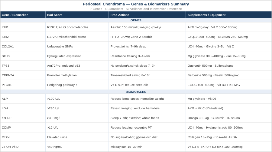
Periosteal chondroma is genuinely benign in most cases, but the molecular landscape around it is more complex and more actionable than a standard post-excision follow-up conversation conveys. IDH1 and IDH2 mutations in tumor tissue inform surveillance frequency and metabolic support strategy. Germline factors affecting COL2A1, SOX9, CDKN2A, TP53, and the hedgehog pathway influence the cartilage environment over a lifetime, and several of these can be partially compensated through targeted supplementation and lifestyle change. Biomarkers — ALP, LDH, hsCRP, COMP, CTX-II, and Vitamin D — provide a measurable, trackable window into whether the underlying biology is moving in the right direction.
The next smart step is to gather a baseline. Request IDH1/IDH2 status from your pathology report if you have not already. Order hsCRP and 25-OH Vitamin D at your next blood draw. If you have access to specialty labs, add COMP and an Omega-3 Index. Review your metabolic markers — fasting insulin and fasting glucose — and share these findings with a physician who understands both orthopedic oncology and metabolic medicine. Better information is not a substitute for clinical care, but it is the foundation on which better decisions are made.
Cancer & Oncology Endocrine & Metabolic
Musculoskeletal: Bone Conditions
Autoimmune: Inflammatory Conditions Connective Tissue Conditions
Cancer & Oncology: Bone Cancer