This article contains AI generated content.
· UpdatedPeriostitis Genes and Biomarkers — 4 Genes and 7 Biomarkers to Track
Introduction
Periostitis is one of those conditions that tends to get addressed at the surface level — rest, reduced training load, ice, and a gradual return to activity. For many people, this is enough to move through an acute episode. But for anyone dealing with recurring pain, slow healing despite adequate rest, or flare-ups that return as soon as training resumes, standard advice quickly runs out of explanations.
What rarely gets examined is the biological layer underneath: the quality of the periosteum itself, how effectively the body regulates bone inflammation, and whether specific measurable markers are quietly sustaining the problem. Two people with identical training loads and the same bone injury can have entirely different recovery timelines — and that difference often reflects something biological, not just behavioral.
This article does not offer a simpler version of the standard advice. Instead, it looks at what you can actually measure and investigate: specific biomarkers that reflect bone turnover, inflammatory status, and healing capacity, and key genetic variants that shape how your periosteum responds to stress and repair. Neither approach replaces clinical care, but both sharpen the picture considerably.
Better information leads to better decisions. Knowing whether your vitamin D is truly optimal, whether your bone resorption is outpacing formation, or whether a collagen gene variant is reducing the tensile strength of your periosteum gives you something concrete to act on — and something meaningful to track over time.
Summary
This article covers four strategies for approaching periostitis with more precision than generic recovery advice allows. The first and primary strategy examines 7 biomarkers — hsCRP, 25-OH vitamin D, P1NP, CTX-I, bone-specific alkaline phosphatase, PTH, and ferritin — and for each one you'll find how to measure it, what an unfavorable result reveals, and what to do about it with and without supplementation. A second section covers 4 genetic variants — COL1A1, VDR, IL-6, and MMP3 — that directly influence periosteal structure and inflammatory response, with practical compensation strategies for each. A third section distills 10 of the most actionable insights from Peter Attia's Outlive, whose biomarker framework maps directly onto bone health and recovery. Finally, five evidence-backed complementary modalities are examined — including photobiomodulation, massage therapy, and biofeedback — chosen specifically for their relevance to periosteal healing.
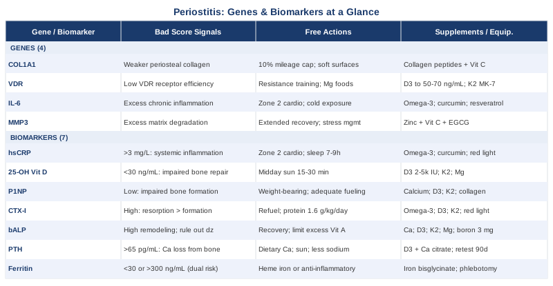
7 Biomarkers to Track for Periostitis
Most people dealing with periostitis are told to rest and wait. But rest without information is just time passing. Tracking the right biomarkers tells you whether your body is genuinely healing, whether inflammation is being kept in check, and whether underlying nutritional or hormonal issues are quietly prolonging the problem. The following seven markers represent the most clinically meaningful data points available for periostitis — ranging from affordable basics to more specialized tests.
1. High-Sensitivity C-Reactive Protein (hsCRP)
Why it matters: hsCRP is the most accessible indicator of systemic inflammation available from a standard blood draw. When periostitis is active, the inflammatory cascade involving cytokines like IL-6 drives hepatic production of CRP. Tracking hsCRP gives you a reliable window into whether inflammatory activity is persisting between episodes — which matters because chronic low-grade inflammation both slows periosteal repair and increases vulnerability to re-injury. Peter Attia consistently frames hsCRP as a first-tier health marker; a level above 3 mg/L is associated with impaired recovery across multiple tissue types, and even in musculoskeletal contexts a persistently elevated result points to a biological environment where healing is inefficient. See related research on PubMed.
How to measure it: Request "high-sensitivity CRP" specifically at your next blood panel — standard CRP is a cruder tool with worse sensitivity at low levels. Available at most primary care labs, cost typically ranges from $20 to $50. Optimal target is below 1 mg/L; 1–3 mg/L is borderline; above 3 mg/L warrants active investigation.
If the score is bad — the plan without supplements: The most powerful non-supplemental interventions for reducing hsCRP are sustained moderate aerobic exercise (zone 2 intensity, not high-impact training during a flare), consistent sleep of 7–9 hours per night, elimination of ultra-processed foods and refined seed oils, and meaningful reduction of training load to allow tissue recovery. Time-restricted eating (10–12 hour eating window) has shown consistent CRP-lowering effects in controlled trials and requires no financial investment. These lifestyle factors are the foundation — supplementation sits on top of them, not in place of them.
If the score is bad — the plan with supplements or equipment: Omega-3 fatty acids (EPA+DHA combined, 2–4 g/day from a triglyceride-form fish oil) have among the strongest natural evidence for hsCRP reduction, with effects confirmed in multiple RCTs. Curcumin with piperine (500 mg twice daily with food, 8-week cycle followed by a 2-week break) and ginger extract (500–1,000 mg/day) add complementary anti-inflammatory signaling. Red light therapy panels in the 660–850 nm range used systemically (20 min/day) have emerging evidence for inflammatory modulation. Main considerations: omega-3s at higher doses may slightly increase bleeding time; curcumin may interact with some medications via CYP enzymes; ginger may affect platelet aggregation. Disclose all supplementation to your physician before any surgical procedure.
2. 25-OH Vitamin D
Why it matters: Vitamin D functions as a hormone-like molecule that regulates osteoblast differentiation, calcium absorption in the gut, and immune modulation — all directly relevant to periosteal healing. Low 25-OH D is one of the best-established modifiable risk factors for bone stress injuries. Multiple prospective studies in military populations and athletes have demonstrated that individuals with the lowest vitamin D levels have significantly higher rates of stress fractures and periosteal reactions. See related research on PubMed. The periosteum contains VDR-expressing cells throughout its cambium layer — the cellular engine of bone repair — meaning that vitamin D signaling directly influences how well the periosteum responds to injury.
How to measure it: A standard 25-OH vitamin D blood test, available at most primary care labs for $30–70. Most conventional labs mark 20 ng/mL as sufficient, but experts including Peter Attia and Rhonda Patrick use 40–60 ng/mL as a functional optimum — the "sufficient" threshold reflects population averages rather than optimal biology. For active individuals with periostitis, targeting the higher end of this range is well-supported.
If the score is bad — the plan without supplements: Direct midday sun exposure on uncovered arms and legs (15–30 minutes, ideally between 10am and 2pm) is the most efficient natural means of raising vitamin D. At latitudes above 37° during winter months, meaningful UVB-mediated vitamin D synthesis becomes limited regardless of time spent outdoors — supplementation is typically necessary. Skin tone, age, and BMI all affect conversion efficiency. Beyond sun exposure, foods like fatty fish, egg yolks, and liver contribute modest amounts.
If the score is bad — the plan with supplements or equipment: Vitamin D3 at 2,000–5,000 IU daily is a reasonable starting dose for most adults with confirmed deficiency; retest in 90 days to assess response. Pair with vitamin K2 in MK-7 form (100–200 mcg/day) to direct calcium toward bone rather than soft tissues — this pairing is mechanistically important, not optional. Magnesium glycinate or malate (300–400 mg at night) is a required enzymatic cofactor for vitamin D conversion; deficiency in magnesium caps how far vitamin D can respond even with supplementation. At doses above 5,000 IU, monitor serum calcium to avoid hypercalcemia. These three supplements are generally taken continuously with blood monitoring every 90 days.
3. P1NP (Procollagen Type 1 N-Terminal Propeptide)
Why it matters: P1NP is the gold-standard serum marker of bone formation. It reflects the synthetic activity of osteoblasts — the cells responsible for depositing new bone matrix. In periostitis, elevated P1NP during active repair is actually a positive signal: it confirms that the periosteum is attempting to rebuild. Chronically suppressed P1NP, however, indicates impaired formation capacity, which can perpetuate injury cycles by leaving periosteal tissue in a state of unresolved damage. Clinicians like Thomas Dayspring who use advanced cardiometabolic marker panels have noted that bone turnover markers are systematically underused outside of osteoporosis management — yet they provide dynamic information that static imaging simply cannot offer. See related research on PubMed.
How to measure it: P1NP is measured from a fasting morning blood draw for consistency across timepoints. Cost typically ranges from $50 to $120 and may require a specialist referral in some healthcare systems. An endocrinologist or sports medicine physician can contextualize results against appropriate reference ranges for age and sex. Track it over time — absolute values matter less than whether the trend is moving in the right direction.
If the score is bad — the plan without supplements: Weight-bearing exercise is the most potent stimulus for osteoblast activity and P1NP elevation. Bone responds to mechanical loading through mechanosensing osteocytes — this is the biological basis for progressive loading even during periostitis rehabilitation rather than extended rest alone. A carefully graded return-to-load protocol (e.g., aqua jogging progressing to walking before running) drives bone formation signals without creating the high-impact loads that caused injury in the first place. Adequate caloric intake and sleep are necessary co-conditions: underfueling and sleep restriction both suppress osteoblastic activity.
If the score is bad — the plan with supplements or equipment: Calcium (1,000–1,200 mg/day from food or supplement in two divided doses), vitamin D3, and vitamin K2 form the core trio for supporting osteoblast function. Collagen peptides (10 g/day, marine or bovine hydrolyzed) taken with 250–500 mg vitamin C approximately 30–60 minutes before a weight-bearing training session have shown promising effects on connective tissue and bone matrix synthesis in emerging RCTs. Whole-body vibration platforms (25–45 Hz, 10–20 minutes three times per week) are used in some clinical rehabilitation protocols to stimulate osteogenesis when high-impact loading is contraindicated. Side effects of calcium supplementation at standard doses are mild; avoid exceeding 2,500 mg/day total.
4. CTX-I (C-Terminal Telopeptide of Type I Collagen)
Why it matters: Where P1NP measures bone building, CTX-I measures bone breakdown — specifically the activity of osteoclasts as they resorb bone matrix. In healthy remodeling, formation and resorption stay tightly coupled. In periostitis, excessive CTX-I relative to P1NP signals that breakdown is outpacing repair — a net loss of periosteal structural integrity. This imbalance is particularly common in athletes who are under-recovering, underfueling, or experiencing relative energy deficiency in sport (RED-S), and in women in low-estrogen phases where osteoclast activity is normally less suppressed. See related research on PubMed.
How to measure it: CTX-I requires a fasting morning blood draw — diurnal variation is significant, and food intake measurably suppresses the marker, making fed-state results unreliable. Labs typically charge $50–120. Ordering CTX-I alongside P1NP provides the most informative picture: a high CTX with low P1NP is the most concerning pattern, indicating active breakdown with impaired repair.
If the score is bad — the plan without supplements: The primary driver of excessive bone resorption in otherwise healthy individuals is underfueling — inadequate caloric intake relative to training demands, low protein intake, and insufficient dietary calcium. Reducing training volume temporarily while meaningfully increasing caloric density and protein (1.6–2.2 g protein per kg bodyweight per day) can shift the CTX-P1NP ratio toward a more favorable balance. Sleep quality is also a direct variable: bone resorption increases measurably under conditions of sleep deprivation, partly through elevated cortisol.
If the score is bad — the plan with supplements or equipment: Pharmaceutical-grade CTX suppression (bisphosphonates) is reserved for clinical osteoporosis under physician supervision and is not appropriate for self-management in this context. A functional approach uses omega-3 fatty acids, vitamin D3, vitamin K2, and adequate calcium collectively to support osteoclast regulation through physiological pathways. Red light therapy devices (660–850 nm range) applied over the affected bone area have emerging evidence — primarily from animal models with early human data — for favorably modulating bone turnover. Strontium ranelate, which has dual formation-promoting and resorption-suppressing effects, is available by prescription in some countries but not the United States.
5. Bone-Specific Alkaline Phosphatase (bALP)
Why it matters: Total alkaline phosphatase is a routine liver and bone panel test, but it mixes signals from both organs. Bone-specific ALP (bALP) isolates the osteoblast signal, making it a cleaner marker of periosteal and bone remodeling activity. Elevated bALP during active periostitis reflects the body's bone-building response — which is generally a positive healing indicator. Chronically elevated bALP without a clear remodeling trigger points toward systemic conditions including hyperparathyroidism, Paget's disease, or hypervitaminosis A — each of which can independently cause or worsen periosteal inflammation and deserves investigation before self-managing. See related research on PubMed.
How to measure it: Bone-specific ALP requires a specific test order beyond a standard metabolic panel. Cost ranges from $30 to $80. Total ALP from a standard metabolic panel is a free starting point — if elevated, request the bone-specific fraction to isolate the source. Contextualizing bALP alongside PTH, 25-OH D, and vitamin A levels is necessary for accurate interpretation.
If the score is bad — the plan without supplements: Unexplained chronically elevated bALP warrants a referral to rule out metabolic bone disease or endocrine dysfunction before any self-directed intervention. If the elevation is clearly exercise-related — a common and benign finding in athletes — the priority is ensuring adequate recovery periods and caloric intake to allow the remodeling process to complete rather than remaining perpetually activated. Excessive vitamin A supplementation (retinol form above 5,000 IU/day) is a rare but documented dietary cause of periosteal reactions and elevated bALP; this should be reviewed if relevant.
If the score is bad — the plan with supplements or equipment: When elevated bALP reflects high remodeling demand without sufficient raw materials, the support protocol mirrors the P1NP approach: calcium, vitamin D3, vitamin K2, magnesium, and adequate protein. Boron (3 mg/day from a glycinate or citrate form) is a lesser-known mineral that appears to positively influence bone ALP and has supportive data from small human trials. Avoid calcium carbonate forms in individuals with low stomach acid — calcium citrate absorbs reliably regardless of acid status and is generally better tolerated.
6. PTH (Parathyroid Hormone)
Why it matters: PTH regulates blood calcium homeostasis. When vitamin D is insufficient or dietary calcium is inadequate, PTH rises — the parathyroid glands signal bones to release calcium into the bloodstream to compensate. This secondary hyperparathyroidism is a common and under-recognized driver of increased bone resorption and impaired periosteal repair, particularly in athletes and active individuals who have borderline-adequate but not optimal vitamin D and calcium status. It is a critical piece of context that gives meaning to otherwise difficult-to-interpret vitamin D or CTX results. See related research on PubMed.
How to measure it: Intact PTH is a standard blood test, typically $40–80. Optimal range is generally 15–65 pg/mL; chronically elevated PTH above 65 pg/mL in conjunction with low or low-normal vitamin D is a key red flag. The test should be ordered alongside serum calcium, albumin, phosphate, and 25-OH vitamin D to be interpreted correctly. An isolated PTH result without these co-markers is difficult to contextualize.
If the score is bad — the plan without supplements: Increasing dietary calcium through whole food sources — dairy, sardines with bones, leafy greens — and improving vitamin D through sun exposure are the foundational non-supplemental steps. Reducing high-sodium dietary patterns (which increase urinary calcium excretion) and high-caffeine intake (which slightly increases urinary calcium loss) are underappreciated supporting adjustments. Regular weight-bearing exercise has a modest PTH-lowering effect through improved calcium retention.
If the score is bad — the plan with supplements or equipment: Vitamin D3 supplementation (starting at 2,000–4,000 IU/day, titrated to target) with calcium citrate (500 mg twice daily, spread across meals) directly addresses the most common driver of secondary hyperparathyroidism. Retest PTH alongside vitamin D after 90 days of consistent supplementation. If PTH remains elevated despite normalized vitamin D, primary hyperparathyroidism requires exclusion by an endocrinologist — this is not a condition to self-manage past that point. Magnesium remains essential as a cofactor throughout this process.
7. Ferritin
Why it matters: Ferritin serves a dual purpose as a biomarker that is almost always under-discussed in periostitis contexts. Low ferritin (below 30–40 ng/mL) is common in endurance athletes, especially females, and is strongly associated with increased bone stress injury rates — iron deficiency impairs red blood cell function, reduces oxygen delivery to healing periosteal tissue, and disrupts collagen synthesis. High ferritin (above 200–300 ng/mL in the absence of iron supplementation) functions as an acute-phase reactant — a signal of systemic inflammation — and its presence alongside elevated CRP suggests that periostitis is occurring in an already-inflamed biological environment that may prolong recovery. Both ends of the spectrum are clinically relevant and point toward very different interventions. See related research on PubMed.
How to measure it: Ferritin is included in most standard iron panels; cost ranges from $20 to $50. A full iron panel (ferritin, serum iron, TIBC, transferrin saturation) provides richer context and is recommended for anyone with low-to-borderline ferritin. Target range for most active individuals is 50–150 ng/mL. Athletes with high training loads often benefit from targeting ferritin above 70 ng/mL. A result above 300 ng/mL without supplementation warrants investigation.
If the score is bad — the plan without supplements: For low ferritin: heme iron from red meat, organ meats, and dark poultry is the most bioavailable dietary source. Consuming vitamin C-rich foods alongside iron-rich meals meaningfully enhances non-heme iron absorption. Avoiding tea, coffee, and calcium supplements within two hours of iron-containing meals prevents competitive absorption inhibition — this single adjustment can make a meaningful difference. Cooking in cast iron provides a small but real iron contribution. For elevated ferritin driven by inflammation, addressing the inflammatory load through the CRP-focused interventions above is the appropriate direction rather than directly restricting iron.
If the score is bad — the plan with supplements or equipment: For confirmed iron deficiency: iron bisglycinate (25–50 mg elemental iron) is significantly better tolerated than ferrous sulfate, with comparable absorption and far fewer GI side effects. Take on an empty stomach if tolerated, otherwise with a light meal. Retest ferritin every 60–90 days and adjust dose to avoid overshooting into excess — iron overload carries its own health risks. For high ferritin reflecting genuine iron overload (hemochromatosis) rather than inflammation, therapeutic phlebotomy under physician supervision is the appropriate intervention, not dietary or supplemental self-management.
---
With the measurable layer established, it is worth examining the genetic layer that shapes how well any given person manages bone inflammation and periosteal repair in the first place — because some people are working with less favorable starting conditions than others, through no behavioral fault of their own.
4 Genes That Shape Periosteal Health
Genetic testing through consumer platforms or clinical providers can surface variants in genes that affect periosteal architecture, inflammatory sensitivity, and remodeling efficiency. The four variants below represent the most relevant genetic factors for periostitis based on current human research — though most evidence comes from association studies rather than interventional trials. They should inform your approach, not determine it.
COL1A1 — The Collagen Architecture Gene
Type I collagen is the structural backbone of both bone and periosteum. COL1A1 encodes the alpha-1 chain of this collagen, and specific polymorphisms — particularly the Sp1 binding site variant (rs1800012, the G/T substitution) — have been associated with reduced bone mineral density, increased stress fracture risk, and altered mechanical properties of connective tissue in multiple human studies. The T allele (heterozygous GT or homozygous TT) is associated with weaker collagen cross-linking and potentially less resilient periosteal tissue under repetitive mechanical loading. See related research on PubMed. The practical implication is straightforward: individuals with this variant may have periosteum that responds less robustly to mechanical stress and requires more careful load management to avoid injury cycles.
If the gene is bad — the plan without supplements: The most direct non-supplemental response to a collagen-weakening gene variant is progressive mechanical loading at conservatively managed volumes. The periosteum adapts structurally to load through collagen turnover and osteoprogenitor activity in the cambium layer — but this process requires time. Mileage build-up in running should follow a no more than 10% weekly increase with mandatory recovery weeks every 3–4 weeks. Cross-training modalities (cycling, swimming, pool running) maintain aerobic capacity while limiting cumulative periosteal stress on bone. Footwear selection and surface choice matter more for this genotype: softer running surfaces and well-cushioned footwear reduce peak tibial impact loads meaningfully. Stretching protocols targeting the posterior chain (calf, soleus, tibialis posterior) reduce the tensile pull on periosteal attachment points.
If the score is bad — the plan with supplements or equipment: Hydrolyzed collagen peptides (10–15 g/day, marine or bovine) taken with 250–500 mg vitamin C approximately 45 minutes before a weight-bearing session have shown measurable effects on connective tissue synthesis in emerging RCTs. Vitamin C is a required cofactor for the hydroxylation of proline and lysine residues in collagen — without it, supplemental collagen peptides are less effective. This protocol is continuous, with no defined cycling requirement. Whole-body vibration platforms (25–45 Hz, 3× weekly, 10–20 minutes) may stimulate periosteal adaptation without high mechanical loading when pain is limiting conventional weight-bearing exercise. Side effects are minimal — a small minority experience GI sensitivity with collagen peptides, which resolves with taking them with food.
VDR — The Vitamin D Receptor Gene
Even with optimal serum vitamin D levels, the downstream effectiveness of vitamin D depends on the VDR gene. The vitamin D receptor mediates virtually all genomic effects — from calcium absorption in the gut to immune modulation to osteoblast differentiation in the periosteum. Four common polymorphisms (BsmI, FokI, ApaI, TaqI) have been studied extensively in bone health contexts. The FokI variant (rs2228570) is particularly relevant: it affects the length and transcriptional efficiency of the VDR protein — individuals with the ff genotype produce a less active receptor and may need higher circulating vitamin D to achieve the same tissue-level response as FF carriers. See related research on PubMed.
Gary Brecka and other researchers working in personalized health have highlighted VDR variants as an underappreciated reason why some individuals fail to respond adequately to standard vitamin D supplementation despite technically normal serum levels. This is especially relevant to periostitis because periosteal cells are VDR-expressing targets.
If the gene is bad — the plan without supplements: The upstream synthesis of 25-OH vitamin D from UVB is not significantly blunted by VDR variants — production proceeds normally. What changes is the receptor-level response. High-load resistance training stimulates VDR upregulation in muscle tissue, which partially compensates for reduced baseline receptor sensitivity — this is an argument for incorporating resistance training early in periostitis rehabilitation rather than delaying until fully healed. Ensuring adequate dietary magnesium through whole foods (pumpkin seeds, dark leafy greens, legumes) supports the enzymatic conversion steps of vitamin D into its most active form.
If the score is bad — the plan with supplements or equipment: For individuals with unfavorable VDR variants, targeting higher 25-OH D serum levels (50–70 ng/mL rather than the standard 40 ng/mL floor) is a practical compensation strategy — more circulating substrate can partially offset reduced receptor efficiency. Vitamin D3 dosing should be titrated until this target range is achieved and confirmed on retest every 90 days. Vitamin K2 (MK-7 form, 100–200 mcg/day) works synergistically with VDR-mediated bone calcium metabolism and should be included as a standard pairing. Magnesium supplementation (glycinate or malate, 300–400 mg at night) remains essential as a conversion cofactor. These three are used continuously with regular monitoring; no cycling is required.
IL-6 Gene — The Inflammatory Thermostat
IL-6 is a pleiotropic cytokine that plays both a pro-inflammatory role in the acute phase of periosteal injury and an essential role in initiating bone healing and callus formation. The problem arises when genetic variants in the IL-6 promoter region lead to chronically elevated IL-6 expression, sustaining inflammation beyond the acute healing window. The most studied variant, -174G>C (rs1800795), influences baseline IL-6 transcription. GG homozygotes tend to produce higher IL-6 in response to inflammatory triggers than CC carriers, and are associated with greater post-exercise inflammatory responses and potentially slower resolution of periosteal inflammation. See related research on PubMed. This does not make injury inevitable, but it does mean that inflammatory load management deserves heightened attention in GG carriers.
If the gene is bad — the plan without supplements: Sustained low-intensity aerobic exercise (zone 2 training at 60–70% of maximum heart rate, 3–5 hours per week) is one of the best-documented lifestyle interventions for reducing baseline IL-6 through anti-inflammatory myokine release — particularly IL-10 and irisin, which are released during sustained low-intensity muscle contraction. Cold exposure protocols (3–5 minutes at 10–15°C via cold shower or ice bath, 3–5× weekly) have shown IL-6 modulation effects in several controlled trials, though evidence specific to periostitis remains limited. Sleep quality is a direct driver of IL-6 variability — poor sleep is independently associated with elevated next-day IL-6 across multiple longitudinal studies.
If the score is bad — the plan with supplements or equipment: Omega-3 fatty acids (EPA+DHA, 3–4 g/day combined) have robust human trial evidence for reducing IL-6 production. Curcumin with piperine (500 mg twice daily with food, on an 8-week cycle with a 2-week break) and resveratrol (250–500 mg/day taken with a fat-containing meal for better absorption) both show IL-6-modulating effects in human trials. Quercetin (500–1,000 mg/day) has emerging but promising evidence. Important: monitor for drug interactions if taking anticoagulants; curcumin at high doses affects CYP enzyme metabolism and may interact with several pharmaceuticals. Disclose supplementation to your prescribing physician.
MMP3 — The Tissue Remodeling Gene
Matrix metalloproteinase 3 (stromelysin-1) is an extracellular enzyme that degrades structural proteins in the periosteum and surrounding connective tissue — collagen, fibronectin, and proteoglycans. MMP3 plays a critical role in remodeling the extracellular matrix during injury repair. The 5A/6A promoter polymorphism (rs3025058) significantly affects MMP3 expression: the 5A allele drives higher MMP3 transcription, meaning more active matrix degradation. While this supports initial tissue clearance during repair, chronically elevated MMP3 activity can undermine periosteal healing and increase the proportion of scar tissue rather than organized collagen deposition during recovery from repeated injury episodes. See related research on PubMed.
If the gene is bad — the plan without supplements: The key non-supplemental countermeasure for a high-MMP3 genotype is ensuring the repair phase after periostitis episodes is not cut short. Returning to full training before periosteal remodeling is complete is the exact context in which elevated MMP3 activity becomes counterproductive — the tissue is still organizing its new collagen matrix when renewed loading begins breaking it down again. Conservative return-to-load protocols graded over 6–8 weeks rather than 3–4 weeks are warranted. Sleep consistency matters here because MMP activity has circadian variation, and chronic sleep disruption appears to disregulate this rhythm. Reducing psychological stress (cortisol directly upregulates MMP expression) through evidence-based stress management is a legitimate and often overlooked lever.
If the score is bad — the plan with supplements or equipment: Zinc (15–25 mg/day as zinc picolinate or glycinate) and copper (1–2 mg/day, always paired with zinc to prevent zinc-induced copper depletion) are essential cofactors that regulate MMP activity — zinc deficiency upregulates uncontrolled MMP expression and impairs tissue repair. Vitamin C (500–1,000 mg/day) supports collagen synthesis as a structural counterbalance to MMP-driven matrix degradation. EGCG from green tea extract (400–800 mg/day, standardized catechins) is a natural MMP inhibitor with some human evidence for connective tissue and joint benefit. Cycle EGCG on 8 weeks on / 2 weeks off; take with food to reduce GI irritation. High-dose, long-term EGCG use warrants monitoring of liver enzymes — this is a documented concern at doses above 800 mg/day.
---
The genetic and biomarker information above frames periostitis recovery in measurable terms. The next section takes a different angle — synthesizing one of the most practically actionable frameworks for bone health and longevity medicine, which applies with surprising directness to the periostitis recovery challenge.
What Outlive by Peter Attia Reveals About Bone Health
Outlive: The Science and Art of Longevity (2023) is primarily framed around healthspan and performance in later life, but its chapters on musculoskeletal health, inflammation, and biomarker strategy are among the most practically useful resources available on bone biology for non-specialists. Attia's "Medicine 3.0" framework — proactive, data-driven, and oriented toward trajectories rather than symptoms — maps directly onto how to approach periostitis before it becomes a recurring or degenerative condition.
1. Chronic Inflammation Costs You Years of Tissue Quality
Attia argues that most people do not track inflammatory markers until something is already wrong — and by then, low-grade chronic inflammation has been degrading tissue quality for years. He treats hsCRP as a first-tier annual marker for everyone, not just those with obvious symptoms. Periostitis patients sitting at 2–3 mg/L chronically are operating in an inflammatory environment that significantly delays repair.2. The 20 ng/mL Vitamin D Threshold Is Not an Optimal Target
Attia is explicit that conventional "sufficient" thresholds for vitamin D reflect population distribution, not optimal physiology. He targets 40–60 ng/mL and has observed that many patients with bone-related issues are technically "normal" by lab reference ranges while substantially suboptimal by functional standards. This framing is directly applicable to athletes with recurrent periosteal injuries.3. Bone Density Is Built Early and Cannot Be Fully Rebuilt Later
One of the book's more sobering conclusions is that peak bone mass — largely set by the mid-20s — cannot be fully recovered after the fact; it can only be preserved or lost more slowly. For young athletes with recurring periostitis, every episode of inadequate recovery may represent a long-term bone integrity cost that does not fully compound back. This is an argument for investing in optimal recovery biology early, not waiting until density loss becomes visible on a DEXA scan.4. Bone Turnover Markers Provide Information That DEXA Cannot
Attia advocates using P1NP and CTX-I alongside bone density scans rather than relying on static imaging alone. Two people with identical T-scores can have dramatically different fracture risk depending on whether remodeling favors formation or resorption. This insight is directly applicable to periostitis — tracking these markers over time reveals dynamic biology that a yearly scan misses entirely.5. Zone 2 Aerobic Training Is the Most Powerful Systemic Anti-Inflammatory Tool Available
Attia dedicates significant discussion to zone 2 training as the most consistent evidence-backed lifestyle intervention for lowering resting inflammation, improving mitochondrial efficiency, and reducing metabolic disease risk. For periostitis specifically, it is the exercise modality most compatible with maintaining cardiovascular fitness during a flare without adding to periosteal mechanical stress.6. Protein Adequacy Is Non-Negotiable During Repair
Attia recommends a minimum of 1.6 g/kg/day of protein for regularly active individuals and higher during tissue repair phases. The majority of periostitis patients he profiles are well below this threshold. Inadequate protein directly suppresses P1NP, elevates CTX, and extends recovery timelines — the biomarker picture worsens in predictable ways.7. Sleep Is the Most Underestimated Recovery Variable in Musculoskeletal Medicine
Attia cites substantial research linking sleep deprivation to impaired bone remodeling, elevated cortisol (which directly increases osteoclast activity and CTX-I), and upregulated IL-6. He recommends treating sleep as a performance priority equal to training load — 7.5–9 hours, cool room, consistent timing. For periostitis, this is not soft advice: it is a direct modulator of the exact biomarkers discussed above.8. Muscle Is the Best Structural Protection for Bone
Muscle and bone communicate via myokines. Muscle contraction stimulates osteocytes through mechanotransduction and releases irisin, which directly promotes bone formation. Attia's consistent recommendation to prioritize resistance training as early as is safely possible in rehabilitation applies here: maintaining or rebuilding calf and posterior tibial muscle mass around an inflamed tibial periosteum accelerates structural healing and reduces recurrence risk.9. Nutritional Tracking Reveals What Intuition Misses
Attia is consistently skeptical of the idea that subjective "healthy eating" translates automatically to meeting micronutrient needs for tissue repair. He advocates structured dietary logging for short periods to identify specific gaps — particularly calcium, magnesium, and vitamin C — that are easy to miss on varied modern diets. Three to four days of accurate logging frequently surfaces patterns that subjective recall cannot.10. Track Biomarker Trajectories, Not Single Values
The Medicine 3.0 framework emphasizes trend lines over snapshots. A single hsCRP result tells you where you are today; three results over 12 months tell you whether interventions are working. For periostitis, this means running a panel at baseline, after 90 days of any significant change, and after any return to full training load — building a longitudinal picture rather than waiting for the next injury to prompt testing.---
The biomarker, genetic, and educational frameworks above all work most effectively when combined with physical approaches that directly address tissue healing and pain management. The following modalities have meaningful clinical evidence for relevance to periostitis.
Complementary Approaches with Clinical Evidence
Low-Level Laser Therapy / Photobiomodulation
Photobiomodulation uses specific wavelengths of light — typically in the 630–1,000 nm red and near-infrared range — to stimulate cellular energy production via cytochrome c oxidase in the mitochondria. For periostitis, its relevance spans multiple mechanisms: enhanced ATP production in osteoblasts, reduced expression of pro-inflammatory cytokines including IL-1β and IL-6, and accelerated collagen remodeling in the extracellular matrix. Multiple animal studies have demonstrated increased periosteal cell proliferation following LLLT application; human clinical trials have extended these findings to bone stress reactions and soft tissue healing in athletes. See related research on PubMed.
A systematic review published in Photomedicine and Laser Surgery examined LLLT across musculoskeletal conditions and found consistent reductions in pain and inflammatory markers alongside improvements in tissue repair timelines. Protocols studied for bone stress injuries typically apply 4–6 J/cm² at 810–830 nm, administered daily or every other day over 2–4 weeks to the affected bone region. Clinical evidence for periostitis specifically remains limited in scope but is mechanistically coherent with the broader bone-healing literature.
Practically, LLLT is available through sports medicine clinics and physiotherapy practices equipped with Class IV lasers — more powerful than consumer devices and capable of reaching deeper periosteal tissue. For acute tibial or foot periostitis, clinical administration is preferred during the first 2–4 weeks. Home devices in the red and near-infrared range (such as Joovv or Mito Red panels) can be used for maintenance and systemic anti-inflammatory support. Sessions of 10–20 minutes over the affected area at 5 times per week represent a reasonable maintenance protocol. No significant side effects at standard clinical doses; avoid direct, unshielded eye exposure.
Massage Therapy
Massage therapy addresses the muscular and fascial components of periostitis rather than the periosteum directly — and this targeting is mechanistically appropriate. In the most common presentation of tibial periostitis, excessive tensile loading on the periosteum arises from tight posterior tibial and soleus musculature pulling at their periosteal attachment points. Regular massage of the lower leg — specifically the posterior compartment — reduces this tensile stress, improves local circulation and lymphatic drainage, and may reduce inflammatory mediator concentration in the surrounding tissue bed. See related research on PubMed.
A study published in the Journal of Athletic Training found that soft tissue manipulation of the lower leg significantly reduced pain scores in athletes with medial tibial stress syndrome, with benefits maintained at four-week follow-up. Deep tissue massage of the posterior compartment (4–6 sessions over 2–3 weeks) followed by regular maintenance (every 3–4 weeks during high training loads) represents a practical protocol for both recovery and recurrence prevention.
For practical application: sessions every 1–2 weeks during active recovery and monthly during peak training periods are a reasonable frequency. Self-massage with a foam roller or IASTM (instrument-assisted soft tissue mobilization) tool over the posterior calf and soleus for 5–10 minutes daily is a valid daily complement, though not equivalent in depth to manual therapy. One important caution: direct pressure over the tender bone surface itself should be avoided during active inflammation — massage should target the surrounding musculature, not the inflamed periosteal site.
Biofeedback
Biofeedback uses real-time physiological monitoring — typically heart rate variability, skin conductance, or EMG — to train individuals to voluntarily regulate their stress response. For periostitis, its primary utility is chronic pain management and interrupting the cortisol-driven inflammatory amplification loop. Chronic musculoskeletal pain activates the hypothalamic-pituitary-adrenal axis, elevating cortisol — which directly suppresses osteoblast activity and increases bone resorption, raising CTX-I. Biofeedback trains the autonomic nervous system toward parasympathetic dominance, reducing the cortisol burden and creating a more favorable bone healing environment over time. See related research on PubMed.
HRV biofeedback using resonance frequency breathing (approximately 0.1 Hz, or 5–6 breaths per minute) has the strongest evidence base among biofeedback modalities for pain and stress reduction. A review in Applied Psychophysiology and Biofeedback documented consistent improvements in pain perception and quality of life across chronic musculoskeletal conditions with HRV biofeedback protocols. This mechanism is relevant to periostitis whenever pain is becoming chronic or stress physiology is amplifying the recovery timeline.
For self-directed application, HRV biofeedback is accessible using a Polar H10 chest strap paired with the Elite HRV app, or purpose-built devices like Lief or Garmin HRV monitoring. A 20-minute daily session of resonance breathing for 8–12 weeks is a validated protocol. Evidence specific to periostitis is limited — this is a pain management and systemic recovery tool, not a direct periosteal healing intervention. It is most useful when pain chronicity or psychological stress response appears to be prolonging what should be a more straightforward recovery.
Progressive Muscle Relaxation
Progressive muscle relaxation (PMR) involves systematically tensing and then releasing muscle groups throughout the body to induce physical and psychological relaxation. For periostitis recovery, its value lies in pain modulation, sleep quality improvement, and reduction of sympathetically driven muscle guarding around the injured area. Pain-related muscle guarding in the lower leg perpetuates the same tensile periosteal forces described in the massage section — and breaking this pattern through systematic relaxation training is a legitimate, low-cost adjunct to other interventions. See related research on PubMed.
PMR has been validated in multiple RCTs for reducing pain intensity and improving sleep quality in musculoskeletal conditions. A meta-analysis in Sleep Medicine Reviews confirmed its efficacy for sleep onset and quality across clinical populations — directly relevant given the role of sleep in bone turnover that Outlive documents. PMR is one of the few evidence-backed interventions that simultaneously addresses pain psychology, sleep quality, and physical muscle tension.
Practically, PMR sessions take 15–20 minutes and are most effective when performed nightly before sleep. Free guided audio sessions are available via the NHS and apps including Insight Timer. No equipment is required and the technique has no known adverse effects. For athletes with periostitis, tailoring the sequence to include a deliberate focus on the hip flexors, quadriceps, gastrocnemius, and soleus — the muscles most directly responsible for periosteal tensile loading — adds practical specificity to a general technique.
Breathing-Based Therapies
Controlled breathing protocols — particularly slow diaphragmatic breathing and CO2 tolerance training — influence both systemic inflammation and local tissue oxygenation through distinct but complementary mechanisms. Slow breathing at 4–6 breaths per minute shifts autonomic balance toward parasympathetic dominance, lowering resting CRP and IL-6 production through vagal anti-inflammatory pathways. The Bohr effect — whereby elevated CO2 shifts the oxyhemoglobin dissociation curve rightward — is particularly relevant to tissue healing: improved oxygen delivery to the periosteal repair site directly supports cellular regeneration in the cambium layer. See related research on PubMed.
Patrick McKeown's Oxygen Advantage protocol — which encompasses nasal breathing habituation and progressive CO2 tolerance exercises — has been adopted in elite sports recovery settings. Several case reports and small trials support improved athletic recovery with consistent nasal breathing training, though randomized controlled trial evidence specific to periostitis is not yet available.
Practically, 10 minutes of nasal-only diaphragmatic breathing at approximately 5 breaths per minute (4 seconds inhale, 8 seconds exhale) performed before sleep and after any low-load training session is a realistic daily protocol with essentially no barrier to entry. Breath-hold training for CO2 tolerance should be introduced gradually — McKeown's resources provide progressive protocols starting from very mild holds. This is best used as a low-risk systemic complement to the biomarker and nutritional strategies detailed above, rather than as a primary intervention. Evidence is promising but limited; do not substitute it for clinically indicated treatment.
Conclusion
Periostitis rarely has a single cause and rarely responds to a single fix. What the biomarker and genetic layers examined in this article reveal is that recovery trajectory varies considerably between individuals — and much of that variation is measurable. Whether the underlying issue is chronically elevated hsCRP, a suboptimal vitamin D-PTH axis, a bone resorption/formation imbalance reflected in CTX-I and P1NP, or a COL1A1 variant that quietly weakens periosteal collagen architecture, each of these factors can be identified, tracked, and meaningfully addressed.
The goal of this framework is not to replace medical care — it is to sharpen it. A sports medicine physician or orthopedic specialist working with a patient who has already tracked these biomarkers and explored their genetic context starts from a substantially more informed position than one operating on symptoms alone. That information gap is where recurrence often hides.
The next practical step is to start simply: request hsCRP and 25-OH vitamin D at your next blood panel, review your ferritin, and if you have access to a consumer genetic report, look up the variants covered in the genetics section. Identify the one or two markers most out of optimal range and address them systematically using the protocols above before adding complexity. Better data drives better decisions — and better decisions, consistently applied, compound over time.
Musculoskeletal: Bone Conditions Sports Injuries
Autoimmune: Inflammatory Conditions Connective Tissue Conditions