This article contains AI generated content.
Peripatellar Adhesion Syndrome: 6 Genes and 7 Biomarkers to Track
A stiff, tight, "glued-together" feeling behind the kneecap after surgery or a significant knee injury is one of the more frustrating recoveries in orthopedics. You did the physical therapy. You iced, elevated, and pushed through the exercises. And yet the range of motion plateaus, the front of the knee feels tethered, and the standard advice — "keep stretching, give it time" — starts to feel thin against what your knee is actually telling you.
That advice isn't wrong, but it's incomplete. It treats every knee the same, when the underlying biology of scar tissue formation is not the same from one person to the next. Some people mount a short, contained inflammatory response after knee surgery and glide back to full motion. Others develop an exaggerated, prolonged fibrotic response — excess collagen, overactive fibroblasts, and adhesions that fill the suprapatellar pouch and peripatellar gutters — and end up with a second surgery just to release the scar tissue. That difference is not random. It has measurable biological signatures.
This article looks at those signatures directly: the inflammatory and fibrotic biomarkers that track with adhesion risk, and the genetic variants that appear to predispose certain people to an overactive scarring response after knee trauma or surgery. Some of this evidence comes straight from arthrofibrosis research; some is borrowed, carefully and with that caveat stated, from the closely related biology of frozen shoulder and tendon fibrosis, where the genetics have been studied more thoroughly.
None of this is a guarantee or a cure. But knowing which biomarkers are worth watching, and which genetic tendencies might be working against you, turns a vague "stretch more" into a concrete plan — one with actual levers you can pull, and realistic expectations about what those levers can and cannot do.
Summary
Peripatellar adhesion syndrome doesn't come from nowhere — it comes from a biological cascade that starts with inflammation, escalates through a handful of fibrosis-driving proteins, and ends with collagen deposited faster than your body can remodel it. This article walks through the seven biomarkers most worth tracking around a knee surgery or injury — from a basic hs-CRP panel you can order for the cost of a lab visit, to research-grade markers like TGFBR1 that predict stiffness with striking accuracy. It then looks at six genes, including TGFB1, MMP3, and WNT7B, that shape how aggressively your body scars in response to joint trauma, with practical, low-risk ways to work around an unfavorable variant. Beyond the biology, you'll find ten evidence-based takeaways from a leading flexibility and connective-tissue podcast episode, and a look at which complementary therapies — massage, photobiomodulation, mindfulness, biofeedback — actually have human trial data behind them for post-surgical knee stiffness. By the end, the goal is simple: replace "just keep stretching" with a specific, personalized plan.
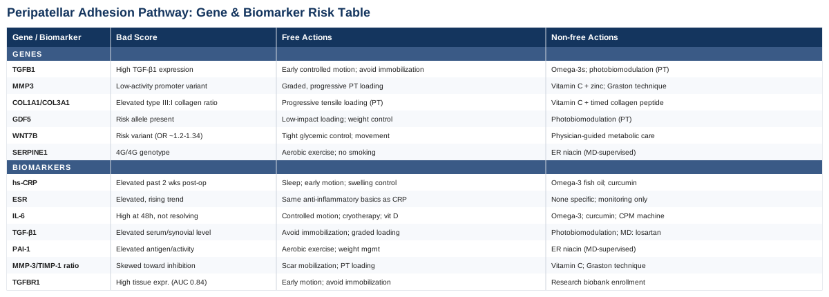
The 7 Biomarkers Worth Tracking If Your Knee Is Prone to Adhesions
Peripatellar adhesions form through a fairly well-mapped sequence: an injury or surgical trauma triggers inflammation, inflammation activates synovial fibroblasts, and those fibroblasts differentiate into myofibroblasts that lay down excess collagen. Each stage of that sequence leaves a fingerprint in the blood or joint fluid. Tracking those fingerprints doesn't just confirm what you already feel in the knee — it tells you which stage of the process is misbehaving, which changes what you should actually do about it.
1. High-sensitivity CRP (hs-CRP)
CRP is the most accessible marker of systemic inflammation, and it has a well-documented trajectory after knee surgery: it typically peaks around day three post-op and returns to near-baseline within about two weeks under a normal recovery course. When CRP stays elevated well past that window, it's a signal that the inflammatory phase driving fibroblast activity hasn't resolved on schedule.
How to measure it: A standard blood draw at any lab or through your surgeon's office; hs-CRP typically costs between 15 and 30 dollars out of pocket, or is bundled into routine post-op bloodwork.
If the score is bad, the plan without supplements: Prioritize sleep (inflammation resolves poorly on short sleep), keep early controlled motion going rather than resting the knee into stiffness, and manage swelling aggressively with elevation and compression in the first two to three weeks.
If the score is bad, the plan with supplements or equipment: Omega-3 fish oil (2 to 3 grams combined EPA/DHA daily) and curcumin with piperine (500 to 1,000 mg daily) both have modest anti-inflammatory evidence. Run either for 8 to 12 weeks post-injury, then reassess CRP rather than continuing indefinitely. Side effects: fish oil at these doses can cause mild GI upset and, at higher doses, increased bleeding risk; curcumin can cause GI upset and interacts with blood thinners, so check with your surgeon if you're on anticoagulants.
2. ESR (erythrocyte sedimentation rate)
ESR moves more slowly than CRP and is useful mainly as a trend-confirmation marker and to help rule out an occult infection driving prolonged inflammation, which is an important distinction — infected stiffness needs a different workup entirely than fibrotic stiffness.
How to measure it: Included in most standard post-op inflammatory panels; roughly 10 to 20 dollars on its own.
If the score is bad, the plan without supplements: Same anti-inflammatory basics as above — sleep, gentle consistent motion, swelling control — since ESR isn't independently supplement-responsive the way CRP can be.
If the score is bad, the plan with supplements or equipment: There's no strong equipment or supplement lever specific to ESR; treat it as a monitoring tool that confirms whether your CRP-directed plan is working, checked every few weeks rather than daily.
3. IL-6 (Interleukin-6)
IL-6 is one of the earliest and most dramatic responders after knee surgery, and a cohort study following total knee arthroplasty patients found that a distinct early cytokine profile — with IL-6 prominent among it — measured in just the first two postoperative days was associated with stiffness at six weeks out (Postoperative Serum Cytokine Levels Are Associated With Early Stiffness After Total Knee Arthroplasty). Under a normal recovery course, IL-6 peaks around 48 hours and falls back to baseline within roughly two weeks (Normal trajectory of IL-6 and CRP after total knee arthroplasty).
How to measure it: Not a routine test; it requires a specialty or research lab ELISA, typically 50 to 150 dollars, and is more often ordered by a sports medicine or rheumatology specialist than a general practitioner.
If the score is bad, the plan without supplements: Early, protected, controlled motion (rather than immobilization) appears to blunt the inflammatory cascade; cryotherapy in the first two weeks; and correcting a vitamin D deficiency if one exists, since low vitamin D status tracks with higher inflammatory cytokine levels.
If the score is bad, the plan with supplements or equipment: Omega-3s and curcumin again apply here; a continuous passive motion (CPM) machine in the first two to three weeks post-op is a reasonable equipment-based lever some surgeons already use. Cycle supplements through the acute window (first 6 to 8 weeks), not longer — there's no evidence that indefinite high-dose anti-inflammatory supplementation adds benefit, and it adds unnecessary GI and bleeding-risk exposure.
4. TGF-β1 (Transforming Growth Factor Beta 1)
TGF-β1 is arguably the single most important driver in this entire process. In an animal model, simply delivering the TGF-β1 gene into a joint was enough, on its own, to induce arthrofibrosis and abnormal cartilage-like tissue in the synovium (Gene delivery of TGF-β1 induces arthrofibrosis and chondrometaplasia of synovium in vivo). In human knee fibroblasts taken from total knee arthroplasty patients, TGF-β1 stimulation directly triggered the collagen production (COL1A1 and COL3A1) responsible for scar tissue buildup (Human outgrowth knee fibroblasts undergo myofibroblastogenesis upon TGFβ1 stimulation).
How to measure it: Serum or synovial fluid TGF-β1 ELISA, generally 100 to 200 dollars, available mainly through research-affiliated labs rather than a standard clinic order.
If the score is bad, the plan without supplements: Avoid immobilizing the knee beyond one to two weeks; mechanical loading through supervised, graded motion appears to modulate TGF-β signaling favorably, whereas prolonged rest does the opposite.
If the score is bad, the plan with supplements or equipment: There's no supplement that reliably lowers TGF-β1 with good human safety data, so this is one to discuss with your surgeon rather than self-treat — some fibrosis research has looked at prescription options like losartan for their TGF-β-blunting effect, but that's a physician decision, not a supplement aisle one. On the equipment side, photobiomodulation (covered below) has some tissue-level evidence of modulating fibrotic signaling and is a lower-risk option to raise with your physical therapist.
5. PAI-1 (Plasminogen Activator Inhibitor-1)
PAI-1 blocks the enzyme system that would otherwise break down excess fibrin and matrix protein. A systematic review of postsurgical knee arthrofibrosis biomarkers identified the gene encoding PAI-1, SERPINE1, as preferentially expressed in arthrofibrotic tissue, working alongside TGF-β signaling to keep matrix breakdown suppressed (Biochemical markers of postsurgical knee arthrofibrosis: A systematic review).
How to measure it: Plasma PAI-1 antigen or activity assay through a coagulation specialty lab, roughly 50 to 100 dollars.
If the score is bad, the plan without supplements: Aerobic exercise has some of the best human evidence for lowering PAI-1 levels; weight management and smoking cessation both matter here too, since PAI-1 is closely tied to metabolic health.
If the score is bad, the plan with supplements or equipment: Extended-release niacin has evidence for lowering PAI-1, but it needs physician supervision — cycle it in an 8 to 12 week block with liver enzyme and lipid monitoring, not as a standing supplement. Expect flushing as a near-universal side effect; at higher doses, watch for GI upset and liver enzyme elevation. Omega-3s again have a smaller, secondary effect here.
6. MMP-3 / TIMP-1 ratio
Matrix metalloproteinase-3 breaks down collagen; its tissue inhibitor, TIMP-1, keeps that breakdown in check. When the ratio tips too far toward inhibition, collagen accumulates faster than it's cleared. A polymorphism in the MMP3 gene has also been linked to increased risk of frozen shoulder, a fibrotic joint condition that shares much of its underlying biology with knee arthrofibrosis (A Study of IL-1β, MMP-3, TGF-β1, and GDF5 Polymorphisms and Their Association with Primary Frozen Shoulder).
How to measure it: A research-grade assay, not a standard clinical order; roughly 80 to 150 dollars through a specialty or rheumatology-affiliated lab.
If the score is bad, the plan without supplements: Manual scar mobilization and progressive tensile loading through physical therapy both stimulate more balanced matrix remodeling than rest alone.
If the score is bad, the plan with supplements or equipment: Vitamin C (500 to 1,000 mg daily) is a genuine cofactor for proper collagen cross-linking rather than just raw collagen bulk, and is a reasonable, low-risk addition. Avoid loading up on collagen peptide powders here specifically — the concern with an already skewed remodeling ratio is feeding more raw material into a system that isn't clearing it efficiently. Instrument-assisted soft tissue mobilization (the Graston technique) is an equipment-based option with some evidence for affecting scar tissue remodeling.
7. TGFBR1 tissue expression (advanced marker)
This is the most predictive biomarker identified so far, and also the least accessible. In a combined animal and human study, TGFBR1 expression in synovial tissue predicted arthrofibrosis with strong accuracy (an AUC of 0.838), and correlated closely with both pain scores and loss of range of motion before and after revision surgery (Identification of novel biomarkers for arthrofibrosis after total knee arthroplasty).
How to measure it: Currently requires a tissue or synovial fluid sample, typically only available if you're already undergoing a procedure or enrolled in a research protocol at an academic sports medicine center — there's no standalone clinical order for this yet.
If the score is bad, the plan without supplements: The same TGF-β-pathway lifestyle levers described above (early controlled motion, avoiding prolonged immobilization) apply directly, since TGFBR1 sits downstream in the same signaling chain.
If the score is bad, the plan with supplements or equipment: Nothing supplement-based currently targets TGFBR1 directly with good human safety data. Treat this one as a marker to watch emerge in clinical practice over the next several years, and ask your surgeon if a research biobank or registry is available if you're facing revision surgery — contributing a sample can help refine this marker for the next patient, including possibly yourself.
With the measurable side of the picture in place, it's worth asking why some people run hotter on these markers in the first place. That's where genetics comes in — not as a separate story, but as the upstream reason some knees produce more TGF-β1 or PAI-1 than others to begin with.
What Your Genes May Be Telling You About Adhesion Risk
Researchers like Ali Torkamani, who has spent years studying how genetic variants translate into real clinical risk, and Gary Brecka, who has popularized using genetic and biomarker panels to personalize health decisions, both make a similar point: a "bad" gene is rarely a life sentence — it's a tendency that shifts how much lifestyle and clinical attention a given system needs. Peripatellar adhesion syndrome doesn't have its own dedicated genetics literature yet, but the fibrotic joint condition it most closely resembles, frozen shoulder, does — and the overlap in fibroblast biology makes that evidence a reasonable, if not perfect, guide.
TGFB1
Variants in the gene coding for TGF-β1 itself affect how much of this fibrosis-driving protein your body produces after an injury. The clearest human data comes from tendinopathy rather than knee fibrosis directly: a study on lateral elbow tendinopathy found that TGFB1 polymorphisms were associated with both pain symptoms and how well patients responded to platelet-rich plasma treatment (Association of TGFB1 Gene Polymorphisms with Pain Symptoms and PRP Effectiveness) — evidence worth taking seriously, but early when extrapolated to the knee.
If the gene is bad, the plan without supplements: Controlled early mobilization is the single most evidence-backed lever for anyone running a high-TGF-β1 profile — avoid prolonged immobilization past one to two weeks post-injury or post-op.
If the gene is bad, the plan with supplements or equipment: Omega-3s for general anti-inflammatory support, cycled 8 to 12 weeks; photobiomodulation as an equipment option discussed with your physical therapist. Avoid over-supplementing here — there's no supplement shown to directly override a TGFB1 variant, and the honest answer is that mechanical rehab strategy matters more than anything in a bottle.
MMP3
The promoter polymorphism in MMP3 changes how much matrix-remodeling enzyme your fibroblasts produce, and it was significantly associated with frozen shoulder risk in the same Chinese Han population study referenced above (IL-1β, MMP-3, TGF-β1, and GDF5 Polymorphisms in Primary Frozen Shoulder).
If the gene is bad, the plan without supplements: Graded, progressive loading through physical therapy — not rest — is what stimulates healthy remodeling in someone whose enzyme system runs low.
If the gene is bad, the plan with supplements or equipment: Vitamin C and zinc at RDA-level doses (avoid zinc megadosing above 40 mg/day, which risks copper deficiency); Graston or similar instrument-assisted soft tissue work as an equipment-based complement, applied two to three times weekly during active rehab, not indefinitely.
COL1A1 and COL3A1
These genes govern the ratio of type I to type III collagen your body lays down during healing. A higher proportion of type III collagen produces weaker, more disorganized, more fibrotic-prone tissue, and this is exactly the shift seen in TGF-β1-stimulated knee fibroblasts from arthroplasty patients (Human outgrowth knee fibroblasts and TGFβ1-driven collagen expression).
If the gene is bad, the plan without supplements: Progressive tensile loading protocols in physical therapy are the main tool known to shift the collagen ratio toward the stronger, better-organized type I over time.
If the gene is bad, the plan with supplements or equipment: Vitamin C paired with light collagen peptide intake taken 30 to 60 minutes before loading sessions has some tendon-healing evidence for supporting collagen synthesis timing; side effects are minimal at standard doses, mostly mild GI upset.
GDF5
Growth differentiation factor 5 is a broadly studied joint-health gene, and its polymorphisms were also associated with frozen shoulder risk in the same population study cited above.
If the gene is bad, the plan without supplements: Favor joint-friendly, lower-impact loading during rehab and control body weight, since GDF5 variants are also linked more broadly to osteoarthritis susceptibility.
If the gene is bad, the plan with supplements or equipment: No supplement directly corrects GDF5 expression; photobiomodulation has modest, early tissue-signaling evidence and is a reasonable low-risk equipment option to discuss with a physical therapist.
WNT7B
This is the strongest single genetic signal identified for frozen shoulder, and it came out of a genome-wide association study covering over two thousand cases, with the lead variant showing an odds ratio of roughly 1.2 to 1.34 depending on the cohort. The same study also found that diabetes and elevated HbA1c were causal risk factors, and that several of the same loci overlap with Dupuytren's disease, another fibroblastic condition (A genome-wide association study identifies 5 loci associated with frozen shoulder and implicates diabetes as a causal risk factor).
If the gene is bad, the plan without supplements: Since diabetes and elevated blood sugar are directly implicated in this same fibrotic pathway, tight glycemic control — regular movement, minimizing refined sugar, adequate sleep — is the most directly evidence-linked lever available, whether or not you carry the variant.
If the gene is bad, the plan with supplements or equipment: There's no supplement targeting WNT signaling with good human safety data for this use. If you're prediabetic, this is a conversation for your physician about metabolic management rather than a self-directed supplement plan.
SERPINE1
This gene codes for PAI-1, and the 4G/4G genotype is associated with higher PAI-1 production and reduced fibrinolysis — directly relevant given that SERPINE1 was flagged as preferentially expressed in arthrofibrotic knee tissue in the systematic review cited earlier.
If the gene is bad, the plan without supplements: Aerobic exercise, weight management, and not smoking are the best-supported non-supplement levers for lowering PAI-1 output, regardless of genotype.
If the gene is bad, the plan with supplements or equipment: Extended-release niacin, cycled 8 to 12 weeks with physician-supervised liver and lipid monitoring, has genuine PAI-1-lowering evidence; expect flushing as the main side effect, with liver enzymes as the thing to actually watch.
Genes and biomarkers explain the biology, but they don't tell you how to actually move a stiff knee day to day — for that, it's worth looking at what the flexibility and rehabilitation research world has converged on.
What a Leading Flexibility Research Podcast Gets Right About Stiff, Scarred Tissue
Andrew Huberman's episode "Improve Flexibility with Research-Supported Stretching Protocols" is built around the physiology of joint range of motion, and while it isn't written specifically for post-surgical knees, its core findings map unusually well onto what's needed to work through peripatellar adhesions. Here are the ten most useful takeaways.
1. Flexibility has three separate components
Range of motion is governed by your nervous system, your muscles, and your connective tissue — three different systems with three different timelines for change. A stiff knee after surgery is rarely just "tight muscle"; the connective tissue component, which includes the exact scar tissue behind the kneecap this article is about, changes far more slowly than the neural component.2. Connective tissue changes are the slow variable
Neural adaptations to stretching (your nervous system allowing more range) can show up within a single session. Actual structural changes in connective tissue take weeks of consistent input. If you're expecting adhesion-related stiffness to resolve in days, the biology simply doesn't work that fast — patience is a physiological requirement, not just a mindset.3. Consistency beats intensity
Frequent, moderate stretching sessions produce more durable range-of-motion gains than infrequent, aggressive ones. For an adhesion-prone knee, this argues for short daily mobility work over sporadic, painful, all-out stretching sessions that risk re-triggering the inflammatory cascade described earlier in this article.4. Working near — not through — discomfort is the target zone
Effective stretching protocols train just past the point of mild discomfort, not into sharp pain. Pushing through sharp pain in a knee with active adhesions risks re-inflaming the tissue you're trying to calm down, which can paradoxically feed more fibrotic signaling.5. The nervous system actively limits your range of motion as a protective mechanism
Muscle spindles and other proprioceptive systems constantly adjust how much stretch your body "allows," independent of actual tissue length. This is part of why range of motion can vary day to day even without any tissue change — useful context for not over-reading a single bad day as a setback.6. Fascia is a distinct, underappreciated tissue layer
The episode specifically calls out fascia as deserving its own deep dive, separate from muscle and joint capsule. Peripatellar adhesions often involve this same connective tissue layer gluing structures together that should glide independently — the suprapatellar pouch and peripatellar gutters that lose their normal sliding function.7. Temperature affects tissue extensibility
Warmed tissue stretches more easily and with less risk of micro-injury than cold tissue. Practically, this supports doing mobility work after a brief warm-up or heat application rather than cold, first-thing-in-the-morning stretching on a stiff post-surgical knee.8. Static and dynamic approaches serve different goals
Static holds and dynamic, movement-based mobility work aren't interchangeable — they train different aspects of range of motion. A well-rounded adhesion-recovery routine benefits from both rather than relying on one exclusively.9. Breath and nervous system state influence how much range you can access
A calmer nervous system state allows more range of motion in the same tissue, since much of the resistance you feel is neurally mediated rather than purely mechanical. This is a reasonable, low-cost argument for combining slow breathing with mobility work rather than treating them as unrelated.10. Range of motion is a trainable, ongoing skill — not a one-time fix
Flexibility gains fade without continued input, meaning a peripatellar adhesion recovery plan doesn't end when you hit an acceptable range of motion — some ongoing maintenance work is what keeps that range from slowly regressing.These principles are about how to move the knee. The next question is what other, non-mechanical therapies have actual trial evidence behind them for exactly this kind of post-surgical stiffness.
Complementary Approaches Worth Considering
Massage therapy
Massage works directly on the tissue layer where peripatellar adhesions form, aiming to improve local circulation, reduce pain-driven muscle guarding around the knee, and mechanically mobilize fascia and scar tissue that restrict the normal gliding of the peripatellar gutters.A systematic review and meta-analysis of eleven randomized controlled trials covering 940 total knee arthroplasty patients found that massage produced significantly greater pain relief at 7, 14, and 21 days post-surgery compared with standard care alone (Massage for rehabilitation after total knee arthroplasty: a systematic review and meta-analysis). A separate randomized dose-finding trial in knee osteoarthritis found meaningful improvements in pain and function with 60-minute sessions, though notably no significant change in range of motion itself (Massage Therapy for Osteoarthritis of the Knee: A Randomized Dose-Finding Trial).
Realistically, this means massage is best used as a pain- and guarding-reduction tool alongside active rehab, not a standalone fix for adhesions — book sessions in the early-to-mid recovery window (roughly weeks 2 through 8) and pair each session with active range-of-motion work while the tissue is warm and less guarded.
Low-level laser therapy / photobiomodulation
Photobiomodulation uses specific wavelengths of red or near-infrared light aimed at reducing inflammation and modulating cellular signaling in healing tissue, which makes it directly relevant to the TGF-β-driven inflammatory cascade described earlier in this article.A randomized controlled study following total knee arthroplasty patients found that a laser therapy group (650 nm, low fluence, 12 sessions over six weeks) achieved better range of motion outcomes than standard rehab alone (Effect of Low-Level Laser Therapy on Knee Range of Motion and Functional Abilities After Total Knee Arthroplasty). A separate three-arm trial found consistently higher range of motion in the laser group at three months (116.8 degrees versus 104.0 and 92.3 in the comparison groups) along with reduced swelling.
This is generally delivered by a physical therapist or sports medicine clinic rather than at home, typically two to three sessions per week for 4 to 6 weeks; it's low-risk, with no significant reported side effects in these trials, making it one of the more reasonable add-ons if it's available and covered.
Mindfulness meditation / MBSR
Chronic post-surgical pain and guarding behavior can themselves slow rehab progress by limiting how much a patient is willing to load and move a healing knee — mindfulness-based stress reduction targets that pain-avoidance loop directly rather than the tissue itself.A randomized controlled trial of an eight-week MBSR program before total joint arthroplasty found greater improvements in pain and physical function at 12 months compared to usual care. A broader systematic review and meta-analysis of mindfulness-based interventions in hip and knee arthroplasty patients, covering 299 patients across two trials, described the evidence as promising but still early, calling for more randomized trials to confirm the size of the effect (Mindfulness-Based Interventions for the Reduction of Postoperative Pain in Hip and Knee Arthroplasty Patients: A Systematic Review and Meta-Analysis).
Practically, an eight-week structured MBSR course started before a planned surgery — or as early as possible after an unplanned one — is the format with actual trial support; free or low-cost app-based programs are a reasonable substitute if a structured clinical program isn't accessible, with the caveat that the strongest evidence comes from the guided, in-person format.
Biofeedback
Surface EMG biofeedback gives real-time visual or audio feedback on muscle activation, most often used around the knee to retrain the quadriceps, which tend to shut down (arthrogenic muscle inhibition) after knee surgery and can indirectly worsen stiffness by reducing active, tissue-mobilizing movement.A randomized controlled trial in ACL reconstruction patients found that adding EMG biofeedback for the vastus medialis muscle in the first postoperative week significantly improved passive knee extension and quadriceps activation at six weeks compared to standard rehab alone (The influence of electromyographic biofeedback therapy on knee extension following anterior cruciate ligament reconstruction).
This is typically delivered through a physical therapist with surface EMG equipment, starting as early as the first week post-op and continuing two to three sessions weekly for the first six weeks; it carries essentially no downside risk and directly supports the "early controlled motion" theme that runs through nearly every biomarker and gene discussed above.
Conclusion
Peripatellar adhesion syndrome isn't just bad luck or insufficient effort in physical therapy — it's a biological process with identifiable drivers, from inflammatory markers like CRP and IL-6 to fibrotic signals like TGF-β1, PAI-1, and the genes that set their baseline output. Knowing where your own biology sits on that spectrum turns a generic stretching routine into a targeted plan: which markers to ask your surgeon or physical therapist about, which supplements are genuinely worth a supervised trial, and which complementary therapies — massage, photobiomodulation, mindfulness, biofeedback — have real trial evidence behind them rather than just popularity.
The next concrete step is a simple one: if you're recovering from knee surgery or a significant injury and progress has stalled, ask your surgeon or physical therapist about an inflammatory panel (CRP, ESR) at your next follow-up, and bring this list of biomarkers to that conversation. Track your range of motion weekly rather than relying on memory. And if adhesions are confirmed or strongly suspected, treat the mechanical rehab work — early, consistent, moderate-intensity motion — as the foundation everything else here supports, not a replacement for it.
Musculoskeletal: Joint Conditions Sports Injuries
Autoimmune: Connective Tissue Conditions