This article contains AI generated content.
· UpdatedPeroneal Nerve Entrapment at the Fibular Head — 5 Genes and 6 Biomarkers to Track
Introduction
If you have been dealing with peroneal nerve entrapment at the fibular head — the numbness creeping down the outer shin, the weakness that makes lifting your foot feel uncertain, or the nagging discomfort after sitting with your legs crossed — you already know that most explanations you find online stop at "avoid compression and do some physical therapy." That is not wrong, but it is incomplete. For many people, the nerve does not recover as expected, or symptoms return despite doing everything right mechanically. There is usually a reason for that, and it often starts below the surface.
The common peroneal nerve is the most frequently compressed nerve in the lower limb precisely because it wraps around the fibular head with almost no protective padding. But whether that compression becomes a temporary inconvenience or a lingering, recurrent problem often depends on the internal environment of the body — how well the nerve can remyelinate, how inflamed the surrounding tissue is, how efficiently nutrients are delivered to nerve fibers, and whether inherited vulnerabilities make the nerve unusually fragile under mechanical stress.
Generic advice does not account for these differences. Two people with identical imaging findings and identical compression histories can have completely different trajectories because their metabolic profiles, inflammatory states, and genetic backgrounds differ significantly. This article is built on that premise: that tracking the right biological signals gives you far more traction than following a one-size-fits-all recovery plan.
What follows is a structured look at two complementary approaches. The primary section covers six biomarkers — measurable numbers that reveal how your body is managing the nerve injury and what may be slowing recovery. A second section examines five genes that influence nerve vulnerability and regeneration capacity. Together, they offer a clearer picture of what is actually happening and, more importantly, what you can do about it.
---
6 Biomarkers to Track for Peroneal Nerve Entrapment Recovery
Biomarkers are the closest thing to a feedback loop in chronic nerve conditions. They do not diagnose entrapment, but they reveal the terrain in which your nerve is trying to heal. The six below were chosen because each one has a documented, mechanistic relationship with peripheral nerve health, demyelination risk, or axonal repair capacity — not just general wellness.
Biomarker 1 — High-Sensitivity C-Reactive Protein (hs-CRP)
Why it matters: Nerve entrapment is not a purely mechanical event. When the peroneal nerve is compressed at the fibular head, the resulting microtrauma triggers a local and systemic inflammatory response. Schwann cells — the cells responsible for building and maintaining the myelin sheath — are particularly vulnerable to prolonged inflammatory signaling. Elevated hs-CRP reflects a systemic inflammatory burden that can impair myelin repair, slow axonal regeneration, and increase pain sensitivity through central sensitization. If your hs-CRP is elevated while you are trying to recover from a nerve compression injury, you are essentially trying to rebuild a road while a fire is still burning nearby.
How to measure it: hs-CRP is a standard blood test available at any commercial lab. Cost typically ranges from $10 to $40 depending on whether it is ordered through a physician or directly. Unlike standard CRP, the high-sensitivity version can detect low-grade chronic inflammation in the range of 1–3 mg/L, which is the clinically meaningful zone for nerve and vascular health. Optimal for nerve recovery purposes is below 1 mg/L; values above 3 mg/L consistently correlate with slower peripheral nerve healing in the clinical literature.
If the score is bad, the plan without supplements: The most powerful non-supplemental tools for lowering hs-CRP are sleep quality and physical movement. Aerobic exercise performed at moderate intensity for 30 to 45 minutes, four to five days per week, reduces systemic inflammation significantly over eight to twelve weeks. This is not walking slowly — it means reaching a genuine cardiovascular effort. Sleep duration and quality also directly regulate inflammatory cytokines; less than seven hours of sleep per night independently raises CRP. Removing ultra-processed foods, refined oils, and high-fructose corn syrup from the diet removes major upstream drivers. Anti-inflammatory dietary patterns (Mediterranean-style, whole food emphasis) consistently lower hs-CRP by 25 to 40% over three months in randomized trials.
If the score is bad, the plan with supplements or equipment: Omega-3 fatty acids at 2–4 grams of combined EPA and DHA per day have the strongest evidence for reducing systemic inflammation and specifically support nerve membrane health. Take with a fat-containing meal for optimal absorption. Cycling is not strictly necessary but re-evaluating the dose quarterly is reasonable. High-dose fish oil above 4 grams per day can mildly thin blood — relevant if you take anticoagulants. Curcumin with piperine (500–1000 mg of standardized curcumin, twice daily with food) has demonstrated hs-CRP reductions in multiple trials. Side effects at these doses are generally limited to mild GI discomfort in sensitive individuals. Magnesium glycinate (300–400 mg at night) reduces inflammatory signaling and supports sleep quality simultaneously — low risk, high utility.
---
Biomarker 2 — Vitamin B12 and Methylmalonic Acid (MMA)
Why it matters: Myelin is not static tissue — it is continuously maintained by Schwann cells, and that maintenance requires adequate vitamin B12. Cobalamin serves as a cofactor in the methylation reactions that produce the phospholipids forming the myelin sheath. When B12 is insufficient, myelin production slows, repair after compression injury becomes incomplete, and axonal conduction velocity drops. What makes B12 status particularly deceptive is that serum B12 levels can appear normal while functional deficiency exists at the cellular level. This is why measuring methylmalonic acid (MMA) alongside serum B12 is critical — MMA rises when B12 is functionally inadequate, even if serum levels look acceptable. Peter Attia has discussed this discrepancy extensively in his work on longevity biomarkers, noting that MMA is the more accurate measure of what the cell can actually use.
How to measure it: Serum B12 costs $20–50; MMA adds $50–100. Both can be ordered directly or through a physician. Serum B12 below 400 pg/mL warrants attention in a nerve recovery context, even though many lab reference ranges go as low as 200 pg/mL. MMA above 0.4 µmol/L suggests functional insufficiency regardless of serum B12 levels.
If the score is bad, the plan without supplements: Dietary B12 is found exclusively in animal products. For those eating a mixed diet, emphasizing organ meats, shellfish (especially clams and oysters), and eggs increases intake meaningfully. For those following plant-based diets, fortified foods are the primary option, though absorption can be variable. Reducing proton pump inhibitor use (if applicable) is important because PPIs significantly impair B12 absorption by reducing gastric acid. Metformin similarly reduces B12 absorption, a well-documented drug-nutrient interaction worth discussing with a physician if you take it.
If the score is bad, the plan with supplements or equipment: Methylcobalamin is the preferred form for nerve conditions — it is the active form used directly in the central and peripheral nervous system and has been shown in studies to accelerate nerve regeneration compared to cyanocobalamin. Typical therapeutic dose: 1,000–2,000 mcg daily, sublingual for optimal absorption, at least for the first 90 days. After repletion, 500–1,000 mcg daily as maintenance. Side effects are negligible. Methylcobalamin injections (intramuscular) are an option for those with absorption issues and are used routinely in Japan and some European countries specifically for peripheral neuropathy recovery. A well-cited randomized controlled trial published in the Journal of the Neurological Sciences demonstrated nerve conduction velocity improvement with methylcobalamin supplementation in diabetic peripheral neuropathy — a condition that shares mechanistic overlap with compressive neuropathy recovery.
---
Biomarker 3 — 25-OH Vitamin D
Why it matters: Vitamin D receptors are expressed on Schwann cells, neurons, and macrophages — all key players in nerve injury response and repair. Vitamin D modulates the inflammatory response after nerve compression, supports nerve growth factor (NGF) synthesis, and influences the balance between pro-inflammatory and anti-inflammatory macrophage activity at the injury site. Low vitamin D status is particularly common in people with recurrent or poorly resolving nerve entrapment, and epidemiological data consistently shows an association between deficiency and peripheral neuropathy severity. This is not speculative — the mechanistic pathway from vitamin D insufficiency to impaired Schwann cell function is documented at the cellular level.
How to measure it: 25-OH vitamin D blood test, $30–80. Optimal range for nerve health purposes is 50–80 ng/mL (125–200 nmol/L) — above the deficiency cutoff of 20 ng/mL used in standard lab reports, and closer to the functional range preferred by clinicians like Rhonda Patrick and Peter Attia who emphasize tissue-level adequacy rather than just avoiding frank deficiency.
If the score is bad, the plan without supplements: Direct sun exposure to large skin surface areas (arms, legs, torso) for 15–30 minutes near solar noon, four to five days per week, can raise vitamin D levels by 10–20 ng/mL over 8–12 weeks in fair-skinned individuals. This is highly latitude- and season-dependent. For those living above 35–40°N latitude in winter, meaningful sun-derived synthesis is not realistically available from November through March. Dietary sources (fatty fish, egg yolks, fortified dairy) contribute modestly but are rarely sufficient to correct established deficiency.
If the score is bad, the plan with supplements or equipment: Vitamin D3 (cholecalciferol) at 2,000–5,000 IU daily, taken with a fat-containing meal, is the standard corrective dose for insufficiency. Co-supplementing with vitamin K2 (100–200 mcg of MK-7 form) is important when doses exceed 2,000 IU to support appropriate calcium distribution and prevent arterial calcification. Retest at 90 days to adjust. Toxicity is possible but rare below 10,000 IU daily in adults without underlying conditions. Magnesium is needed to convert vitamin D to its active form — magnesium deficiency can blunt the response to supplementation entirely.
---
Biomarker 4 — Total Homocysteine
Why it matters: Homocysteine is an amino acid produced in normal methionine metabolism, but when it accumulates — due to B vitamin deficiencies, MTHFR variants, or poor diet — it becomes directly neurotoxic. Elevated homocysteine damages the endothelium of small blood vessels (vasa nervorum) that supply the nerve with oxygen and nutrients, impairs myelin synthesis, and promotes oxidative stress within axons. For peroneal nerve entrapment specifically, a nerve that is already mechanically compressed at the fibular head is heavily dependent on intact microvascular supply for any chance of recovery. High homocysteine compromises precisely that supply. Thomas Dayspring, one of the foremost lipidology experts, has written extensively about homocysteine as an underappreciated vascular biomarker with broad tissue implications beyond cardiovascular disease.
How to measure it: Plasma homocysteine, $30–60. Optimal for nerve health: below 8 µmol/L. Values between 10–15 µmol/L are considered moderately elevated; above 15 µmol/L is clinically significant. Many labs flag only values above 15 µmol/L, which means someone at 11 µmol/L may be told they are "normal" when their nerve health is already affected.
If the score is bad, the plan without supplements: The primary non-supplemental intervention is dietary: reducing high-methionine animal proteins (particularly red meat and poultry eaten in very large quantities) while increasing folate-rich vegetables (leafy greens, legumes). Alcohol significantly raises homocysteine — reduction or elimination is one of the most direct interventions. Regular aerobic exercise also lowers homocysteine modestly. Avoiding excessive protein powder supplementation with methionine-heavy profiles (like casein and whey at very high doses) can help.
If the score is bad, the plan with supplements or equipment: The B vitamin trio — methylfolate (400–800 mcg), methylcobalamin (1,000 mcg), and pyridoxal-5-phosphate or P5P (the active form of B6, 25–50 mg) — is the first-line supplemental approach. These three work together in the remethylation and transsulfuration pathways that clear homocysteine. Using methylated forms is important for individuals with MTHFR variants (see the genetics section below) who cannot efficiently convert synthetic folic acid. Betaine (trimethylglycine, TMG) at 1,000–2,000 mg daily provides an alternative remethylation route independent of folate cycling and can lower homocysteine by 10–20% on its own — useful when B vitamin response is partial. Retest at 90 days. Side effects at these doses are minimal; high-dose B6 (above 100 mg daily long-term) can cause peripheral neuropathy paradoxically, so keeping P5P below 50 mg is prudent.
---
Biomarker 5 — HbA1c and Fasting Glucose
Why it matters: Insulin resistance and elevated blood glucose are among the strongest drivers of peripheral neuropathy risk and poor nerve recovery. Glucose molecules attach non-enzymatically to proteins through a process called advanced glycation — myelin proteins and axonal proteins are not exempt. Glycation stiffens and damages nerve tissue, reduces nerve conduction velocity, and impairs the function of ion channels critical to normal signaling. Even pre-diabetes (HbA1c between 5.7 and 6.4%) is associated with detectable peripheral nerve damage in population studies — meaning you do not need a diabetes diagnosis for metabolic dysregulation to be actively harming the nerve you are trying to rehabilitate. HbA1c reflects average blood glucose over the prior 2–3 months, making it more informative than a single fasting glucose reading.
How to measure it: HbA1c: $20–40. Fasting glucose: $10–20. Optional additional depth: fasting insulin ($20–40), which reveals insulin resistance earlier than HbA1c. HOMA-IR (calculated from fasting glucose and fasting insulin) below 1.5 is ideal; above 2.5 indicates meaningful insulin resistance. Peter Attia recommends tracking HOMA-IR alongside HbA1c for anyone with nerve or vascular concerns.
If the score is bad, the plan without supplements: Time-restricted eating (compressing meals into a 8–10 hour window) consistently reduces HbA1c and fasting insulin in clinical trials without requiring caloric restriction. Zone 2 aerobic exercise (the intensity at which you can still hold a conversation, typically 60–70% of max heart rate) for 45–60 minutes, four to five days per week, improves insulin sensitivity at the muscle level more effectively than any drug other than metformin. Resistance training two to three days per week adds glycogen storage capacity and further improves glucose disposal. Reducing refined carbohydrates and liquid calories (juice, sweetened drinks) produces measurable HbA1c reductions within 12 weeks.
If the score is bad, the plan with supplements or equipment: Berberine at 500 mg two to three times daily with meals is one of the best-studied natural compounds for insulin sensitivity, with effects in clinical trials comparable to low-dose metformin. Cycling is recommended: 8 weeks on, 4 weeks off, as prolonged continuous use may alter gut flora composition. Alpha-lipoic acid (ALA) at 600 mg per day has specific evidence for peripheral nerve recovery in diabetic neuropathy — it is both an antioxidant and a mitochondrial cofactor. It can mildly lower blood glucose, so those on glucose-lowering medication should monitor. Continuous glucose monitoring (CGM) devices, now available without prescription in many countries, provide 24-hour feedback on how specific foods, sleep, and exercise affect glucose — a powerful behavioral tool that often produces significant behavior change independently of any supplement.
---
Biomarker 6 — Nerve Conduction Velocity (NCV) and Electromyography (EMG)
Why it matters: Unlike blood biomarkers, nerve conduction velocity is a direct functional measure of the peroneal nerve itself. NCV testing measures how fast and how efficiently an electrical signal travels through the nerve segment crossing the fibular head — exactly the site of entrapment. A slowed conduction velocity or a reduced amplitude of the evoked response tells you whether the damage is primarily to the myelin sheath (demyelination, which tends to recover faster) or to the axon itself (axonal loss, which recovers more slowly and incompletely). EMG adds information about the muscles the peroneal nerve innervates — if there is active denervation, the degree of axonal injury can be quantified. This distinction is clinically critical because it determines whether conservative management is likely to succeed or whether surgical decompression warrants discussion.
How to measure it: NCV and EMG are performed by a neurologist or physiatrist specializing in electrodiagnostic medicine. Cost ranges from $200 to $600 in the US depending on insurance coverage and clinic setting. The test is mildly uncomfortable but not painful for most people. A baseline study at diagnosis and a repeat study at 3–6 months documents whether the nerve is recovering, stable, or declining — information that neither symptoms nor physical exam alone can reliably provide.
If the score is bad, the plan without supplements: Mechanical decompression is the foundational intervention — removing whatever is compressing the nerve (habitual leg crossing, prolonged squatting, poorly fitted knee bracing, or occupation-related postures). Ankle-foot orthoses (AFO) unload the weakened muscles and protect against secondary injury from foot drop while recovery occurs. Targeted physiotherapy focusing on neuromuscular re-education, gait retraining, and progressive strengthening of the tibialis anterior and peroneal muscles is the standard of care. Evidence supports early active rehabilitation over extended rest.
If the score is bad, the plan with supplements or equipment: Low-level laser therapy (photobiomodulation) applied to the fibular head region has demonstrated measurable improvements in NCV parameters in clinical trials — see the complementary approaches section for detail. Acetyl-L-carnitine (ALC) at 1,500–3,000 mg daily has the most robust evidence among nutritional supplements for axonal regeneration in peripheral neuropathy — multiple randomized trials (including work cited in the journal Diabetes Care) show NCV improvements with 6–12 months of use. Side effects are minimal; fishy odor possible at higher doses. Electrical stimulation devices — both TENS and neuromuscular electrical stimulation (NMES) applied to the peroneal nerve distribution — are used in clinical settings to maintain muscle activation and potentially support nerve reinnervation signaling, though the evidence for the latter remains preliminary.
---
What Your Genes May Be Telling You About Peroneal Nerve Vulnerability
Genetics does not determine your fate in nerve entrapment, but it can explain why some people are disproportionately susceptible to compression injury, why recovery is slower than expected, or why the condition is recurrent despite apparently good mechanical management. The following five genes are the most clinically relevant for understanding individual biological risk in this condition.
Gene 1 — PMP22 (Peripheral Myelin Protein 22)
What it affects: PMP22 encodes a structural protein that makes up roughly 50% of peripheral myelin. A deletion of one copy of this gene causes Hereditary Neuropathy with Liability to Pressure Palsies (HNPP) — a condition in which the myelin sheath is structurally fragile and vulnerable to compression at typical anatomical bottlenecks, including the fibular head. People with HNPP often present with recurrent peroneal nerve palsies triggered by positions or activities that would not affect most people. The condition is frequently undiagnosed because many physicians do not think to test for it in someone presenting with a first or second peroneal nerve episode.
Genetic testing (via peripheral blood) can identify the PMP22 deletion, typically through chromosomal microarray or MLPA. This is not a standard first-line test but is worth considering when entrapment is recurrent, bilateral, or triggered by minimal mechanical stress.
If the gene is bad, the plan without supplements: There is no gene therapy available for PMP22 deletion. Management is entirely behavioral and symptomatic. This means proactively padding pressure points (custom padding or silicone inserts at the fibular head during any activity that involves prolonged compression), avoiding at-risk positions categorically (not just "trying to be careful"), and using ankle-foot orthoses during high-risk activities. People with HNPP should discuss their genetic status with any surgeon, anesthesiologist, or physical therapist, since even tourniquet use during surgery can precipitate a palsy.
If the gene is bad, the plan with supplements or equipment: No supplement corrects the PMP22 deletion, but supporting myelin health broadly through the biomarker strategies above (B12, vitamin D, omega-3s, reduced inflammation) provides the best possible environment for the fragile myelin to resist further injury. Some researchers are investigating progesterone receptor modulators that might regulate PMP22 expression, but these remain experimental. Custom-fabricated peroneal nerve protectors — medical-grade silicone sleeves or orthopedic pads designed specifically for the fibular head — are commercially available and represent the most practical protective equipment for daily use.
---
Gene 2 — MTHFR (Methylenetetrahydrofolate Reductase)
What it affects: MTHFR is the enzyme that converts dietary and supplemental folate into 5-methyltetrahydrofolate — the active form the body uses for DNA methylation, myelin synthesis, and homocysteine clearance. Two common variants — C677T and A1298C — reduce this enzyme's efficiency by 30 to 70%. About 40–60% of the general population carries at least one variant copy; being homozygous for C677T (TT genotype) reduces MTHFR function by approximately 70%.
The nerve relevance is direct: impaired MTHFR leads to elevated homocysteine (as discussed in the biomarkers section), reduces the methyl groups available for myelin maintenance, and may impair the cell signaling cascades needed for Schwann cell proliferation after compression injury. Ali Torkamani and Gary Brecka have both spoken about MTHFR as a foundational genetic factor in neurological vulnerability, noting that its effects are manageable once identified.
If the gene is bad, the plan without supplements: The most impactful dietary shift is emphasizing natural food folate — leafy dark greens, legumes, asparagus, liver — rather than relying on folic acid from fortified foods or most standard multivitamins. Folic acid (synthetic) requires MTHFR to process it; natural folate and methylfolate do not. Eliminating alcohol, which depletes folate stores, is particularly important for MTHFR carriers. Homocysteine should be measured and tracked as a functional proxy for how well the variant is being compensated.
If the gene is bad, the plan with supplements or equipment: Switch to fully methylated B vitamins: methylfolate (5-MTHF, 400–800 mcg daily) and methylcobalamin (1,000 mcg daily) instead of folic acid and cyanocobalamin. These bypass the impaired conversion step entirely. Add trimethylglycine (TMG, 1,000–2,000 mg daily) as a methyl donor independent of the MTHFR pathway. Recheck homocysteine at 90 days. Riboflavin (B2) at 100 mg daily specifically supports MTHFR enzymatic function — a frequently overlooked addition. This combination is low-risk and can be sustained indefinitely. Avoid high-dose folic acid (above 800 mcg of the synthetic form) as it may accumulate unmetabolized in MTHFR carriers and potentially interfere with folate receptor signaling.
---
Gene 3 — TNF-α (Tumor Necrosis Factor Alpha, rs1800629)
What it affects: TNF-α is a master regulator of the inflammatory response. The -308 G>A polymorphism (rs1800629) increases TNF-α gene expression and has been associated with heightened inflammatory responses to tissue injury, including peripheral nerve injury. Carriers of the A allele mount a more robust inflammatory response after nerve compression — which can be beneficial acutely (clearing debris, initiating repair) but becomes problematic when it persists, as chronic TNF-α signaling actively inhibits remyelination and promotes neuropathic pain sensitization through central mechanisms.
If the gene is bad, the plan without supplements: Anti-inflammatory lifestyle interventions take on heightened priority for TNF-α rs1800629 A-allele carriers. This means consistent aerobic exercise (which directly suppresses TNF-α production from adipose tissue), sleep optimization (TNF-α is elevated by sleep deprivation acutely), and strict avoidance of dietary TNF-α promoters: trans fats, refined sugars, and excessive saturated fat. Cold water immersion (10–15 minutes at 10–15°C) has documented anti-inflammatory effects partly mediated through reduced TNF-α — a low-cost tool if tolerated.
If the gene is bad, the plan with supplements or equipment: Omega-3 fatty acids (EPA/DHA, 3–4 g daily) suppress TNF-α through competitive inhibition of arachidonic acid signaling. Curcumin with piperine (1,000 mg of 95% curcuminoids with black pepper extract, twice daily) directly inhibits NF-κB, the transcription factor that drives TNF-α production. Boswellia serrata extract (400–500 mg of 65% AKBA form, twice daily) is a complementary anti-inflammatory with NF-κB inhibition independent of curcumin, useful when response to curcumin alone is partial. These three can be combined without significant interaction risk; GI tolerance should be monitored, and cycling every 3 months is reasonable to assess ongoing need.
---
Gene 4 — BDNF (Val66Met, rs6265)
What it affects: Brain-derived neurotrophic factor (BDNF) is the primary growth signal for neurons and is critically involved in peripheral nerve regeneration after injury. The Val66Met polymorphism (rs6265) reduces activity-dependent secretion of BDNF — meaning carriers of the Met allele release less BDNF in response to exercise and neural activity. This has direct relevance to peroneal nerve recovery: BDNF promotes Schwann cell proliferation, axonal sprouting, and the formation of new synaptic connections in denervated muscles. Lower activity-dependent BDNF release means a weaker regenerative signal after compression injury.
If the gene is bad, the plan without supplements: The most powerful non-supplemental BDNF booster is high-intensity interval exercise — short bursts of intense effort followed by recovery, which produce acute BDNF surges even in Val66Met carriers. Aerobic exercise (particularly running and cycling) remains effective, but intensity matters more for Met allele carriers than for Val/Val individuals. Deliberate physical practice of the affected movements — ankle dorsiflexion exercises, balance training, gait drills — also stimulates activity-dependent BDNF release specifically in the motor circuits involved. Intermittent fasting (16+ hours) transiently raises BDNF levels through ketone-mediated signaling.
If the gene is bad, the plan with supplements or equipment: Lion's mane mushroom (Hericium erinaceus) extract at 500–1,000 mg twice daily contains hericenones and erinacines that stimulate nerve growth factor (NGF) synthesis — a complementary pathway to BDNF. Multiple small clinical trials in humans have shown nerve function benefits. Omega-3 DHA (specifically, not EPA) at 1–2 g daily supports BDNF receptor sensitivity. Magnesium threonate (preferred form for CNS and peripheral nerve penetration, 1,500–2,000 mg daily) enhances BDNF receptor expression. These can be combined safely; cycle lion's mane every 8–12 weeks to assess continued benefit. Avoid very high doses of exogenous DHA without physician guidance in those with clotting concerns.
---
Gene 5 — SOD2 (Ala16Val, rs4880)
What it affects: SOD2 encodes manganese superoxide dismutase, the primary antioxidant enzyme protecting mitochondria in every cell — including neurons and Schwann cells. The Ala16Val polymorphism (Val/Val genotype) reduces the efficiency with which this enzyme reaches the mitochondrial matrix, leading to greater mitochondrial oxidative stress during metabolic demand. In the context of nerve recovery, this is significant because peripheral nerve repair is energy-intensive and heavily dependent on mitochondrial function. Higher mitochondrial oxidative stress in Schwann cells impairs their ability to synthesize myelin lipids, maintain ion gradients, and sustain the metabolic demands of nerve regeneration.
If the gene is bad, the plan without supplements: Mitochondrial health is significantly improved by Zone 2 aerobic training — sustained moderate-intensity effort that forces mitochondrial adaptation without overwhelming antioxidant capacity. Sauna use (15–20 minutes at 80–90°C, three to four times per week) upregulates heat shock proteins and mitochondrial biogenesis independently of exercise. Restricting refined carbohydrates reduces mitochondrial oxidative load from glucose-driven electron transport. Time-restricted eating activates mitophagy — the clearance of dysfunctional mitochondria — which complements new mitochondrial generation from exercise.
If the gene is bad, the plan with supplements or equipment: MitoQ (mitoquinone) is a mitochondria-targeted antioxidant with clinical evidence for reducing mitochondrial oxidative stress — available as a supplement at 10–20 mg daily. Coenzyme Q10 (ubiquinol form, 200–300 mg daily with food) supports the electron transport chain and is well-tolerated. N-acetylcysteine (NAC, 600–900 mg daily) replenishes glutathione — the master cellular antioxidant — and has specific evidence supporting peripheral nerve protection. PQQ (pyrroloquinoline quinone, 10–20 mg daily) stimulates mitochondrial biogenesis through CREB/PGC-1α pathways. These compounds work at different points in the mitochondrial pathway and can be combined; NAC may cause GI discomfort in some individuals and should be taken with food.
---
Andrew Huberman's Framework for Nerve Recovery — 10 Things That Change the Game
Andrew Huberman's podcast, the Huberman Lab, has covered neuroplasticity, peripheral nerve repair, and recovery science across multiple episodes drawing on peer-reviewed research. The following ten principles synthesize the most impactful insights directly applicable to peroneal nerve entrapment recovery — and several of them challenge conventional clinical thinking.
1. Neuroplasticity Requires Attention, Not Just Repetition
Nerve repair is not passive. Huberman cites research showing that focused, deliberate attention to the affected movement dramatically accelerates neural rewiring compared to rote repetition. When doing ankle dorsiflexion exercises, deliberate attention to sensation and effort quality produces measurably different neural adaptation than going through the motions.2. Brief, Intense Bouts of Exercise Spike BDNF More Than Long Gentle Sessions
Based on work by Wendy Suzuki and others, Huberman emphasizes that BDNF — the nerve growth signal discussed in the BDNF gene section — rises most sharply after intense exercise. A 5-minute cycling sprint before nerve rehabilitation exercises may prime the nervous system for greater plasticity during the subsequent recovery-focused work.3. Sleep Is When Nerve Repair Actually Happens
Huberman has repeatedly cited the primacy of sleep over all other recovery interventions. During deep non-REM sleep, the glymphatic system clears inflammatory debris, myelin repair occurs, and motor memory consolidation happens. Inadequate sleep — below seven hours — effectively reverses much of the recovery work done during waking hours.4. Cold Exposure Improves Nerve Conduction After the Rewarming Phase
Contrary to the instinct that cold might harm a compromised nerve, controlled cold exposure (cold water immersion, cold showers) followed by rewarming increases norepinephrine by up to 300%, which has documented neuroprotective and anti-inflammatory effects. Huberman recommends rewarming through endogenous heat (movement) rather than external sources for the greatest adrenergic benefit.5. Visual Rehearsal Activates Motor Pathways in Denervated Muscles
Mental imagery of foot dorsiflexion activates the same motor cortex regions as actual movement. For people with foot drop where physical execution is limited, Huberman references studies showing that sustained mental practice of the movement — imagining performing it vividly — maintains motor circuit activity and may slow denervation atrophy pending physical recovery.6. Omega-3s Are Not Optional for Nerve Regeneration
Huberman cites DHA in particular as foundational for membrane fluidity in neuronal repair — not a supplement for general wellness, but a structural necessity for the myelin repair process. He recommends at minimum 2 grams of combined EPA and DHA daily for anyone managing nerve injury.7. Deliberate Heat Exposure Upregulates Heat Shock Proteins That Protect Schwann Cells
Regular sauna use (based on Finnish cohort data Huberman frequently references) upregulates heat shock proteins HSP70 and HSP90, which protect Schwann cells from thermal and oxidative stress and support protein folding accuracy in myelin synthesis — a mechanism not widely discussed in peripheral neuropathy clinical guidelines.8. Stress Hormones Actively Inhibit Nerve Repair
Chronic cortisol elevation — from psychological stress, poor sleep, or overtraining — directly suppresses BDNF production and inflammatory resolution. Huberman emphasizes physiological sighing (double inhale through the nose, extended exhale) as the fastest documented tool for acutely reducing cortisol and shifting the autonomic state toward repair-permissive parasympathetic dominance.9. Photobiomodulation Has Documented Neurological Effects
Huberman has discussed red and near-infrared light therapy in relation to mitochondrial function and nerve tissue — citing studies showing that light in the 630–1000 nm range penetrates tissue and stimulates cytochrome c oxidase in mitochondria, increasing ATP production in nerve and Schwann cells. This is the mechanism behind the low-level laser therapy discussed in the complementary approaches section.10. The Nervous System Responds to Novel Challenge, Not Comfortable Repetition
Huberman summarizes a core principle of plasticity research: the nervous system prioritizes resources for novel, challenging tasks. Recovery exercises that push slightly past current capacity — balance challenges that are genuinely difficult, coordination tasks that require real concentration — generate stronger regenerative signals than exercises that have become comfortable and automatic.---
Complementary Approaches With Clinical Evidence for Nerve Entrapment
Low-Level Laser Therapy / Photobiomodulation
Low-level laser therapy (LLLT), also called photobiomodulation, applies red and near-infrared light at non-thermal intensities to stimulate cellular metabolic activity. For peripheral nerve entrapment, the mechanism is specific: photons in the 630–1000 nm range are absorbed by cytochrome c oxidase in neuronal and Schwann cell mitochondria, increasing ATP production, reducing oxidative stress, and stimulating the release of growth factors including NGF and VEGF that support nerve repair and microvascular regeneration around the fibular head.
A randomized controlled trial published in Lasers in Surgery and Medicine (Rochkind et al.) demonstrated measurable improvements in nerve conduction velocity and clinical function in patients with peripheral nerve injuries treated with LLLT — one of the more methodologically rigorous studies in the field. Additional evidence comes from systematic reviews showing that photobiomodulation accelerates axonal regeneration in animal models and reduces neuropathic pain markers in human peripheral neuropathy studies.
For practical application at the fibular head: use a device delivering 10–50 mW of power at 630–850 nm wavelength. Apply the probe to the fibular head and surrounding peroneal nerve distribution for 60–120 seconds per point, three to five times per week, for at least 6–8 weeks before evaluating response. Handheld consumer devices are available at $150–600; clinical devices used by physical therapists deliver higher-accuracy dosimetry. This is a low-risk intervention with no documented adverse effects at therapeutic intensities when used as directed.
Massage Therapy
Manual soft tissue therapy applied to the area surrounding the fibular head addresses one of the underappreciated contributors to peroneal nerve entrapment: fascial restriction and myofascial tension in the peroneus longus, tibialis anterior, and iliotibial band that increase compression force on the nerve mechanically. When the surrounding connective tissue is tight, normal fascial glide is impaired — meaning the nerve cannot move freely within its tissue plane, increasing friction and compression during movement even when the primary mechanical cause has been addressed.
A 2017 study in the Journal of Orthopaedic & Sports Physical Therapy demonstrated that neurodynamic mobilization combined with soft tissue massage produced significant improvement in pain and function for lower extremity nerve compression syndromes. The mechanism includes improved nerve excursion (the ability of the nerve to slide longitudinally during movement), reduced fascial adhesion, and improved local circulation through the vasa nervorum.
Practically: sessions should target the lateral lower leg, peroneal compartment, and iliotibial band rather than the fibular head directly (direct pressure over an acutely inflamed nerve should be avoided). Neuromuscular technique, myofascial release, and instrument-assisted soft tissue mobilization (IASTM) are all applicable. Self-massage with a foam roller along the iliotibial band and lateral lower leg (avoiding direct pressure on the fibular head) can be performed daily between professional sessions. A course of 6–8 weekly sessions with a licensed massage therapist or physiotherapist familiar with nerve conditions is a reasonable starting protocol.
Biofeedback
Biofeedback uses real-time physiological monitoring — surface EMG, temperature, heart rate variability — to teach patients to consciously regulate bodily processes normally outside conscious control. For peroneal nerve entrapment with associated foot drop, surface EMG biofeedback over the tibialis anterior is one of the most clinically validated rehabilitation tools, providing patients with visual or auditory feedback on muscle activation that helps them retrain motor patterns when nerve-muscle communication is reduced. The signal is subtle — the device amplifies tiny motor unit potentials and makes them perceivable — allowing the patient to attempt activation that they cannot otherwise feel.
Multiple clinical studies and rehabilitation guidelines support EMG biofeedback for foot drop rehabilitation. A controlled study published in Archives of Physical Medicine and Rehabilitation (Cozean et al.) demonstrated that EMG biofeedback combined with functional electrical stimulation produced significantly better tibialis anterior activation and gait improvement compared to conventional therapy alone in foot drop patients — findings applicable to peroneal nerve entrapment recovery.
In practice, biofeedback for peroneal nerve entrapment is typically delivered by a physiotherapist or physiatrist using clinical EMG biofeedback equipment. Consumer-grade devices exist but lack the sensitivity for weak motor unit detection. Sessions of 30–45 minutes, twice weekly, over 6–12 weeks represent a realistic protocol. The approach is entirely safe, non-invasive, and compatible with all other interventions discussed in this article. It requires an intact residual nerve-muscle connection — if complete axonal loss is confirmed by EMG/NCV, the nerve must regenerate before biofeedback can meaningfully engage the muscle.
Progressive Muscle Relaxation
Progressive muscle relaxation (PMR) is a structured technique involving systematic tensing and releasing of major muscle groups, developed to reduce overall sympathetic nervous system tone and muscular tension. Its relevance to peroneal nerve entrapment is indirect but meaningful: chronic sympathetic activation from pain, anxiety, or poor sleep increases muscle tension in the lower extremity, worsens peripheral circulation, and activates inflammatory cytokine production — all of which impede nerve healing. PMR directly counters these processes by inducing sustained parasympathetic dominance and reducing the cortisol burden that blunts BDNF and nerve repair.
A Cochrane-reviewed meta-analysis and multiple individual RCTs have documented PMR's effectiveness in reducing neuropathic pain intensity and improving sleep quality in peripheral neuropathy patients. Though most studies focus on diabetic neuropathy, the autonomic and inflammatory mechanisms are not condition-specific — any peripheral neuropathy in a stress-amplified physiological environment benefits from interventions that restore parasympathetic balance.
The standard PMR protocol involves 15–20 minutes of sequential muscle tensing and releasing from feet to face, performed nightly before sleep. Audio-guided sessions are widely available and appropriate for self-administration. Results accumulate over 4–6 weeks of regular practice. PMR is completely safe and has no documented adverse effects. When combined with the sleep improvements discussed in the Huberman section, it creates a recovery-permissive physiological state that amplifies the benefit of every other intervention in this article.
---
Conclusion
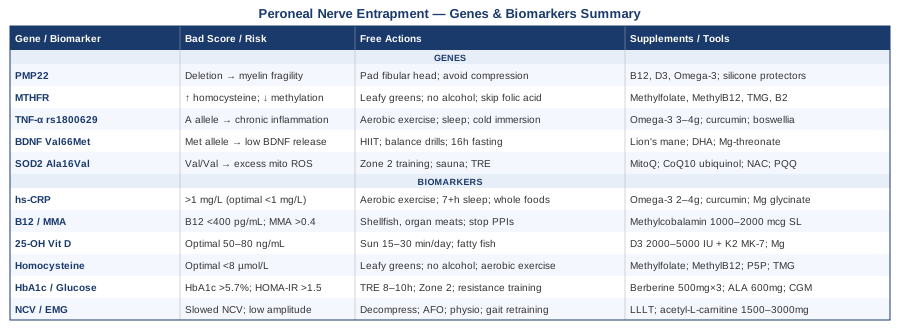
Peroneal nerve entrapment at the fibular head is a condition where biology and mechanics intersect — and where biology is often the missing half of the recovery conversation. Tracking six biomarkers (hs-CRP, B12 and MMA, vitamin D, homocysteine, HbA1c, and nerve conduction velocity) gives you a clear, actionable picture of what is supporting or obstructing your nerve's ability to heal. Understanding your genetic profile — particularly PMP22, MTHFR, TNF-α, BDNF, and SOD2 — adds another layer of precision that explains why some people struggle despite doing everything else right.
None of this requires you to become a medical specialist. It requires you to ask the right questions, order the right tests, and work with the information you receive rather than waiting passively. Start with the biomarkers: they are affordable, widely available, and immediately actionable. Then consider genetic testing if recurrence, slow recovery, or disproportionate severity suggests underlying vulnerability. Layer in the complementary approaches — particularly photobiomodulation and EMG biofeedback — where clinical evidence supports them. And bring this information to the conversation with your neurologist, physiatrist, or physiotherapist as a starting point for a more personalized plan.
Better information consistently leads to better decisions. The nerve is capable of recovery — give it the best possible environment to do so.
Musculoskeletal: Sports Injuries
Neurological: Nerve Conditions Movement Disorders
Endocrine & Metabolic: Diabetes & Blood Sugar
Autoimmune: Inflammatory Conditions