This article contains AI generated content.
· UpdatedPeroneal Nerve Palsy — 7 Genes And 7 Biomarkers To Track
Introduction
If you are living with peroneal nerve palsy — dealing with foot drop, a dragging gait, weakness when lifting the front of your foot, or numbness along the outer shin and top of the foot — you have probably been told to wait, brace, and hope for the best. Sometimes that is the right clinical advice. But it rarely addresses the underlying biological terrain that determines how quickly and completely you recover. Two people can have the same injury graded identically on nerve conduction studies and recover at completely different speeds. The reason often lives in their metabolic profile and genetic background.
The standard workup for peroneal nerve palsy focuses on mechanism (compression, stretch, laceration) and anatomical severity. It does not typically ask whether your homocysteine is damaging your nerve vasculature, whether your vitamin D is low enough to impair Schwann cell function, whether a common genetic variant has been quietly reducing your myelin quality for years, or whether your inflammatory load is high enough to slow the Wallerian regeneration your nerve needs to complete. These factors do not change the diagnosis, but they profoundly shape recovery.
Generic advice — eat well, rest, do physical therapy — is not wrong. It is just incomplete. The research on peripheral nerve health increasingly points to specific, measurable variables: biomarkers that can be tested with a routine blood draw, and genetic variants that can be identified with consumer-grade DNA testing. Getting this information does not require a specialist. Acting on it does not always require a prescription. It mostly requires knowing what to look for.
This article covers two layers of that picture. First, seven biomarkers that are directly relevant to nerve health, practical to measure, and actionable when they come back abnormal. Then, seven gene variants with meaningful implications for nerve vulnerability and recovery, along with concrete protocols for each. Neither section offers a cure. Both offer a more precise, more useful frame for understanding what is happening in your body — and what you can reasonably do to support it.
7 Biomarkers That Directly Affect Nerve Health and Recovery
Biomarkers give you a metabolic fingerprint. For peroneal nerve palsy, the most useful ones share a common thread: they each influence either nerve regeneration rate, myelin integrity, inflammatory burden, or nerve vulnerability to compression. The seven below are consistently recommended by clinicians focused on peripheral nerve and longevity medicine — including Peter Attia, Thomas Dayspring, and integrative neurologists who routinely run expanded labs on patients with slow or incomplete nerve recovery.
1. Homocysteine
Homocysteine is an amino acid intermediate produced during methionine metabolism. Under normal circumstances, it is rapidly recycled with the help of folate, B6, and B12. When those nutrients are depleted — or when genetic variants (particularly MTHFR) reduce recycling efficiency — homocysteine accumulates and becomes neurotoxic. Elevated homocysteine damages the endothelium of the vasa nervorum (the small blood vessels that supply peripheral nerves), impairs myelin synthesis, and promotes oxidative stress in axonal tissue. Research published in PubMed-indexed studies consistently associates elevated homocysteine with peripheral neuropathy independent of other risk factors.
How to measure it: A standard plasma homocysteine test is available through most GPs or direct-to-consumer labs such as LabCorp or Quest. Cost: $20–$60. Optimal range for nerve health: below 8 µmol/L. Above 10 µmol/L carries meaningful risk; above 15 µmol/L indicates high-grade elevation requiring investigation of root causes.
If the score is bad, the plan without supplements: The dietary foundation involves increasing natural folate (dark leafy greens, lentils, liver, asparagus), B6 (poultry, potatoes, bananas), and B12 (animal products, especially liver, sardines, and eggs). Reduce or eliminate alcohol, which depletes all three B-vitamins and raises homocysteine significantly. Prioritize sleep — sleep deprivation disrupts methionine cycle efficiency. These changes alone can reduce elevated homocysteine by 20–30% within 8–12 weeks.
If the score is bad, the plan with supplements or equipment: - Methylfolate (5-MTHF): 400–800 mcg/day. Critical to use the active methyl form, not folic acid, especially in MTHFR carriers. No cycling needed. - Methylcobalamin (active B12): 500–1,000 mcg/day sublingually. Superior absorption over cyanocobalamin in many individuals. - Pyridoxal-5-phosphate (P5P, active B6): 25–50 mg/day. Do not exceed 100 mg/day chronically — high-dose B6 can paradoxically cause peripheral neuropathy. - TMG (trimethylglycine): 500–1,000 mg/day. Provides an alternative methylation pathway (betaine cycle) that lowers homocysteine even when the folate pathway is suboptimal. No significant side effects at these doses. Retest homocysteine every 3 months until stable below 8 µmol/L.
2. 25-OH Vitamin D
Vitamin D is far more than a bone mineral regulator. Its nuclear receptor (VDR) is expressed in peripheral nerve cells and Schwann cells — the specialized glial cells that produce and maintain myelin around peripheral axons. Low vitamin D impairs Schwann cell proliferation and function, reduces nerve conduction velocity, and blunts the anti-inflammatory signaling needed for nerve tissue repair. Epidemiological and mechanistic data consistently link deficiency with both increased peripheral neuropathy risk and slower recovery from nerve injuries.
How to measure it: Serum 25-OH vitamin D, the standard clinical test. Cost: $30–$70. Optimal range for neurological function: 50–80 ng/mL. Below 30 ng/mL is frank deficiency; below 20 ng/mL is severe and warrants immediate correction. The NIH Office of Dietary Supplements provides current clinical reference ranges.
If the score is bad, the plan without supplements: 15–30 minutes of midday sun exposure on arms and legs (without sunscreen) generates 1,000–2,000 IU of vitamin D3 in lighter skin types; darker skin requires longer. Increase dietary sources: oily fish (salmon, mackerel, sardines), egg yolks, cod liver oil. Weight loss improves vitamin D levels by reducing sequestration in adipose tissue.
If the score is bad, the plan with supplements or equipment: - Vitamin D3: 2,000–5,000 IU/day for maintenance; up to 10,000 IU/day short-term for correction under clinical supervision. Always pair with vitamin K2 (MK-7 form, 100–200 mcg/day) to prevent ectopic calcium deposition in arteries. - Retest 25-OH vitamin D after 8–12 weeks. Side effects are rare at supplementation doses; risk of hypercalcemia emerges at very high doses (above 10,000 IU/day without monitoring). No cycling needed for maintenance doses.
3. Vitamin B12 and Methylmalonic Acid
B12 is arguably the single most critical nutrient for peripheral nerve integrity. It is essential for myelin synthesis, axonal cytoskeletal maintenance, and the production of the phospholipids that form nerve cell membranes. B12 deficiency produces a peripheral neuropathy that is clinically indistinguishable from other etiologies — meaning it is frequently missed. The key nuance: serum B12 can appear normal while functional B12 deficiency exists at the cellular level. Methylmalonic acid (MMA) rises before serum B12 falls into the deficient range and is therefore the more sensitive marker. Together they give the complete picture.
How to measure it: Request both tests simultaneously — serum B12 alone is insufficient. Serum B12 cost: $20–$50. Methylmalonic acid (urine or serum): $40–$80. Optimal serum B12: above 500 pg/mL; many integrative practitioners aim for 700–900 pg/mL. MMA should be below 0.28 µmol/L; elevated MMA confirms functional B12 deficiency even when serum B12 appears borderline normal. The NIH Vitamin B12 Fact Sheet outlines absorption mechanisms and risk groups.
If the score is bad, the plan without supplements: B12 exists only in meaningful quantities in animal products. Increase intake of liver, clams, beef, sardines, and eggs. Identify and address absorption blockers: chronic use of proton pump inhibitors (PPIs) and metformin both significantly impair B12 absorption and are among the leading causes of overlooked deficiency. Older adults over 50 absorb B12 less efficiently due to declining stomach acid and intrinsic factor.
If the score is bad, the plan with supplements or equipment: - Methylcobalamin sublingual: 1,000–5,000 mcg/day. Sublingual absorption bypasses the intrinsic factor pathway, making it effective even in those with absorption issues. - B12 injections (methylcobalamin, 1,000 mcg intramuscular): Weekly for 4–8 weeks, then monthly — the gold standard for severe deficiency or confirmed pernicious anemia. No toxicity ceiling; B12 is water-soluble. - If MMA remains elevated after 12 weeks of supplementation, investigate for intrinsic factor antibodies. Retest serum B12 and MMA after 8 weeks of intervention.
4. HbA1c and Fasting Glucose
Chronic hyperglycemia is one of the most potent drivers of peripheral nerve damage. Even pre-diabetes — HbA1c between 5.7% and 6.4% — substantially increases peripheral neuropathy risk through multiple mechanisms: formation of advanced glycation end products (AGEs) that stiffen nerve membranes, oxidative stress from excess glucose, and microvascular damage that starves nerves of oxygen and nutrients. In the context of peroneal nerve palsy, poor metabolic health makes the nerve more susceptible to compression injury in the first place — and dramatically slows regeneration after the fact.
How to measure it: Fasting glucose and HbA1c are standard tests available through any GP or direct-to-consumer lab. Combined cost: $25–$60. Target fasting glucose: 70–90 mg/dL. Target HbA1c: below 5.5% for optimal nerve health — not just below 6.5%, which is simply the threshold for diagnosing diabetes. Peter Attia routinely targets sub-5.3% HbA1c in patients at risk for neuropathic conditions.
If the score is bad, the plan without supplements: Time-restricted eating (eating within an 8–10 hour window), consistent Zone 2 cardio (30–45 minutes at a conversational pace, 4–5 times/week), and resistance training 2–3 times/week all improve insulin sensitivity meaningfully. Reducing refined carbohydrates and ultra-processed foods is non-negotiable. Consistent Zone 2 training alone has been documented to reduce HbA1c by 0.5–1.5% within 12 weeks in insulin-resistant individuals.
If the score is bad, the plan with supplements or equipment: - Berberine: 500 mg, 2–3 times/day with meals. Multiple randomized controlled trials have found berberine comparable to metformin for fasting glucose and HbA1c reduction. Cycle: 8 weeks on, 2–4 weeks off (prolonged use without cycling may affect gut microbiome composition). - Continuous Glucose Monitor (CGM): Devices such as Abbott FreeStyle Libre or Dexcom G7 provide real-time blood glucose feedback. Cost: $50–$80/month. Highly effective for identifying glycemic spikes from specific foods and guiding behavioral changes. Peter Attia considers this one of the highest-ROI health tools for anyone with borderline metabolic markers. - Alpha-lipoic acid (ALA): 600 mg/day. Has specific published evidence for diabetic peripheral neuropathy — reduces oxidative stress in nerve tissue, improves nerve conduction velocity. No cycling needed. Side effects minimal; may cause mild nausea on an empty stomach.
5. High-Sensitivity CRP (hs-CRP)
Inflammation is not merely a background condition in peripheral nerve injury — it is an active determinant of recovery pace and quality. Pro-inflammatory cytokines including TNF-alpha, IL-6, and IL-1β impair Schwann cell proliferation, slow the formation of the regeneration tube (Büngner bands) that guides axonal regrowth, and create a hostile biochemical environment for remyelination. hs-CRP is the most widely accessible systemic inflammation marker. While it is not nerve-specific, elevations above 1.0 mg/L signal an inflammatory state that will almost certainly impede recovery.
How to measure it: High-sensitivity CRP specifically — standard CRP lacks sensitivity at low levels. Cost: $20–$50. Target: below 0.5 mg/L for optimal tissue healing environment. The range of 1–3 mg/L indicates moderate systemic inflammation; above 3 mg/L warrants investigation for chronic infection, autoimmune disease, or significant metabolic dysfunction.
If the score is bad, the plan without supplements: The highest-impact anti-inflammatory interventions are behavioral. Eliminate ultra-processed foods and refined seed oils (linoleic acid-heavy vegetable oils are potent NF-kB activators). Prioritize 7–9 hours of quality sleep — even two nights of poor sleep can raise CRP measurably. Consistent aerobic exercise lowers CRP through anti-inflammatory myokine release. Address obesity if present, as adipose tissue is a significant source of pro-inflammatory cytokines.
If the score is bad, the plan with supplements or equipment: - Omega-3 fatty acids (EPA+DHA): 2–4 g/day combined. Among the most evidence-supported anti-inflammatory supplements available. No cycling needed; take with meals. - Curcumin (high-bioavailability form): 500–1,000 mg/day of BCM-95 or Meriva formulation. Standard curcumin has poor absorption — formulation matters. Cycle: 12 weeks on, 4 weeks off. Side effects rare; may interact with blood thinners. - Regular sauna use: Finnish population data shows 4 sessions/week of 20 minutes at 170–185°F is associated with significantly reduced CRP and improved vascular health. Commercial gym saunas cost nothing extra. Home infrared units: $1,500–$4,000.
6. Omega-3 Index
The Omega-3 Index measures the percentage of EPA and DHA in red blood cell membranes — a far more accurate reflection of tissue-level omega-3 status than a plasma measurement, which fluctuates hour-to-hour with meals. Peripheral nerve cell membranes are rich in long-chain omega-3 fatty acids; low omega-3 status increases membrane rigidity, elevates nerve inflammation, worsens neuropathic pain outcomes, and slows nerve regeneration. Thomas Dayspring has consistently identified the Omega-3 Index as an underutilized but clinically important marker in standard metabolic panels.
How to measure it: Omega-3 Index testing requires a specialized finger-prick test — OmegaQuant is the clinical standard and widely recognized. Cost: $50–$100. Target: above 8%. The average American tests at 4–5%, which is associated with significantly higher inflammatory and cardiovascular risk. Above 8% is associated with favorable neurological and cardiac outcomes; elite athletes sometimes test at 10–12%.
If the score is bad, the plan without supplements: Consume fatty cold-water fish — salmon, mackerel, sardines, herring, anchovies — at minimum 3–4 times per week. Simultaneously reduce omega-6 intake (seed oils, processed and fried foods) to improve the ratio. Dietary-only correction of a low Omega-3 Index takes 3–6 months to reflect meaningfully in testing.
If the score is bad, the plan with supplements or equipment: - High-quality triglyceride-form EPA+DHA supplement: 2–4 g/day combined. For peroneal nerve palsy, a higher EPA ratio (at least 2:1 EPA to DHA) is preferable for anti-inflammatory action. Take with the largest meal of the day for optimal absorption. NIH Omega-3 Fact Sheet summarizes current evidence on formulations. No cycling needed. - Side effects: mild fishy aftertaste (use enteric-coated), occasional loose stools at high doses. Retest Omega-3 Index after 4–6 months.
7. RBC Magnesium
Magnesium participates in over 300 enzymatic reactions, including those governing nerve conduction, neuromuscular transmission, ATP production, and the synthesis of myelin components. The critical measurement point: serum magnesium represents only 1% of total body magnesium and is tightly regulated — it can remain normal while intracellular stores are depleted. RBC magnesium measures magnesium inside red blood cells and reflects tissue-level status far more accurately. Low RBC magnesium increases nerve excitability, contributes to muscle cramping in peroneal palsy presentations, and impairs mitochondrial energy production needed for axonal transport and regeneration. Estimates suggest 50–70% of adults in Western countries are insufficient.
How to measure it: Request RBC magnesium specifically — not serum magnesium. Cost: $50–$80, often available through direct-to-consumer labs. Optimal range: above 5.5 mg/dL. Below 4.5 mg/dL represents functional insufficiency. The NIH Magnesium Fact Sheet provides reference ranges and absorption data by form.
If the score is bad, the plan without supplements: Increase dietary magnesium through pumpkin seeds, dark chocolate (85%+), dark leafy greens, almonds, cashews, and avocado. Reduce alcohol and excess coffee (both increase urinary magnesium loss). Correct vitamin D deficiency, which impairs intestinal magnesium absorption. Reduce phytate intake (soaking grains and legumes improves mineral availability).
If the score is bad, the plan with supplements or equipment: - Magnesium glycinate: 200–400 mg elemental magnesium/day, taken in the evening (supports sleep quality and muscle recovery simultaneously). Glycinate is the best-tolerated form for those with sensitive digestion. - Magnesium malate: preferred by some practitioners for daytime use, as malate supports mitochondrial energy production directly — a relevant combination for nerve repair metabolism. - Avoid magnesium oxide: it has less than 4% absorption and is primarily a laxative. - Transdermal magnesium oil (applied directly to the calf/shin area near the peroneal nerve pathway): limited direct evidence but low-cost and low-risk adjunct to oral magnesium. No cycling needed for standard doses. Retest RBC magnesium after 8–12 weeks.
With those seven biomarkers as a measurable foundation, it is worth adding a second layer of context: the genetic background that shapes how efficiently your body processes these nutrients and responds to nerve injury. Genes do not override biology, but they do explain why the same intervention works dramatically better for one person than another.
7 Genetic Variants That Shape Nerve Vulnerability and Regeneration
Genetic testing is now accessible and affordable. Consumer platforms like 23andMe provide raw data that can be analyzed through tools like Genetic Lifehacks or SelfDecode. Clinical genetic panels ordered through a neurologist are more comprehensive. The following seven variants have the most meaningful evidence for peripheral nerve health — both for understanding vulnerability and for tailoring recovery strategies.
1. PMP22 — Peripheral Myelin Protein 22
PMP22 is the gene most directly relevant to peroneal nerve palsy. It encodes a structural protein essential to compact myelin in peripheral nerves. Deletion of one copy causes Hereditary Neuropathy with Liability to Pressure Palsies (HNPP) — a condition in which peripheral nerves are structurally weaker and abnormally susceptible to compression injury and minor mechanical stress. Many people with HNPP experience recurrent peroneal nerve palsies, ulnar nerve palsies, and carpal tunnel syndrome across years before anyone thinks to run a genetic test. NCBI GeneReviews provides a complete clinical summary of HNPP, including diagnostic criteria and management. Duplication of PMP22 causes Charcot-Marie-Tooth type 1A — a different but related pathology. Testing: a targeted MLPA or chromosomal microarray test, typically ordered by a neurologist.
If the gene is bad, the plan without supplements: Compression avoidance is the primary management strategy. This means never crossing legs for extended periods, avoiding prolonged squatting or kneeling, using fibular head padding during sleep and exercise, and wearing protective bracing during activities that load the peroneal region. Lightweight ankle-foot orthosis (AFO) during activities with high fall risk. Occupational therapy assessment of workstation ergonomics. Weight management reduces adipose tissue pressure on nerve beds at vulnerable anatomical sites.
If the score is bad, the plan with supplements or equipment: No supplement modifies PMP22 gene expression, but myelin-supporting nutrients reduce secondary damage: methylcobalamin (1,000 mcg/day), phosphatidylcholine (1–2 g/day, provides structural lipids for myelin membranes), and omega-3s (3–4 g/day EPA+DHA). Wearable pressure-monitoring insoles and activity trackers can help HNPP patients identify and avoid high-risk loading patterns. Genetic counseling for family members is warranted given autosomal dominant inheritance.
2. MTHFR — Methylenetetrahydrofolate Reductase (C677T and A1298C)
MTHFR is the rate-limiting enzyme in converting dietary folate to its bioactive form (5-methyltetrahydrofolate, or 5-MTHF), which is required to recycle homocysteine and support cellular methylation. The C677T variant reduces enzyme activity by approximately 40% in heterozygous carriers and 70% in homozygous carriers. A1298C further reduces activity when combined with C677T. These variants are among the most common in the human genome — affecting 40–60% of the population at varying degrees — yet remain widely underappreciated in clinical neurology. The primary consequence for nerve health is elevated homocysteine and impaired production of the methyl groups needed for myelin maintenance. Gary Brecka and functional medicine practitioners have drawn significant public attention to MTHFR's broad health implications.
If the gene is bad, the plan without supplements: Shift all folate intake to natural food folate — not folic acid from enriched grains or many standard supplements, which requires the defective enzyme to convert. Prioritize leafy greens, liver, lentils, and avocado. Eliminate alcohol (dramatically inhibits folate cycling). Test and monitor homocysteine as a functional readout of MTHFR impact. Reduce stress, which increases methylation demand.
If the score is bad, the plan with supplements or equipment: - Methylfolate (5-MTHF): 400–1,000 mcg/day. Bypasses the defective enzyme entirely. Start at a low dose — some MTHFR individuals are sensitive to methyl donors and experience anxiety, irritability, or insomnia when starting too high. - Methylcobalamin: 500–1,000 mcg/day. Works synergistically with methylfolate in the homocysteine recycling pathway. - P5P (active B6): 25–50 mg/day supports the transsulfuration pathway, the secondary homocysteine clearance route. - TMG: 500–1,000 mg/day as a backup methyl donor via the betaine pathway. This combination typically normalizes homocysteine within 8–12 weeks. Retest and adjust dose accordingly.
3. APOE — Apolipoprotein E (E2/E3/E4)
APOE is best known for its association with Alzheimer's risk, but its role in peripheral nerve biology is equally important. APOE proteins transport cholesterol and phospholipids to sites of myelin repair — meaning APOE genotype directly shapes how efficiently your nerve rebuilds its myelin sheath after injury. Studies in nerve injury models show that APOE4 carriers display significantly slower and less complete peripheral nerve regeneration compared to APOE2 and APOE3 carriers. APOE4 is present in approximately 25% of the population. It also confers hyper-responsiveness to dietary saturated fat, raising LDL cholesterol more aggressively than other genotypes — which has downstream effects on nerve vascular supply.
If the gene is bad (APOE4 carrier), the plan without supplements: Zone 2 aerobic exercise (4–5 sessions/week) is uniquely beneficial for APOE4 individuals, improving lipid metabolism and APOE4-related metabolic dysregulation. Reduce saturated fat intake (APOE4 individuals respond more aggressively to dietary saturated fat than other genotypes). Prioritize sleep — APOE4 carriers show greater disruption to glymphatic clearance when sleep is impaired, affecting neurotrophic factors in the CNS that also influence peripheral nerve repair. Time-restricted eating (10-hour eating window) reduces APOE4-associated inflammatory signaling.
If the score is bad, the plan with supplements or equipment: - Phosphatidylserine: 100–300 mg/day. Supports neuronal membrane integrity and lipid transport relevant to myelin repair. - Omega-3 DHA+EPA: 3–4 g/day. Particularly important for APOE4 individuals — DHA specifically supports neurotrophin production and reduces the inflammatory effects of APOE4-associated lipid metabolism. - Lion's Mane mushroom extract (standardized): 500–1,000 mg/day. Promotes nerve growth factor (NGF) synthesis and emerging evidence supports peripheral nerve repair applications. Cycle: 8 weeks on, 2–4 weeks off. Side effects rare; some report mild digestive upset.
4. BDNF — Brain-Derived Neurotrophic Factor (Val66Met, rs6265)
BDNF is a neurotrophin that promotes the survival, growth, and regeneration of neurons — including peripheral motor and sensory fibers. The Val66Met polymorphism reduces activity-dependent BDNF secretion by approximately 25–35%. In practice, this means that the acute BDNF response to exercise and behavioral stimuli is blunted — the very mechanism the nervous system uses to drive axonal sprouting and reinnervation after peripheral nerve injury. Approximately 20–30% of the population carries at least one copy of the Met allele. Andrew Huberman has discussed the Val66Met variant and its implications for neuroplasticity and recovery in multiple Huberman Lab contexts.
If the gene is bad, the plan without supplements: Exercise is the most powerful known driver of BDNF upregulation. High-intensity interval training (HIIT) produces acute BDNF spikes 2–3x greater than sustained moderate exercise. Protocol: 2–3 sessions/week of 20 minutes (4–6 rounds of 30–60 seconds all-out effort followed by 2–4 minutes recovery). Cold water immersion (2–5 minutes at 14–15°C, 3–5 times/week) raises BDNF through norepinephrine release. Learning new motor skills — as in physical therapy after nerve injury — also drives BDNF in an activity-dependent fashion. Fasting (16+ hours) upregulates BDNF expression.
If the score is bad, the plan with supplements or equipment: - Lion's Mane mushroom: 500–1,000 mg/day of an hericenone/erinacine-standardized extract. Promotes NGF synthesis, which shares downstream signaling pathways with BDNF relevant to peripheral axon maintenance. - Magnesium L-threonate: 1.5–2 g/day (as sold — roughly 144 mg elemental magnesium). One of very few magnesium forms that reliably crosses the blood-brain barrier; supports synaptic density and BDNF-related neuroplasticity pathways. Cycle: 12 weeks on, 4 weeks off. Some individuals report vivid dreams or mild headache initially. - DHA supplementation: Specifically DHA (not just EPA) supports BDNF gene expression in animal models; aim for at least 1 g/day DHA within your total omega-3 intake.
5. TNF-alpha — Tumor Necrosis Factor Alpha (−308 G>A, rs1800629)
TNF-alpha is a master pro-inflammatory cytokine. The −308 G>A polymorphism places the A allele carrier into a hyper-inflammatory phenotype — elevated TNF-alpha production in response to injury, infection, or metabolic stress. In peripheral nerve injury, elevated TNF-alpha accelerates Wallerian degeneration and inhibits the Schwann cell response required for remyelination. Carriers experience more inflammation-driven secondary damage from the same mechanical nerve insult as non-carriers. This variant is present in approximately 15–20% of European-ancestry populations.
If the gene is bad, the plan without supplements: Anti-inflammatory lifestyle interventions are not optional for −308 A allele carriers — they are a direct therapeutic strategy. Eliminate refined seed oils (strong NF-kB activators), ultra-processed foods, and excess alcohol. Consistent aerobic exercise modulates TNF-alpha through anti-inflammatory myokine release (particularly IL-6 from active muscle, which has anti-inflammatory properties at moderate exercise intensity). Prioritize 7–9 hours of sleep. Address psychological stress, as chronic cortisol upregulates TNF-alpha transcription.
If the score is bad, the plan with supplements or equipment: - Curcumin (BCM-95 or Meriva form): 500–1,000 mg/day. A direct NF-kB inhibitor. Cycle: 12 weeks on, 4 weeks off. Side effects rare at standard doses; may enhance blood thinner effects. - Omega-3 EPA+DHA: 3–4 g/day. EPA in particular competes with arachidonic acid in the inflammatory cascade. - Resveratrol: 250–500 mg/day with a fat-containing meal (dramatically improves absorption). Inhibits NF-kB and downregulates TNF-alpha signaling. Cycle: 8–12 weeks on, 4 weeks off to prevent adaptation. Mild estrogen-like activity at high doses — note if hormonally sensitive.
6. SOD2 — Superoxide Dismutase 2 (Ala16Val, rs4880)
SOD2 encodes the mitochondrial form of superoxide dismutase, the primary antioxidant defense inside mitochondria. The Ala16Val polymorphism reduces the efficiency of SOD2 protein import into the mitochondrial matrix, leaving nerve cell mitochondria more vulnerable to oxidative damage under metabolic stress. Peripheral axons are exceptionally long and energetically demanding — they rely heavily on efficient mitochondrial function for axonal transport, membrane potential maintenance, and the ATP-driven processes of regeneration. Reduced SOD2 activity means nerve tissue accumulates more oxidative damage during and after injury.
If the gene is bad, the plan without supplements: Minimize dietary oxidative load: eliminate processed vegetable oils high in linoleic acid, reduce charred and heavily processed meats, and increase antioxidant-rich plant foods (berries, dark greens, cruciferous vegetables). Paradoxically, regular moderate-intensity exercise improves mitochondrial antioxidant capacity over time through hormetic adaptation — brief oxidative stress from exercise upregulates endogenous SOD2 expression. Avoid excessive caloric intake, which creates mitochondrial electron transport overflow.
If the score is bad, the plan with supplements or equipment: - Alpha-lipoic acid (ALA): 600 mg/day. Both mitochondria-targeted and directly recycles glutathione. Specific published evidence for diabetic peripheral neuropathy with improvements in nerve conduction. No cycling needed. - Ubiquinol (CoQ10): 100–200 mg/day. The active reduced form of CoQ10, supporting mitochondrial electron transport and reducing oxidative spillover. More bioavailable than ubiquinone for individuals over 40 or with metabolic compromise. No cycling needed. - Manganese: SOD2 requires manganese as its catalytic cofactor. Ensure adequate dietary intake (2–5 mg/day from food or a multimineral with 2 mg manganese). Excessive supplemental manganese is neurotoxic — use dietary and multimineral sources only, not isolated high-dose supplements.
7. COMT — Catechol-O-Methyltransferase (Val158Met, rs4680)
COMT degrades catecholamines — dopamine, norepinephrine, epinephrine — in the prefrontal cortex and elsewhere. The Val158Met polymorphism creates a spectrum from Val/Val ("warrior," fast COMT, low synaptic catecholamines) to Met/Met ("worrier," slow COMT, high synaptic catecholamines). Met/Met individuals have significantly higher pain sensitivity and tend to experience more severe neuropathic pain from the same nerve injury. This is not a difference in injury severity — it is a difference in how pain signals are processed and amplified. Understanding this variant can fundamentally change how a peroneal nerve palsy patient approaches pain management during recovery.
If the gene is bad (Met/Met, slow COMT), the plan without supplements: Mind-body approaches are directly mechanistically relevant, not just adjuncts: mindfulness-based stress reduction (MBSR), biofeedback, and paced breathing directly modulate catecholamine tone through the same pathways affected by slow COMT. Consistent aerobic exercise clears excess catecholamines and reduces pain amplification over time. Avoid prolonged psychological stress, which floods the catecholamine system and worsens pain in slow COMT individuals. Sleep deprivation specifically worsens catecholamine dysregulation in Met/Met carriers.
If the score is bad, the plan with supplements or equipment: - Avoid high-dose methyl donor overload (very high SAMe, excessive methylfolate) — these can flood the COMT pathway in Met/Met individuals and worsen anxiety and pain sensitivity. - Magnesium glycinate: 300–400 mg/day. Magnesium is a required COMT cofactor; deficiency reduces COMT activity further. - Riboflavin (B2): 50 mg/day. Supports COMT enzymatic activity. - EGCG (green tea extract): 400–600 mg/day of a standardized extract. Acts as a gentle catechol-O-methyltransferase substrate competitor, dampening excessive catecholamine signaling without completely blocking it. Cycle: 8 weeks on, 2–4 weeks off (liver safety consideration at higher doses). Take with food to minimize gastrointestinal effects.
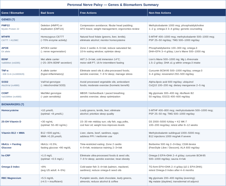
With all 14 variables mapped, the summary table below provides a quick-reference overview that can be useful for tracking and communicating with a healthcare provider.
Summary Table: All Genes and Biomarkers at a Glance
Beyond targeted supplementation and genetic awareness, there is a broader framework for nerve recovery that synthesizes much of the underlying science — one that has been making its way into mainstream health conversation through evidence-based podcast content.
What Neuroscience Research Reveals About Peripheral Nerve Recovery: 10 Things Worth Knowing
The Huberman Lab podcast has, across dozens of episodes, assembled one of the most coherent and research-grounded frameworks for understanding how the nervous system heals, adapts, and regenerates. While no single episode addresses peroneal nerve palsy directly, the mechanistic principles discussed — drawn primarily from peer-reviewed animal and human studies — map directly onto the biology of peripheral nerve recovery. The following ten insights synthesize the most actionable content from that body of work.
1. BDNF Is the Master Switch — and Exercise Turns It On
BDNF (brain-derived neurotrophic factor) drives both central and peripheral nerve regeneration. It supports axonal sprouting, promotes Schwann cell proliferation, and accelerates reinnervation of denervated muscle. The critical practical finding: high-intensity interval training generates a BDNF response 2–3x larger than moderate continuous exercise. Two to three sessions per week of 20 minutes HIIT is not just cardiovascular training for peroneal nerve palsy patients — it is a targeted neurotrophin delivery protocol. The intensity matters; a casual walk does not produce the same signal.
2. Cold Exposure Triggers Norepinephrine — and Norepinephrine Drives Nerve Repair Signaling
Cold water immersion (2–5 minutes at 14–15°C) increases norepinephrine by 200–300%. Norepinephrine acts on adrenergic receptors in Schwann cells and promotes their survival and myelination activity. Andrew Huberman's synthesis of cold exposure research emphasizes the importance of the shiver response as the physiological trigger — meaning cold enough to cause shivering produces a different and more potent neurochemical response than cool-but-comfortable exposure. For peroneal nerve palsy, cold foot baths or lower-limb immersion may provide localized benefits alongside systemic effects.
3. Sleep Is Non-Negotiable for Nerve Regeneration
The majority of peripheral nerve growth factor synthesis, axonal transport activity, and myelin production occur during slow-wave (deep) sleep. Huberman's synthesis of sleep science makes clear that this is not a passive state — the nervous system is actively executing repair programs during NREM sleep. Even moderate sleep restriction (6 hours rather than 8 for two weeks) meaningfully impairs neurotrophic signaling. For peroneal nerve palsy recovery, protecting sleep quality and duration is arguably the most leverage-rich intervention available — and completely free.
4. Non-Sleep Deep Rest (NSDR) Accelerates Neuroplasticity
Research on non-sleep deep rest protocols — including yoga nidra and structured body-scan relaxation — shows that even 20 minutes of NSDR following a learning or rehabilitation session increases the expression of genes associated with neural repair and synaptic consolidation. Applied to peroneal nerve palsy, performing NSDR after physiotherapy sessions (particularly after exercises targeting ankle dorsiflexion and foot eversion) may improve the retention of motor learning and the rate of cortical remapping that accompanies peripheral nerve reinnervation.
5. Photobiomodulation Directly Stimulates Mitochondria in Nerve Tissue
Red and near-infrared light (620–850 nm) penetrates tissue to a depth of several centimeters and is absorbed by cytochrome c oxidase in mitochondria, boosting ATP production and reducing oxidative stress in nerve cells. Huberman has cited multiple studies demonstrating photobiomodulation benefits for tissue repair and neurological function. For the common peroneal nerve — which runs superficially around the fibular head — this mechanism is anatomically accessible. Dedicated panels or targeted LLLT devices cost $150–$1,500 depending on power output.
6. Deliberate Heat Exposure Stimulates Growth Hormone and Improves Blood Flow to Peripheral Nerves
Sauna sessions of 20 minutes at 170–185°F, performed 3–4 times weekly, have been documented to increase growth hormone levels by 200–300% in the hours following exposure. Growth hormone is a critical signal for peripheral tissue repair, including nerve regeneration. Additionally, the dramatic vasodilation induced by sauna increases blood flow to the peripheral vascular bed — including the vasa nervorum supplying the peroneal nerve — delivering oxygen, nutrients, and neurotrophic factors to the healing site.
7. Focused Attention Guides Neural Plasticity — Use It in Rehabilitation
One of the most consistently replicated findings in neuroplasticity research is that attention directed to a body region during motor training significantly increases the magnitude of cortical remapping and axonal sprouting in that area. For peroneal nerve palsy patients doing ankle dorsiflexion exercises, this means the quality of focused attention during the exercise matters — not just the mechanical repetition. Distracted physiotherapy is measurably less effective than mindful physiotherapy from a neural reorganization standpoint.
8. The Neuroplasticity Window Opens With Agitation and Closes With Rest
Huberman's synthesis of neuroplasticity timing research reveals that the actual synaptic changes that underpin neural repair occur during rest and sleep — not during the activity itself. Activity (or physiotherapy) opens a plasticity window by creating agitation in neural circuits; the consolidation happens in the subsequent rest period. This has a direct clinical implication: physiotherapy sessions work best when followed by 20–30 minutes of NSDR or structured rest, not immediately by cognitively demanding work.
9. Omega-3 DHA Is a Structural Requirement for Rebuilding Nerve Cell Membranes
Peripheral nerve cell membranes and myelin sheaths are heavily enriched with DHA (docosahexaenoic acid). During nerve regeneration, the demand for DHA rises sharply. Huberman has cited research showing that adequate DHA status improves neurotrophin production and supports structural plasticity. The mechanism is not just anti-inflammatory — DHA is literally a building material for the membranes being rebuilt. This is why Omega-3 Index testing (to confirm actual tissue-level DHA status, not just recent dietary intake) is more clinically meaningful than a one-time plasma measurement.
10. The Visual System Can Be Leveraged to Drive Motor Plasticity in Adjacent Pathways
Huberman's vision-neuroplasticity research documents how brief periods of visual focus training — particularly following smooth pursuit exercises — prime cortical motor areas for enhanced plasticity during subsequent motor rehabilitation. Applied to peroneal nerve palsy, incorporating brief visual tracking exercises immediately before physiotherapy (not during) may increase the learning and motor remapping efficiency of the session. This is a zero-cost intervention requiring only a slowly moving object and 3–5 minutes of attention.
The evidence-based therapeutic approaches that complement these neurological principles are worth examining in their own right — particularly those with direct clinical evidence for nerve-related conditions.
Complementary Approaches With Meaningful Evidence for Peripheral Nerve Recovery
Low-Level Laser Therapy and Photobiomodulation
Low-level laser therapy (LLLT) and broader photobiomodulation (PBM) use specific wavelengths of red and near-infrared light to stimulate mitochondrial function in tissue. In peripheral nerve applications, this mechanism translates into increased ATP production in Schwann cells and axons, reduced local inflammation, and acceleration of the axonal regeneration process. The common peroneal nerve's superficial position at the fibular head makes it an ideal target for this modality — light penetration of 1–5 cm is sufficient to reach it directly.
A 2014 systematic review published in Photomedicine and Laser Surgery (Rochkind et al.) found consistent evidence from human trials that LLLT applied to peripheral nerve injury sites accelerates both electrophysiological recovery and functional motor outcomes. A randomized controlled trial examining LLLT for peripheral nerve repair reported significantly faster nerve conduction velocity recovery in treated versus sham groups. Evidence quality is moderate; most studies are small and heterogeneous in dosing parameters — but the mechanistic rationale is sound and the safety profile is excellent.
For peroneal nerve palsy, a practical protocol involves applying a 630–850 nm laser or LED panel device to the fibular head region for 5–10 minutes per session, 3–5 times per week. Clinical LLLT is available through sports medicine clinics, some physiotherapy practices, and pain management centers. Treatment cost varies from $50–$150 per session clinically, or $200–$1,500 for consumer-grade photobiomodulation devices for home use. Begin cautiously; there is no meaningful risk at standard therapeutic doses, but confirm the device has adequate irradiance (minimum 30 mW/cm²) to produce a therapeutic effect — decorative red-light devices rarely meet this threshold.
Biofeedback
Biofeedback provides real-time electrophysiological feedback — typically from surface electromyography (sEMG) sensors placed over the peroneal muscles — that allows patients to consciously observe and reinforce neuromuscular activation in muscles that are partially denervated and difficult to voluntarily control. In peroneal nerve palsy, the ankle dorsiflexors and foot evertors may have partial motor return during recovery, but voluntary activation is weak and proprioceptive feedback is disrupted. Biofeedback gives patients a visible signal that an activation has occurred — even a very weak one — reinforcing the motor pattern neuroplastically.
A randomized controlled trial by Binder et al. in JAMA (1981) demonstrated superior motor recovery with EMG biofeedback versus conventional physiotherapy alone in foot drop rehabilitation — a directly relevant finding. Subsequent systematic reviews of sEMG biofeedback for peripheral nerve motor rehabilitation confirm its superiority to passive exercise alone for patients who have some preserved motor signal. The mechanism aligns precisely with Huberman's focused-attention neuroplasticity principle: biofeedback ensures that attention is accurately directed to the target muscle, amplifying plasticity of the reinnervating pathways.
In practice, biofeedback for peroneal nerve palsy is most effectively delivered through a trained physiotherapist using clinical sEMG equipment, with sessions of 30–45 minutes, 2–3 times weekly. Some portable home biofeedback units are available (BioStim or Thought Technology systems, $300–$2,000) for between-session practice. Progress should be tracked by monitoring the EMG amplitude threshold required to trigger feedback — as reinnervation proceeds, smaller signals should be sufficient to cross the threshold.
Massage Therapy
Massage therapy applied along the peroneal nerve pathway addresses several contributing factors in peroneal nerve palsy: reducing perineural adhesions and fibrous tissue that may form around an injured nerve, improving local circulation to the vasa nervorum, reducing myofascial tension in the tibialis anterior and peroneal muscles (which can secondarily compress the nerve's distal branches), and improving proprioceptive awareness in a region that has lost normal sensory feedback. While evidence for massage therapy in pure peroneal nerve palsy specifically is limited, evidence for manual therapy in peripheral nerve decompression and post-injury rehabilitation is reasonably established.
A study published in Manual Therapy (Ellis and Hing, 2008) demonstrated significant improvements in neural mobility, pain, and function following neural mobilization techniques (a specific category of manual therapy targeting peripheral nerve gliding) for lower extremity neuropathies. Neurodynamic mobilization — where the clinician performs passive and active movements that slide and tension the peripheral nerve through its anatomical bed — has the strongest evidence in this category, superior to simple soft tissue massage alone.
For peroneal nerve palsy, the most useful massage and manual therapy protocol involves: (1) sciatic nerve neural mobilization in slump or straight-leg-raise positions (because the peroneal nerve is a branch of the sciatic, and neural tension from proximal sites can impede distal regeneration), (2) soft tissue work over the fibular head to address perineural adhesions, and (3) deep tissue work in the anterior and lateral compartments. Seek a physiotherapist or massage therapist with training in neurodynamics. Begin gently — aggressive nerve mobilization in the acute phase of injury can worsen symptoms. Two sessions per week during the active recovery phase is a reasonable starting point.
Yoga
Yoga offers a meaningful combination of benefits for peroneal nerve palsy recovery that extends beyond generic flexibility training. Specific yoga postures — particularly those involving ankle dorsiflexion, single-leg balance, and peroneal muscle engagement — target the precise functional deficits of foot drop and peroneal weakness. Additionally, the proprioceptive challenge of balance-based postures drives cortical remapping of the lower limb in ways that standard physiotherapy exercises may not. Yoga's breath regulation component also activates the parasympathetic nervous system, which supports the physiological environment for nerve repair.
Small randomized controlled trials of yoga-based programs for peripheral neuropathy (particularly diabetic peripheral neuropathy) have documented improvements in balance, proprioception, pain scores, and nerve conduction velocity. A 2019 trial in Complementary Therapies in Medicine found that a 12-week yoga intervention significantly improved nerve conduction velocity and quality-of-life measures in patients with peripheral neuropathy compared to controls. Evidence specific to peroneal nerve palsy is extrapolated from these related populations — the mechanistic case is strong but direct RCT evidence in peroneal palsy specifically is limited.
A practical yoga protocol for peroneal nerve palsy would begin conservatively, given foot drop risk and potential instability. Wall-supported single-leg balance poses (Vriksasana variations with wall support), seated ankle circling and dorsiflexion sequences (Dandasana derivatives), and supine hip and knee mobilization are appropriate early on. Downward-facing dog provides gentle sciatic-peroneal nerve tensioning that complements the neurodynamic mobilization approach. 20–30 minutes, 4–5 times weekly is a reasonable goal, supervised initially by a yoga instructor familiar with neurological rehabilitation. Avoid unsupported balancing poses until functional dorsiflexion and reliable proprioception are restored.
Conclusion
Peroneal nerve palsy sits at the intersection of mechanical injury and systemic biology. The mechanical part — the compression, the stretch, the structural damage — is what gets diagnosed and treated. The systemic biology — your B12 status, your inflammatory load, your genetic predisposition for nerve vulnerability, your Omega-3 Index — is what largely determines how well and how fast you recover. Most people never look at that layer. They wait, brace, and hope the nerve grows back on its own timeline.
The clearest next step is to start measuring. Book a blood panel that includes homocysteine, 25-OH vitamin D, B12 with methylmalonic acid, HbA1c, hs-CRP, and RBC magnesium. If you have access to genetic testing, raw DNA data from 23andMe can be run through Genetic Lifehacks or a functional medicine clinician to identify the variants discussed above. Each abnormal result is a lever. Each lever has a protocol. Not every protocol will apply to you — but the ones that do will make a meaningful difference, and you now have enough information to have a substantially more useful conversation with your neurologist, physiotherapist, or integrative medicine provider.
Neurological Endocrine & Metabolic
Neurological: Nerve Conditions Movement Disorders
Endocrine & Metabolic: Diabetes & Blood Sugar
Autoimmune: Inflammatory Conditions