This article contains AI generated content.
· UpdatedPes Anserine Tendinopathy: 5 Genes and 6 Biomarkers to Track
Introduction
If you have pes anserine tendinopathy, you are probably already familiar with the standard advice: rest, ice, anti-inflammatory medication, maybe a corticosteroid injection if things get bad enough. You may have been told to lose weight, stretch your hamstrings, or strengthen your quadriceps. This advice is not wrong — but it is often frustratingly incomplete, especially when the pain keeps returning or the original trigger was never clearly identified.
Pes anserine tendinopathy involves the medial aspect of the knee, roughly two inches below the joint line, where three tendons converge: the sartorius, gracilis, and semitendinosus. When that insertion zone becomes inflamed or structurally degraded, the result is pain that makes walking, stair climbing, and sleeping on the affected side genuinely difficult. What is less commonly explained is that this condition almost never appears in isolation. It clusters heavily with type 2 diabetes, obesity, genu valgum, and knee osteoarthritis — and that clustering is not a coincidence. It points toward metabolic and inflammatory drivers that standard orthopedic care rarely investigates.
Generic treatment plans are designed for an average patient, and there is no such thing as the average patient. Two people with identical imaging findings can have entirely different underlying drivers of tendon inflammation: one may have persistently elevated blood sugar that glycates and weakens collagen, another may carry a structural collagen gene variant that makes tendon repair inherently slower, and a third may have a vitamin D receptor mutation that blunts their response to supplementation. Each of these requires a different intervention.
This article takes a more specific approach. It covers six measurable biomarkers that can identify and monitor the metabolic and inflammatory factors most directly linked to pes anserine tendinopathy — including what to do when those numbers fall outside the optimal range. It also covers five genetic variants relevant to tendon vulnerability and inflammation regulation, along with the practical implications of each. Neither section offers a cure. But better information about what is actually driving your condition leads to more precise decisions, fewer wasted interventions, and a clearer path forward.
6 Biomarkers That Reveal What Is Driving Your Pes Anserine Pain
Most people with pes anserine tendinopathy are told the problem is mechanical — bad alignment, weak muscles, too much load on the medial knee. Biomechanics do matter. But the tendons, bursa, and connective tissue in this region are also highly sensitive to metabolic and inflammatory signals circulating in the blood. Measuring those signals gives a window into why the pain appeared, why it is slow to resolve, and what type of intervention is most likely to work. Peter Attia, Thomas Dayspring, and other clinicians who think carefully about long-term health consistently emphasize that biomarker data is far more actionable than symptom-level management alone.
The six biomarkers below were selected because they have direct or strongly plausible connections to the pathology of pes anserine tendinopathy, are measurable with standard blood draws, and respond meaningfully to targeted interventions.
Biomarker 1: High-Sensitivity C-Reactive Protein
Why it matters
hs-CRP is the most widely used blood marker of systemic low-grade inflammation. It is produced by the liver in response to cytokine signals — primarily interleukin-6 — and reflects the overall inflammatory tone of the body's tissues. In tendinopathy, persistent inflammation is not simply a side effect of tissue damage. It is one of the central mechanisms by which the tendon becomes stuck in a failed healing response, remodeling poorly and remaining sensitive to loading. When hs-CRP is chronically elevated, it signals that the biological environment is actively hostile to repair.
In pes anserine tendinopathy specifically, elevated hs-CRP commonly accompanies the same conditions that raise risk for the condition itself: obesity, insulin resistance, and osteoarthritis of the knee. Peter Attia regularly emphasizes that hs-CRP above 1.0 mg/L should prompt investigation into its source — and in the setting of recurrent knee tendinopathy, that source is often directly linked to the pain's persistence.
How to measure it
Standard blood draw ordered with or alongside a metabolic panel. Specify high-sensitivity CRP, not standard CRP, which is less precise at clinically meaningful low levels. Cost: $10–50, often covered by insurance. Optimal target: below 1.0 mg/L. Values between 1.0 and 3.0 mg/L indicate moderate risk; above 3.0 mg/L is high-risk territory independent of acute illness.
If the score is bad — the plan without supplements
The lifestyle interventions with the highest effect size on hs-CRP are consistent moderate physical activity (30–45 minutes most days), elimination of ultra-processed foods, substantial reduction of refined carbohydrates, 7–9 hours of quality sleep per night, and active stress management. Each of these can reduce hs-CRP by 0.5–1.5 mg/L over 8–12 weeks. For someone with active pes anserine pain who is avoiding knee loading, low-impact alternatives such as swimming, stationary cycling, or upper body resistance training are appropriate.
If the score is bad — the plan with supplements or equipment
Omega-3 fatty acids (EPA + DHA combined, 2–4 g per day with food) have the strongest evidence of any supplement for reducing hs-CRP. Use continuously; reassess every 3 months. Do not exceed 4 g/day without medical supervision, as higher doses may affect platelet aggregation.
Curcumin as a high-bioavailability formulation (BCM-95 or Meriva, 500–1000 mg daily with food) has multiple human trials supporting anti-inflammatory effects at this dose range. Can be cycled 8 weeks on, 2 weeks off.
Magnesium glycinate (300–400 mg at night) addresses a common deficiency that is associated with elevated inflammatory markers. Side effects for all three are generally mild; curcumin may interact with anticoagulants and should be cleared with your physician if you take blood thinners.
Biomarker 2: HbA1c and Fasting Glucose
Why it matters
Of all the biomarkers listed here, the link between glycemic dysregulation and pes anserine tendinopathy is the most clinically established. Multiple studies have found a striking overrepresentation of diabetic patients among those who develop this condition — some research suggests that the majority of pes anserine bursitis cases occur in individuals with diabetes or significant insulin resistance, even when controlling for BMI. The mechanisms are multiple: advanced glycation end-products (AGEs) stiffen and structurally weaken tendon collagen; elevated glucose promotes pro-inflammatory cytokine release; and the microvascular disease of chronic hyperglycemia impairs tendon oxygenation and nutrient delivery, slowing repair significantly.
Research on pes anserine bursitis and diabetes at PubMed
HbA1c reflects average blood glucose over the prior 2–3 months and is far more informative than a single glucose reading. Fasting glucose adds precision at the lower end of the range, where HbA1c can miss early insulin resistance.
How to measure it
Standard blood draw, ordered with a fasting metabolic panel. HbA1c: $10–40, frequently covered by insurance. Fasting glucose is included in a basic metabolic panel. Optimal targets: HbA1c below 5.4%; fasting glucose below 85 mg/dL, which is where many longevity-focused clinicians including Attia prefer to see it for early metabolic health.
If the score is bad — the plan without supplements
Dietary intervention is the highest-leverage tool. Reducing refined carbohydrates, adding protein-first meal sequencing (eating vegetables and protein before starches at each meal), and implementing time-restricted eating within an 8–10 hour window have all demonstrated meaningful effects on fasting glucose and insulin sensitivity in human trials.
Resistance training is particularly powerful: skeletal muscle is the primary glucose sink in the body, and building even modest muscle mass substantially improves insulin sensitivity independent of weight loss. For someone with active pes anserine pain, hip strengthening, upper body, and core exercises can substitute for direct knee loading while still providing the metabolic benefit. Walking for 10–15 minutes after meals is a simple, evidence-supported method for blunting postprandial glucose spikes.
If the score is bad — the plan with supplements or equipment
Berberine (500 mg, 2–3 times daily with meals) has human trial data showing effects on fasting glucose and HbA1c comparable in some studies to low-dose metformin. Cycle: 8–12 weeks on, then reassess. Side effects: GI discomfort is common initially; start at one dose per day and increase gradually. Not to be combined with diabetes medications without physician supervision.
Magnesium malate or glycinate (200–400 mg daily) supports insulin receptor signaling and is frequently depleted in people with elevated glucose. Continuous use is appropriate.
A continuous glucose monitor (CGM) — available without prescription in many countries — provides personalized, real-world data on which foods and activities spike your glucose. It is the single most educational tool for understanding your individual metabolic pattern and making targeted changes. Not a supplement but arguably more valuable than any of them.
Biomarker 3: Uric Acid
Why it matters
Uric acid is a breakdown product of purines and fructose metabolism. When levels are chronically elevated — hyperuricemia — uric acid can deposit as crystals in joints and periarticular tissues, including the bursa and tendons at the medial knee. Even before crystal formation, elevated uric acid promotes oxidative stress and local inflammatory signaling in soft tissues. Pes anserine tendinopathy and gout or pseudogout can coexist, mimic each other, or share common metabolic ground. More broadly, hyperuricemia is tightly associated with metabolic syndrome, high fructose intake, and insulin resistance — the same cluster of factors that drives pes anserine risk through multiple pathways.
How to measure it
Blood draw, typically included in a comprehensive metabolic panel or specifically ordered. Cost: $10–30, often covered. Target: below 6.0 mg/dL in women, below 6.5–7.0 mg/dL in men. Some functional medicine practitioners prefer levels below 5.5 in patients with any active inflammatory musculoskeletal condition.
If the score is bad — the plan without supplements
The most impactful single dietary change is reducing fructose — particularly high-fructose corn syrup in sodas, sweetened beverages, and processed foods. Alcohol, especially beer, significantly raises uric acid and should be substantially reduced. Adequate hydration (2+ liters of plain water per day) supports renal uric acid excretion. Modest reduction in purine-rich animal foods (organ meats, shellfish) helps in sensitive individuals.
If the score is bad — the plan with supplements or equipment
Tart cherry concentrate (480 mg standardized extract, twice daily) has human trial evidence — particularly from studies in gout patients — showing modest but consistent reductions in serum uric acid and inflammatory markers. Side effects are minimal.
Quercetin (500–1000 mg daily with food) inhibits xanthine oxidase, the enzyme that produces uric acid, through a mechanism similar to allopurinol but much weaker. Early human evidence is promising. Use continuously; recheck uric acid at 3 months.
Vitamin C (500–1000 mg daily) has mild uricosuric effects and is generally well tolerated.
If uric acid remains persistently above 7.0 mg/dL despite dietary and supplement measures, prescription urate-lowering therapy (allopurinol or febuxostat) is highly effective and worth discussing with your physician. This is a case where pharmaceutical intervention is often the most rational next step.
Biomarker 4: 25-OH Vitamin D
Why it matters
Vitamin D deficiency is far more common than most people assume, and its effects on musculoskeletal tissue are broad and specific. Vitamin D receptors are expressed in tendon fibroblasts, skeletal muscle cells, and immune cells — and when circulating vitamin D is insufficient, each of these tissue types functions at a disadvantage. Research has linked low 25-OH vitamin D to increased musculoskeletal pain, reduced muscle strength, impaired tendon healing, and heightened susceptibility to inflammatory conditions. All of these are directly relevant to pes anserine tendinopathy.
A particularly important nuance: the VDR gene (discussed in the genetics section below) encodes the vitamin D receptor, and certain variants reduce receptor sensitivity. This means that two people with identical serum vitamin D levels can experience very different biological effects in their tendons and muscles. This is why measuring levels and targeting them toward the higher end of the optimal range makes sense — especially for people with musculoskeletal conditions.
How to measure it
Blood draw measuring 25-hydroxyvitamin D. Cost: $30–80; often covered with clinical justification. Results in ng/mL or nmol/L. Standard clinical sufficiency is above 30 ng/mL, but most clinicians focused on musculoskeletal and immune health prefer 40–60 ng/mL as the functional optimal range.
If the score is bad — the plan without supplements
Midday sun exposure on large skin surface areas (arms, legs, abdomen) for 15–30 minutes produces meaningful vitamin D in lighter-skinned individuals in favorable latitudes. This is not reliably achievable in winter above 35° latitude or in those with darker skin tones, in which case supplementation becomes necessary. Dietary sources — fatty fish, egg yolks, fortified dairy — provide modest amounts but are generally insufficient to correct a deficiency on their own.
If the score is bad — the plan with supplements or equipment
Vitamin D3 supplementation (2000–5000 IU daily) raises serum levels effectively in most adults. Always pair with vitamin K2 in the MK-7 form (90–180 mcg daily) to support appropriate calcium directionality — D3 increases calcium absorption, and K2 directs it to bone rather than soft tissues and arteries.
Retest 25-OH vitamin D after 8–12 weeks and adjust dose accordingly. Those with VDR variants may need 5000–10,000 IU to reach 50 ng/mL — this must be monitored with periodic blood tests to avoid toxicity. Magnesium (300–400 mg daily) is required for vitamin D conversion to its active form and is frequently depleted in people who fail to raise their vitamin D levels despite supplementing.
Biomarker 5: Interleukin-6
Why it matters
Interleukin-6 (IL-6) is a cytokine that orchestrates the inflammatory cascade at the molecular level. In acute injury, it plays a necessary and helpful role in initiating tissue repair. In chronic tendinopathy, persistently elevated IL-6 sustains the inflammatory environment that prevents proper collagen remodeling and keeps tissues in a dysfunctional state. IL-6 is also the primary stimulus for hs-CRP production by the liver — so hs-CRP is partly a downstream surrogate for IL-6. Measuring IL-6 directly provides additional information, particularly when hs-CRP is borderline or when the clinician wants to understand the upstream driver.
IL-6 has been detected in tendon tissue and peritendinous fluid of patients with chronic tendinopathy in multiple human studies. Elevated circulating IL-6 is also strongly associated with obesity and type 2 diabetes — the two most common comorbidities in pes anserine tendinopathy — making it a particularly relevant marker for this condition.
How to measure it
Serum IL-6 assay via blood draw. Not a standard routine test — must be specifically ordered. Cost: $50–150, rarely covered by standard insurance. Available through functional medicine labs, specialty panels from Quest or LabCorp, or direct-to-consumer testing platforms. Target: below 3.0 pg/mL in a non-acute clinical context. Values above 5–7 pg/mL indicate clinically relevant chronic inflammation.
If the score is bad — the plan without supplements
The lifestyle interventions for IL-6 parallel those for hs-CRP, with particular emphasis on sleep quality. Research consistently shows that insufficient or disrupted sleep drives IL-6 elevation independently of other risk factors, and that improvements in sleep produce measurable reductions in IL-6 within weeks. Aggressive sleep optimization — consistent wake time, no bright light in the final hour before bed, a cool sleeping environment (65–68°F), and blackout conditions — is among the highest-value free interventions available.
Regular moderate-intensity exercise lowers resting IL-6 chronically, even though it causes a transient spike during the session. This transient elevation is normal and part of the repair cascade; the concern is resting, not exercise-induced, elevations.
If the score is bad — the plan with supplements or equipment
Omega-3 fatty acids (EPA/DHA, 2–4 g daily) directly suppress IL-6 production at the cellular level and are the highest-evidence supplement intervention for this marker.
Boswellia serrata extract standardized to 30% AKBA (200–400 mg daily) inhibits inflammatory signaling through mechanisms partly distinct from omega-3 and curcumin, making it a useful addition. Cycle: 8–10 weeks on, 3–4 weeks off. Generally well-tolerated; mild GI effects may occur.
Cold exposure (cool showers or brief cold water immersion, 3–4x per week) has early human evidence for modulating IL-6 through hormetic mechanisms. Apply systemically — not directly to inflamed tissue — and avoid within several hours of resistance training sessions intended to stimulate tissue adaptation.
Biomarker 6: Leptin
Why it matters
Leptin is a hormone secreted by adipose tissue, and in states of excess body fat it becomes chronically elevated. The problem extends beyond the adipose mass itself: elevated leptin promotes a pro-inflammatory state throughout the body, including in periarticular tissues. Leptin receptors are expressed in tendons, cartilage, and synovial tissue, and research has shown that high leptin signaling increases the production of inflammatory cytokines and matrix metalloproteinases — enzymes that degrade the structural proteins that give tendons their mechanical integrity.
This creates a direct biochemical pathway between obesity and pes anserine tendinopathy that goes beyond simple mechanical overloading. Visceral fat tissue is actively secreting signals that compromise tendon quality. This is one reason why patients who lose even 5–10% of body weight often report disproportionate improvements in periarticular pain — the inflammatory signal drops meaningfully even before the mechanical load on the knee changes substantially.
How to measure it
Fasting serum leptin via blood draw. Not a routine test. Cost: $50–100, available through functional medicine or direct-to-consumer labs. Interpret in context: optimal fasting levels in men are toward the lower end of 1–8 ng/mL; in women, 2–15 ng/mL depending on body composition. Leptin resistance — where the signal is chronically high but the hypothalamus stops responding — is often the more clinically relevant condition and is inferred from elevated levels combined with excess adiposity and appetite dysregulation.
If the score is bad — the plan without supplements
Weight loss is the most powerful intervention for elevated leptin. Even modest caloric restriction combined with protein-first eating and regular resistance training can lower leptin significantly within 8–12 weeks. Time-restricted eating has additional evidence for improving leptin sensitivity by resetting hypothalamic receptor responsiveness.
High-intensity interval training (HIIT) — even 20–25 minute sessions 3x per week — has shown specific effectiveness for reducing visceral fat and leptin levels compared to equal-time steady-state cardio. For someone with active pes anserine pain, upper body HIIT circuits or water-based interval training can substitute for knee-intensive exercise.
If the score is bad — the plan with supplements or equipment
No supplement directly replicates the leptin-lowering effect of fat loss. However, several support the underlying physiology:
Zinc (15–30 mg elemental, with food daily) has evidence supporting leptin receptor sensitivity. Do not exceed 40 mg/day long-term without monitoring copper, as zinc at high doses depletes it. Take continuously with periodic reassessment.
Inositol (2–4 g daily in two divided doses) has emerging evidence for supporting insulin and leptin signaling in overweight individuals with insulin resistance. Generally well-tolerated; can be used continuously.
Adequate sleep (see IL-6 section) reduces cortisol chronically, and cortisol dysregulation is closely linked to leptin resistance. Sleep is not optional here — it is mechanistically central to normalizing adipokine signaling.
---
Knowing where your inflammatory and metabolic markers stand gives you a real-time picture of the physiological terrain your tendons are operating in. Understanding the genetic layer beneath that picture explains why some people land there in the first place — and which compensatory strategies are most relevant for their specific biology.
What Your Genes May Tell You About Pes Anserine Risk
Genetics does not determine destiny, but it shapes the baseline from which lifestyle and environment produce outcomes. Researchers like Ali Torkamani at Scripps Genomic Medicine have emphasized that genetic data is most useful not as a label but as a tool to calibrate interventions more precisely. Gary Brecka, whose work has brought gene-environment mismatch thinking into mainstream health conversations, argues that many chronic conditions represent genetically vulnerable individuals meeting modern environments that those variants were never designed to handle.
For pes anserine tendinopathy, the most relevant genetic variants fall into three functional categories: those affecting tendon structural integrity, those affecting inflammatory regulation, and those affecting metabolic predisposition.
Gene 1: COL5A1 (Collagen Type V Alpha 1)
What it affects
COL5A1 encodes a critical component of type V collagen, which regulates the diameter and organizational architecture of collagen fibrils in tendons and ligaments. Common variants — particularly the rs12722 BstUI RFLP polymorphism — have been associated in multiple human studies with increased susceptibility to tendon and ligament injury, including Achilles tendinopathy, anterior cruciate ligament rupture, and general tendon pathology. The mechanism is structural: variant carriers tend to have less organized collagen fibril arrays, producing tendons that are less mechanically efficient and more vulnerable to repetitive stress. Their tendons also appear to remodel more slowly after injury.
Research on COL5A1 and tendon injury at PubMed
If the gene is bad — the plan without supplements
Load management becomes non-negotiable. Eccentric loading programs targeting the sartorius, gracilis, and semitendinosus muscles — performed at moderate intensity with mandatory 48-hour recovery intervals — stimulate controlled collagen remodeling without overwhelming the tissue's adaptive capacity. Progressive tendon loading is the most evidence-supported approach for tendinopathy rehabilitation regardless of genotype; in COL5A1 variant carriers, it needs to be implemented more carefully with slower load progression and more attention to recovery signals (increased pain or swelling after a session indicates excessive load). Frequency: 3x per week with rest days between.
If the gene is bad — the plan with supplements
Hydrolyzed collagen peptides (10–15 g) combined with vitamin C (500 mg), taken 45–60 minutes before a loading session, is a protocol developed and studied by Keith Baar's lab at UC Davis. The rationale: exercise-induced blood flow to tendons creates a brief window for nutrient delivery, and providing collagen precursors plus vitamin C during that window optimizes the anabolic signal. Use 5–6 days per week, paired with loading sessions when possible. Collagen peptides are food-derived and well-tolerated; no significant side effects at this dose range.
Vitamin C (500 mg separately throughout the day, beyond the pre-exercise dose) supports hydroxylation of proline and lysine — the amino acid modifications required to form stable, mature collagen triple helices. For COL5A1 variant carriers where collagen architecture is already compromised, maximizing synthesis efficiency makes sense.
Gene 2: MMP3 (Matrix Metalloproteinase-3)
What it affects
MMP3 encodes a protease that breaks down components of the extracellular matrix, including multiple types of collagen, proteoglycans, and fibronectin. A promoter polymorphism — the 5A/6A variant — influences expression levels, with the 5A/5A genotype associated with higher MMP3 expression and more aggressive matrix degradation. In the context of tendinopathy, overactive MMP3 accelerates the breakdown of an already structurally stressed tendon and impairs the net accumulation of new collagen matrix that repair requires. The result is a tissue that degrades faster than it can rebuild.
If the gene is bad — the plan without supplements
Avoiding chronic NSAID dependence is important for this genotype specifically. While NSAIDs reduce acute pain, long-term use interferes with the mechanical signaling required for tendon adaptation — and for individuals who already have compromised matrix remodeling via MMP3 overexpression, suppressing repair signaling without addressing the degradation pathway may worsen long-term tissue quality.
Reducing systemic oxidative stress — by stopping smoking, eliminating ultra-processed foods, and prioritizing sleep — addresses one of the primary environmental triggers for elevated MMP expression. Oxidative stress is a potent upregulator of MMP3 production.
If the gene is bad — the plan with supplements
Curcumin as BCM-95 or Meriva formulation (500–1000 mg daily with meals) has demonstrated suppression of MMP3 activity in human cell studies and early clinical research. While dedicated tendinopathy RCTs are limited, the mechanistic case is strong. Use daily; can cycle 8 weeks on, 2 weeks off.
Boswellic acids standardized to AKBA (200–400 mg daily) inhibit MMP-3 and MMP-1 through distinct pathways, making them a useful complement to curcumin rather than a replacement. Cycle: 8–10 weeks on, 4 weeks off. Mild GI effects may occur; avoid in pregnancy.
Green tea extract (EGCG, 400–800 mg daily standardized) also demonstrates MMP-3 inhibitory effects in human studies. Can be used continuously at moderate doses; monitor liver enzymes with long-term use at the high end of this range.
Gene 3: GDF5 (Growth Differentiation Factor 5)
What it affects
GDF5 belongs to the TGF-beta superfamily and plays a key role in the development and repair of tendons, ligaments, and cartilage. The rs143384 polymorphism in the GDF5 promoter region is associated with reduced GDF5 expression and has been linked in multiple studies to increased susceptibility to knee osteoarthritis — one of the most common and persistent comorbidities of pes anserine tendinopathy. When GDF5 signaling is attenuated, both tendon and cartilage repair capacity are impaired, creating a more vulnerable periarticular environment.
If the gene is bad — the plan without supplements
GDF5 signaling is upregulated by mechanical loading of connective tissue — specifically compressive load on cartilage and tensile load on tendons. Graduated, consistent weight-bearing activity stimulates GDF5 pathway activity and may substantially compensate for a low-expression variant over time. The principle is that mechanical signaling is the natural inducer of this pathway; the gene variant reduces baseline expression but does not eliminate responsiveness to load. Exercise frequency and load tolerance need to be carefully titrated, but avoiding movement entirely would be counterproductive for this genotype.
If the gene is bad — the plan with supplements
Glucosamine sulfate (1500 mg daily, continuously) has clinical trial evidence specifically in knee osteoarthritis — the condition most closely associated with GDF5 risk variants — and may support cartilage matrix maintenance through pathways related to GDF5 signaling, though the mechanistic link is indirect. Some systematic reviews support modest benefit; use with realistic expectations.
Undenatured type II collagen (UC-II, 40 mg daily on an empty stomach) represents a distinct mechanism from structural collagen peptides: it modulates immune tolerance to cartilage collagen rather than providing building blocks, and has promising human evidence for joint function and pain in osteoarthritis. Use continuously; reassess at 6 months.
Chondroitin sulfate (800–1200 mg daily) is often combined with glucosamine and may further support cartilage proteoglycan structure. Evidence is more mixed than glucosamine but side effects are minimal and long-term continuous use is generally considered safe.
Gene 4: VDR (Vitamin D Receptor)
What it affects
The VDR gene encodes the receptor through which all genomic actions of vitamin D are mediated. Common polymorphisms — including FokI, BsmI, TaqI, and ApaI — alter receptor sensitivity or protein structure. A FokI ff genotype, for example, produces a slightly longer receptor protein with reduced transcriptional activity, meaning that the cellular response to any given level of circulating vitamin D is blunted. This has direct consequences for tendon fibroblast function, immune regulation, and muscle cell behavior — all of which depend on adequate vitamin D receptor signaling.
This is why two people with identical serum 25-OH vitamin D results can have very different tissue-level vitamin D activity. If you have supplemented adequately without noticing improvement in musculoskeletal pain or function, a VDR variant is one plausible explanation.
If the gene is bad — the plan without supplements
Resistance training upregulates VDR expression in muscle tissue — a notable finding, because it suggests that exercise can partially compensate for a receptor-sensitivity deficit by increasing the number of functional receptors. Sunlight exposure also generates UV-related skin signaling that may activate downstream pathways beyond the classical VDR-mediated route, and may be preferable to oral supplementation as a primary source when feasible.
If the gene is bad — the plan with supplements
Higher-dose vitamin D3 (5000–10,000 IU daily) paired with K2-MK7 (180 mcg daily) may be required to achieve sufficient tissue-level D signaling in VDR variant carriers. Target 25-OH levels toward the higher end of the optimal range — 50–60 ng/mL — rather than settling for the standard sufficiency cutoff of 30 ng/mL. Test every 3 months when using doses above 5000 IU to ensure you stay below toxic territory.
Magnesium glycinate or malate (300–400 mg daily) is essential for vitamin D conversion to its active form and is frequently the missing variable when supplementation fails to raise levels. Include it in any vitamin D protocol.
Gene 5: FTO (Fat Mass and Obesity-Associated Gene)
What it affects
The FTO gene contains variants — particularly rs9939609 — that are among the most robustly associated genetic markers for excess body fat and elevated BMI. The AA genotype at this locus is associated with stronger hunger signals, reduced satiety after meals, and a tendency to accumulate fat mass more readily on similar caloric intakes compared to TT carriers. Since obesity drives pes anserine tendinopathy through both mechanical overloading and through adipokine-mediated inflammation (see the leptin section above), an FTO risk variant meaningfully elevates susceptibility and makes weight management genuinely harder on a biological level — not a matter of willpower.
If the gene is bad — the plan without supplements
FTO risk carriers appear to respond better to protein-dominant dietary patterns than to standard low-fat approaches in terms of weight and appetite management. Aiming for 1.6–2.0 g of protein per kg of body weight daily, distributed across meals, activates satiety hormones through pathways that partially counteract FTO-related appetite dysregulation.
Sleep quality is a critical modulator: chronic sleep restriction amplifies FTO-related hunger and fat storage biology. Protecting sleep duration and quality is not optional for this genotype.
High-intensity interval training (HIIT) has shown specific evidence in human studies for blunting the fat mass effects of FTO risk variants — more so than equal-time moderate steady-state exercise. Three sessions per week of 20–30 minutes is the reasonable minimum.
If the gene is bad — the plan with supplements
No supplement directly neutralizes FTO-mediated appetite signaling. The most practical supplement strategy is supporting satiety and metabolic function through protein adequacy (lean protein powder if dietary intake falls short — whey or pea protein, targeting 25–40 g per serving) and glucose management tools as described in the HbA1c section.
Inositol (2–4 g daily in split doses) has evidence for supporting metabolic function in overweight individuals with insulin resistance — a common accompaniment to FTO risk. Well-tolerated for continuous use.
---
Beyond genetics and blood biomarkers, there is a body of translational research-informed protocols that connects laboratory findings to concrete daily habits. Andrew Huberman's work has synthesized several of the most actionable of these in ways directly relevant to tendon recovery and the metabolic conditions that drive pes anserine pain.
10 Research-Backed Insights From the Huberman Lab on Tendon Recovery and Inflammation
The Huberman Lab podcast has addressed tendon biology, inflammation, and metabolic health across dozens of episodes. No single episode targets pes anserine tendinopathy specifically, but the underlying mechanisms — collagen synthesis, cytokine regulation, hormonal context, pain neuroscience — are addressed with depth and consistent reference to primary research. The following represents the most actionable synthesis for someone managing this condition.
1. Collagen Synthesis Has a Timing Window You Can Exploit
Huberman has discussed research from Keith Baar's lab demonstrating that tendons are metabolically most active for 30–60 minutes following a loading session. Taking hydrolyzed collagen (10–15 g) with vitamin C (500 mg) roughly 45–60 minutes before exercise capitalizes on this window by ensuring collagen precursors and synthesis cofactors are circulating at peak tendon metabolic activity. This protocol is simple, inexpensive, and supported by human data showing increased collagen synthesis markers following this approach. For pes anserine rehabilitation, this timing strategy applied to low-impact tendon-loading exercises is directly practical.
2. Sleep Is the Primary Tissue Repair Tool — Not an Optional Recovery Aid
Growth hormone — the dominant driver of soft tissue repair and regeneration — is released predominantly during deep slow-wave sleep. Huberman consistently frames sleep not as passive rest but as the irreplaceable biological window when most repair occurs. For tendinopathy specifically, poor sleep extends recovery timelines significantly by blunting the nightly repair signal. His protocol: consistent wake time (even on weekends), 67–68°F sleeping environment, no bright light within 60–90 minutes of bed, and a complete blackout room. These are free interventions with documented effects on sleep architecture.
3. Acute Versus Chronic Inflammation — Why Blunting Every Flare Can Backfire
A recurring theme in Huberman's discussions of injury recovery is the critical distinction between acute and chronic inflammation. Acute inflammation following injury or exercise is a necessary part of the repair initiation cascade; chronically suppressing it with NSAIDs or aggressive icing at every flare may interrupt the signaling needed to recruit repair cells and begin remodeling. He references human and animal research suggesting that immediate, aggressive anti-inflammatory treatment post-injury can impair long-term tendon healing. The practical implication: NSAIDs and ice are appropriate for acute pain management in the first 24–72 hours; chronic reliance on them during rehabilitation may be counterproductive.
4. Zone 2 Training Reduces Systemic Inflammation Without Loading the Knee
Huberman frequently cites the work of exercise physiologist Iñigo San-Millán on Zone 2 aerobic training — sustained low-to-moderate intensity exercise (conversational pace, roughly 60–70% max heart rate) for 30–60 minutes, 3–4 times per week. This training modality improves mitochondrial efficiency, reduces circulating inflammatory markers, improves insulin sensitivity, and supports fat metabolism — directly addressing multiple biomarkers discussed above. For someone with pes anserine pain, Zone 2 work on a stationary bike, in a pool, or on an elliptical can deliver these systemic benefits without knee joint stress.
5. Cold Exposure: Timing Matters as Much as Temperature
Huberman has addressed a common misconception about icing: cold applied immediately after a training session intended to stimulate tissue adaptation can blunt the very inflammatory signals that drive adaptation and repair. He recommends separating cold exposure from training sessions designed to build tissue by at least 4–6 hours. Cold exposure at other times — particularly morning cold showers or brief cold water immersion — has evidence for reducing resting inflammatory markers through hormetic mechanisms without interfering with training-induced repair signaling.
6. Deliberate Heat Exposure Increases Growth Hormone and Connective Tissue Circulation
Heat, particularly sauna exposure, triggers growth hormone release and improves circulation to connective tissue — both relevant to tendon recovery. Heat shock proteins induced by sauna sessions also help protect structural proteins from misfolding. Huberman's discussed protocol: 3–4 sauna sessions per week, 15–20 minutes at approximately 170–190°F. This is not a direct tendinopathy treatment but supports the systemic hormonal and circulatory conditions that facilitate tendon healing. Separate from cold exposure rather than alternating the same day if possible.
7. Hormonal Context Affects Tendon Biology Significantly
Estrogen influences tendon stiffness and collagen synthesis — this is clinically relevant because pes anserine tendinopathy disproportionately affects post-menopausal women, for whom declining estrogen substantially changes the mechanical properties of connective tissue. Testosterone affects repair speed. Huberman has discussed how optimizing hormonal health through sleep, resistance training, and body composition — and where clinically appropriate, hormone therapy — is part of the full picture of musculoskeletal recovery. Anyone with recurrent tendinopathy who has not had hormone levels assessed may be missing a significant contributor.
8. Morning Light Anchors the Circadian Biology That Governs Repair
Huberman's foundational daily protocol — 5–10 minutes of outdoor light exposure within 30–60 minutes of waking — is not about tendon health per se. But circadian rhythm disruption elevates cortisol at inappropriate times, suppresses overnight growth hormone pulses, and raises resting inflammatory markers. Anchoring the circadian rhythm with morning light stabilizes the hormonal and immune environment in which repair either succeeds or fails. This free, 10-minute daily behavior has outsized downstream effects.
9. Protein Distribution Across Meals Optimizes Synthesis Rates
Huberman references research indicating that distributing protein across multiple meals (rather than concentrating it in one) more consistently activates muscle and connective tissue protein synthesis. The threshold of 1.6 g/kg/day is frequently cited for general tissue maintenance; active rehabilitation warrants 2.0–2.2 g/kg/day. Leucine content per meal matters — approximately 2.5–3 g of leucine per meal is the threshold for triggering the mTOR-mediated synthesis signal. Practical sources: eggs, cottage cheese, Greek yogurt, lean meats, or whey protein.
10. Chronic Pain Has a Neurological Dimension That Requires Direct Attention
When tendinopathy pain persists beyond expected healing timelines, central sensitization becomes increasingly relevant — the nervous system amplifies pain signals independently of ongoing tissue damage. Huberman has discussed graded motor imagery, pain reprocessing therapy, and the cognitive-emotional components of chronic pain. For pes anserine tendinopathy that has persisted for months without structural explanation, addressing this neurological layer alongside the physical — through a pain psychologist, specialized physiotherapist, or MBSR program — is not alternative care. It is evidence-based and increasingly mainstream.
---
The biomarker panel, the genetic context, and the lifestyle protocols above operate primarily at the systemic level. Several targeted modalities offer more localized support for the pes anserine region specifically, with meaningful clinical evidence to justify their use.
Additional Approaches With Meaningful Clinical Evidence
Low-Level Laser Therapy and Photobiomodulation
Low-level laser therapy (LLLT), also called photobiomodulation, applies specific wavelengths of near-infrared or red light (typically 630–1000 nm) to tissue at intensities that produce photochemical rather than thermal effects. At the cellular level, LLLT stimulates cytochrome c oxidase in mitochondria, increasing ATP production, reducing oxidative stress, and modulating inflammatory cytokine production. In tendon fibroblasts specifically, photobiomodulation has been shown to accelerate collagen synthesis and improve cell viability following mechanical stress. The pes anserine insertion is superficially located on the medial knee — well within the penetration depth of near-infrared wavelengths — making it mechanically accessible to this intervention.
The evidence base for LLLT in tendinopathy comes from multiple randomized controlled trials and systematic reviews, including influential work by Bjordal and colleagues that found significant pain reduction and functional improvement compared to sham treatment across several tendinopathy locations. Studies on LLLT and tendinopathy. Direct evidence specific to pes anserine tendinopathy is limited; the evidence base draws from Achilles, patellar, and lateral elbow tendinopathies, with reasonable biological extrapolation.
In practice: 8–12 sessions over 4–6 weeks with a physiotherapist trained in therapeutic laser, or a home-use class 3B laser or high-power LED panel targeting the medial knee. Home devices in the 150–200 mW range at 810 nm or 904 nm are available for $200–600. Apply for 60–120 seconds per point of the medial knee, 3–5x per week. Do not apply directly over acutely inflamed or actively swollen tissue. Meaningful results typically emerge after 4–6 weeks of consistent application.
Massage Therapy
The three muscles forming the pes anserine — sartorius, gracilis, and semitendinosus — originate at the hip and travel distally to the medial tibial insertion, meaning that tension, trigger points, or structural tightness anywhere along their length can increase tensile load at the distal insertion. Massage therapy targeting these muscles reduces myofascial tension, improves local blood flow to muscle-tendon junctions, and addresses neuromuscular contributors to pain that purely local treatments miss. Deep tissue and myofascial release techniques applied to the medial thigh, inner hamstring, and gracilis are most directly relevant.
Clinical evidence supports soft tissue therapy as a component of multimodal care for knee musculoskeletal conditions. A randomized trial in the Journal of Physical Therapy Science (2015) found that massage combined with exercise produced significantly greater improvement in knee osteoarthritis pain and function than exercise alone. Evidence specific to pes anserine tendinopathy comes largely from clinical observation and expert consensus rather than dedicated RCTs, but the anatomical rationale is clear and the risk profile is low.
In practice: work with a licensed massage therapist familiar with sports or orthopedic techniques. Four to six sessions over 3–4 weeks, each targeting the medial thigh, gracilis, and inner hamstring for 45–60 minutes. Between sessions, foam rolling or a massage ball along the medial thigh (3–5 minutes daily) helps maintain tissue mobility. Avoid applying deep pressure directly over the acutely inflamed pes anserine insertion itself; focus manual work proximally along the muscle bellies.
Mindfulness-Based Stress Reduction
MBSR reduces chronic musculoskeletal pain through two mechanistically distinct pathways. First, it reduces cortisol and sympathetic nervous system activity — directly lowering inflammatory cytokine production and improving sleep quality, both of which have been addressed at length in this article as central to tendon recovery. Second, it addresses central sensitization, the process by which persistent pain rewires nervous system processing so that pain signals are amplified independent of actual tissue damage. Central sensitization develops in a meaningful proportion of chronic tendinopathy cases and is one reason why pain can persist after apparent tissue resolution.
A well-known randomized controlled trial published in JAMA Internal Medicine (2016) found that an 8-week MBSR program reduced chronic low back pain and improved function comparably to cognitive behavioral therapy, with effects durable at 52-week follow-up. Cherkin et al., JAMA Internal Medicine (2016). While pes anserine is a different anatomical location, the central sensitization mechanisms driving chronic pain are shared, and this evidence is appropriately extrapolated.
In practice: the standard format is an 8-week MBSR program with weekly 2.5-hour group sessions and daily 30–45-minute home practice. Online formats are accessible and often free (Palouse Mindfulness is a well-regarded no-cost option). If the full program is not accessible, a consistent daily practice of 10–15 minutes of body scan or breath-focused meditation produces measurable neurological effects over 4–8 weeks. Consistency over time — not session intensity — is what produces lasting change in pain processing.
Yoga
Yoga is relevant for pes anserine tendinopathy primarily through its effects on the biomechanical contributors: hip alignment, hamstring and adductor flexibility, and neuromuscular control throughout the lower extremity. The sartorius, gracilis, and semitendinosus are all influenced by hip positioning and medial thigh flexibility. Improving these factors with appropriate yoga postures reduces the chronic tensile load transmitted to the medial knee insertion. There is also a secondary benefit through the parasympathetic activation and cortisol reduction associated with regular yoga practice.
A randomized controlled trial published in the Journal of Rheumatology demonstrated that yoga improved pain and physical function in patients with knee osteoarthritis — the most common comorbidity of pes anserine tendinopathy. Iyengar yoga, which emphasizes precise alignment and prop use, is particularly appropriate for those with joint conditions as it reduces the risk of excessive or poorly aligned loading. Evidence specific to pes anserine is limited, but the anatomical rationale is strong and the risk profile when practiced with appropriate modification is low.
In practice: begin with a yoga-for-knee-health or yoga-for-arthritis program led by an instructor familiar with joint conditions. Appropriate postures include supine hamstring stretches with a strap, reclined bound angle pose (Supta Baddha Konasana), and gentle hip flexor openers. Avoid postures that place significant valgus stress on the medial knee — particularly deep lunges with knee collapse or Warrior II with excessive internal tibial rotation. Practice 3–4x per week for 20–30 minutes, using props for support throughout, and communicate your specific condition to your instructor before beginning.
Progressive Muscle Relaxation
Progressive muscle relaxation (PMR) involves systematically tensing and then releasing muscle groups throughout the body in sequence, producing deep neuromuscular relaxation that reduces resting muscle tone, lowers cortisol, and consistently demonstrates reductions in chronic musculoskeletal pain perception in clinical trials. For pes anserine tendinopathy, the relevance is twofold: the hamstring and adductor muscles that converge at the medial knee often hold chronic tonic tension that increases loading at the pes anserine insertion, and the condition itself — particularly in its chronic phase — creates a pain-tension-guarding cycle that PMR can interrupt at the neurological level.
Multiple controlled trials and a 2012 systematic review in the Clinical Journal of Pain found significant reductions in pain intensity and functional improvement with PMR across musculoskeletal pain conditions. It is among the most accessible interventions in this article — requiring no equipment, no clinical setting, no cost, and no physical exertion. It is appropriate even during periods of acute flare when other interventions are limited.
In practice: perform a full PMR session before bed, 5–7 nights per week. Standard protocol: tense each muscle group moderately (not maximally) for 5–10 seconds, then release completely for 20–30 seconds, moving progressively from feet to face. Total time: 15–20 minutes. A common pitfall is practicing irregularly; the relaxation response is cumulative, and 4–6 weeks of daily practice are needed before the full benefit on resting muscle tone and pain perception becomes apparent. Guided audio sessions are freely available through apps like Insight Timer or hospital stress management programs.
Conclusion
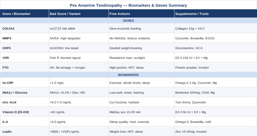
Pes anserine tendinopathy is rarely just a mechanical problem. Its consistent clinical association with diabetes, obesity, and systemic inflammation points toward metabolic and inflammatory drivers that standard orthopedic care does not investigate — and that is precisely where the precision approach described here adds value. The six biomarkers — hs-CRP, HbA1c, uric acid, vitamin D, IL-6, and leptin — give you a measurable picture of the environment your tendons are attempting to heal within. The five genetic variants — COL5A1, MMP3, GDF5, VDR, and FTO — provide a layer of explanation for why you may be more vulnerable than others with similar habits, and point toward specific interventions rather than generic ones.
The most practical next step is a blood draw. Work with your physician to order hs-CRP, HbA1c, uric acid, and 25-OH vitamin D at minimum — this is a basic, affordable panel that most insurers cover with appropriate indication. From there, the results tell you where to focus first. Genetic testing, where accessible, can further sharpen the plan. None of this replaces physical therapy or medical oversight; it informs both of them. The goal is not a perfect protocol — it is a more precise one, grounded in your actual biology rather than a general average.
Musculoskeletal: Joint Conditions Tendon & Ligament Conditions
Endocrine & Metabolic: Diabetes & Blood Sugar Metabolic Syndrome Obesity
Autoimmune: Inflammatory Conditions