This article contains AI generated content.
· UpdatedPes Cavus Genes Biomarkers — 5 Genes And 6 Biomarkers To Track
Introduction
If you have pes cavus — a foot with an unusually high arch — you have probably been told to get custom orthotics, stretch your calves, and strengthen your intrinsic foot muscles. That advice is not wrong, but for a significant proportion of people with high-arched feet, it leaves out the most important question: why does your arch look the way it does? Without that answer, management stays superficial — addressing the shape of the foot while ignoring what is actually driving it.
Pes cavus is not simply a biomechanical quirk that developed independently. In roughly half of all cases, there is an underlying neurological or neuromuscular cause, and in many of those cases, a genetic driver is at the root. A high arch caused by progressive muscle imbalance from a peripheral nerve gene variant responds very differently to intervention than one addressed purely through footwear. Knowing which situation you are in changes what you should actually do and what you should watch for over time.
Generic advice — stretch, support, strengthen — is reasonable when pes cavus is mild and truly idiopathic. But when a gene variant is affecting myelin integrity, axonal transport, or mitochondrial function in peripheral nerves, the interventions that slow progression, protect nerve function, and reduce long-term disability look quite different. The evidence base for genetic-driven pes cavus is growing, and it points toward specific, actionable steps — many of which remain outside the typical orthopedic consultation.
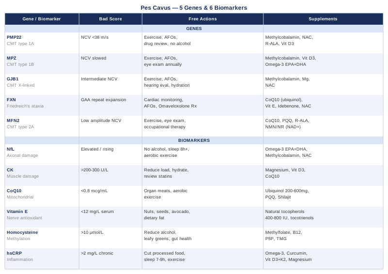
What follows is a look at five of the most clinically relevant genes associated with pes cavus and neuromuscular foot deformity, along with six biomarkers worth tracking to understand where you stand right now. Together, these two angles give you a more complete picture — and a more intelligent starting point for working with a specialist or refining your habits. Genetics tells you about predisposition and mechanism; biomarkers tell you what is actually happening in your body at this moment. Both matter.
What Recent Genetics Research Reveals About Pes Cavus
Pes cavus has a well-recognized genetic architecture, especially when it presents alongside progressive weakness, balance difficulty, or reduced sensation in the feet and legs. The most important genetic context is Charcot-Marie-Tooth disease (CMT), the most common inherited peripheral neuropathy, affecting approximately 1 in 2,500 people. Between 60 and 80 percent of CMT patients develop pes cavus over their lifetime, and in many cases, the foot deformity is the first visible sign of the condition. Beyond CMT, Friedreich's ataxia — a distinct genetic condition with a different mechanism — also causes pes cavus in a majority of those affected.
The genes below represent the most clinically significant and best-studied contributors to neurologically driven pes cavus. For each, the mechanism, the testing approach, and practical plans — with and without supplements — are laid out as concretely as the current evidence allows.
Gene 1: PMP22 — The Most Frequently Implicated Gene
What it is and what it affects: PMP22 encodes Peripheral Myelin Protein 22, a structural protein of the myelin sheath surrounding peripheral nerves. A duplication on chromosome 17p11.2 — not a point mutation — causes CMT type 1A (CMT1A), the most common genetic form of CMT. CMT1A accounts for approximately 50 to 70 percent of all CMT diagnoses and is the most common genetic cause of pes cavus.
When PMP22 is duplicated, Schwann cells overproduce an unstable myelin protein, producing dysfunctional myelination. The result is progressively slowed nerve conduction velocity, distal muscle weakness particularly affecting the feet and ankles, and sensory loss. As the peroneal muscles weaken before the posterior tibial muscle, the foot is pulled into the characteristic high-arched cavus shape. Motor nerve conduction velocities in CMT1A are typically below 38 m/s — a useful diagnostic clue before genetic confirmation.
How to test: MLPA (multiplex ligation-dependent probe amplification), chromosomal microarray, or a dedicated CMT gene panel. A panel is the most cost-efficient first step when CMT is clinically suspected, as it screens for PMP22 duplication alongside other common CMT genes simultaneously.
If the gene is abnormal, the plan without supplements
Exercise is the intervention with the strongest and most consistently demonstrated benefit in CMT1A. Historically, patients were cautioned to limit activity out of concern for overwork weakness — a concern that has not been well-supported by clinical data. A Cochrane review published in 2017 confirmed that moderate aerobic and resistance training can preserve functional capacity without accelerating nerve damage.
Practical protocol: 30 to 45 minutes of moderate-intensity aerobic exercise (cycling, swimming, or walking — low-impact modalities preferred to reduce mechanical load on weakened feet) three to five times per week. Resistance training two to three times per week, focusing on proximal muscle groups — hips, quadriceps, and core — since distal muscles are limited by denervation. Eight to twelve repetitions with progressive overload as tolerated.
Custom ankle-foot orthoses (AFOs) reduce fall risk, improve gait efficiency, and reduce secondary joint stress. Lightweight carbon fiber designs allow more dynamic function than traditional polypropylene models. Physical therapy with a clinician experienced in CMT should include gait retraining, proprioception training, and foot intrinsic muscle work within the limits of innervation.
Medication and exposure review is essential. Numerous drugs are known to worsen peripheral neuropathy in CMT, including vincristine, certain antibiotics (particularly metronidazole at high doses), amiodarone, and some chemotherapy agents. The Charcot-Marie-Tooth Association maintains an updated drug safety list that is worth reviewing with your neurologist. Alcohol must be minimized — it is independently neurotoxic and compounds myelin breakdown.
If the gene is abnormal, the plan with supplements
No supplement has demonstrated robust, statistically significant benefit in CMT1A human trials as of 2024. This is worth stating plainly so expectations are accurate. However, several have biological plausibility and are used thoughtfully in clinical practice:
Vitamin C (ascorbic acid): Early mouse-model studies showed PMP22 overexpression could be suppressed by ascorbic acid, generating significant interest. Two randomized controlled trials — Burns et al. (2009) in Lancet Neurology and Micallef et al. (2009) — found no significant functional benefit in adult CMT1A patients. Not recommended as a primary intervention. Cycling: not established.
Methylcobalamin (active B12): Supports myelin maintenance and axonal transport. Sublingual or injectable forms bypass potential gut absorption issues. Dose: 1,000 to 2,000 mcg/day. Frequency: daily. Side effects: rare; occasional acne-like skin reaction at very high doses.
Vitamin D3: Deficiency is associated with worse neuropathic symptom burden in peripheral neuropathy broadly. Target serum 25-OH-D between 50 and 80 ng/mL. Dose: 2,000 to 5,000 IU/day depending on baseline levels. Side effects: monitor calcium if using high doses long-term.
N-Acetyl Cysteine (NAC): A glutathione precursor that reduces oxidative stress in peripheral nerves. Mouse-model CMT data shows promise; human CMT trial data is limited. Dose: 600 to 1,800 mg/day. Frequency: daily. Cycling: consider 5 days on, 2 days off for long-term use. Side effects: nausea at higher doses, especially on an empty stomach.
R-Alpha Lipoic Acid (R-ALA): A mitochondrial antioxidant and cofactor with published evidence in diabetic peripheral neuropathy. Used empirically in some CMT practices. Dose: 300 to 600 mg/day, taken away from meals for better absorption. Side effects: hypoglycemia risk in diabetics; thiamine depletion with very high doses — consider supplementing B1 if using long-term.
Gene 2: MPZ — The Second Major Myelin Gene
What it is and what it affects: MPZ encodes Myelin Protein Zero (P0), the most abundant protein in compact peripheral myelin. Unlike PMP22, pathology arises from hundreds of different point mutations rather than a single duplication event. These mutations cause CMT type 1B (CMT1B), the second most common demyelinating CMT form.
P0 maintains the compaction of myelin lamellae through homophilic adhesion interactions. When MPZ is mutated, this compaction is disrupted, producing progressive demyelination and secondary axonal loss. The clinical severity varies dramatically depending on the specific mutation — some MPZ mutations cause severe early-onset disease in the first decade of life, while others produce a mild adult-onset picture that is often confused with idiopathic pes cavus for years before the underlying diagnosis is established.
Pes cavus is a consistent feature in moderate-to-severe MPZ phenotypes, often presenting in childhood or adolescence alongside progressive weakness and sensory changes.
How to test: A CMT gene panel with full MPZ sequencing, or whole exome sequencing if the panel is uninformative. Nerve conduction studies showing uniform velocity slowing in the demyelinating range help distinguish MPZ-related CMT from axonal forms.
If the gene is abnormal, the plan without supplements
Exercise and orthotic management are the primary tools, closely mirroring the CMT1A approach. However, given the wider phenotypic variability of MPZ mutations, monitoring needs to be individualized. Patients with early-onset or severe MPZ mutations may need AFO fitting and surgical consultation for foot deformity at a younger age than CMT1A patients.
Ophthalmologic evaluation is warranted in MPZ-related CMT, as some mutations produce Adie's tonic pupil and related pupillary abnormalities. Annual functional review with gait analysis every one to two years is reasonable to track progression.
A thorough medication review remains critical — the same drug safety principles that apply to CMT1A apply here.
If the gene is abnormal, the plan with supplements
The rationale mirrors CMT1A. Methylcobalamin at 1,000 to 2,000 mcg/day and Vitamin D3 optimized to 50 to 80 ng/mL form the baseline. Additionally, Omega-3 fatty acids (EPA + DHA) at 2 to 4 g/day combined are worth including — they support myelin membrane fluidity and reduce systemic neuroinflammation, with a favorable safety profile. Frequency: daily with meals. Side effects: GI upset; mild blood thinning at high doses — caution before surgery or with anticoagulant medications.
Gene 3: GJB1 — The X-Linked Connexin Gap
What it is and what it affects: GJB1 encodes Connexin 32 (Cx32), a gap junction protein expressed in Schwann cells and oligodendrocytes. Mutations in GJB1 cause CMT type X1 (CMTX1), the second most common form of CMT overall, accounting for approximately 10 to 15 percent of all CMT diagnoses.
Cx32 forms channels in the paranodal regions and Schmidt-Lanterman incisures of Schwann cells, facilitating rapid metabolic communication across myelin layers without having to travel all the way around the sheath. When Cx32 is dysfunctional, peripheral nerve repair and Schwann cell homeostasis are impaired. Because GJB1 is X-linked, males (hemizygous for the mutation) are typically more severely affected than females, who often carry the mutation with variable or mild symptoms.
Pes cavus in males with CMTX1 frequently appears in adolescence. Nerve conduction findings are intermediate — neither as severely slowed as CMT1A nor as fast as pure axonal CMT — which can make the diagnosis elusive.
How to test: GJB1 sequencing as part of a CMT gene panel. Women with a family history of CMT predominantly in males, particularly with X-linked inheritance patterns, should be screened as potential carriers.
If the gene is abnormal, the plan without supplements
Physical therapy and orthotic management follow the same principles as other CMT types. Hearing evaluation should be part of routine monitoring in CMTX1, as some GJB1 mutations affect auditory nerve function. Central nervous system white matter lesions have been reported in a subset of CMTX1 patients, particularly triggered by physiological stress such as fever, dehydration, or prolonged hypoxia — making aggressive hydration and prompt fever management more important than in other CMT types.
Hand function receives particular attention in CMTX1, as upper limb involvement is often more prominent and earlier than in CMT1A. Occupational therapy focused on grip strength and fine motor preservation is worth initiating early.
If the gene is abnormal, the plan with supplements
Methylcobalamin at 1,000 to 2,000 mcg/day remains foundational. Magnesium glycinate at 200 to 400 mg/day may provide supporting value — magnesium is involved in gap junction function at the cellular level and contributes to nerve signal stability. Side effects: loose stools at higher doses; take in the evening as it tends to promote relaxation. NAC at 600 to 1,800 mg/day rounds out the antioxidant support protocol.
Gene 4: FXN — The Mitochondrial Iron-Sulfur Driver
What it is and what it affects: FXN encodes frataxin, a mitochondrial matrix protein essential for iron-sulfur cluster assembly. A GAA trinucleotide repeat expansion within intron 1 of the FXN gene silences frataxin expression through heterochromatin formation, causing Friedreich's ataxia (FRDA), an autosomal recessive progressive neurological disease. Pes cavus is a hallmark feature, present in approximately 55 to 80 percent of patients.
Without adequate frataxin, iron accumulates in mitochondria and generates toxic reactive oxygen species via Fenton chemistry. This impairs mitochondrial respiratory chain function, leading to progressive neurodegeneration in the dorsal root ganglia, spinocerebellar tracts, and corticospinal tracts. The resulting motor and sensory imbalance in the foot produces the classic cavus deformity — and the degree of pes cavus often correlates with GAA repeat length, with longer expansions producing earlier and more severe disease.
Friedreich's ataxia typically presents in late childhood or adolescence. It is also associated in approximately 80 percent of patients with hypertrophic cardiomyopathy, making cardiac monitoring non-negotiable.
How to test: FXN trinucleotide repeat expansion analysis via PCR-based sizing, available through most neurological genetic panels. This single test confirms or excludes the diagnosis.
If the gene is abnormal, the plan without supplements
Cardiac monitoring is the highest priority. Annual echocardiography and 12-lead ECG are standard of care. Physical activity should be cleared by a cardiologist given the cardiomyopathy burden. Water-based exercise and stationary cycling are generally well-tolerated and beneficial.
AFOs and assistive devices are important for fall prevention, given both the foot deformity and the cerebellar ataxia component. Speech and swallowing therapy should be initiated early, as dysarthria and dysphagia progress in many FRDA patients.
Omaveloxolone (Skyclarys) is worth discussing with a neurologist — it is the first disease-modifying pharmacological agent approved by the FDA specifically for Friedreich's ataxia (approved February 2023, for patients aged 16 and older). It works by activating the Nrf2 transcription factor, which upregulates antioxidant defense pathways. Side effects as a prescription drug: elevated liver enzymes, fluid retention, nausea — requires monitoring.
If the gene is abnormal, the plan with supplements
Ubiquinol (reduced CoQ10): Directly supports mitochondrial complex I and II function — the electron transfer steps most impaired in frataxin deficiency. A human trial combining CoQ10 and vitamin E in FRDA patients showed modest improvements in cardiac and skeletal muscle bioenergetics. Dose: 300 to 600 mg/day of ubiquinol. Frequency: daily with a fat-containing meal, split into two doses above 300 mg. Cycling: none required. Side effects: very well tolerated; occasional mild GI discomfort.
Vitamin E (natural mixed tocopherols): Protective antioxidant for nerve cell membranes — used in combination with CoQ10 in the above-mentioned trials. Dose: 400 to 1,200 IU/day. Frequency: daily with fat. Side effects: avoid above 2,000 IU/day; increased bleeding time at very high doses.
Idebenone: A short-chain synthetic analogue of CoQ10 that crosses the blood-brain barrier more effectively than standard CoQ10. Used in several European countries for FRDA, with some evidence for cardiac benefit. Dose: 150 to 450 mg/day in divided doses. Frequency: daily. Side effects: elevated liver enzymes in a small proportion of users — monitor liver function every 3 to 6 months.
NAC: 600 to 1,800 mg/day to support glutathione production and reduce oxidative burden on mitochondria. Cycling: 5 days on, 2 off for long-term use.
A note on iron chelation: Despite the mitochondrial iron accumulation that characterizes FRDA, systemic iron chelation without specialist guidance is not recommended. Depleting systemic iron can worsen outcomes — this is a condition where cellular iron handling, not total body iron, is the problem.
Gene 5: MFN2 — The Axonal Mitochondria Fusion Gene
What it is and what it affects: MFN2 encodes Mitofusin 2, a GTPase protein located in the outer mitochondrial membrane that is essential for mitochondrial fusion. Mutations cause CMT type 2A (CMT2A), the most common axonal form of CMT. Unlike CMT1A, which damages the myelin sheath, CMT2A preferentially destroys the axon itself.
In peripheral axons — which can extend a meter or more from the neuronal cell body — mitochondrial dynamics are especially critical. The distal portions of these long axons depend entirely on anterograde transport of mitochondria from the cell body. MFN2 mutations impair mitochondrial fusion and transport, effectively starving the most distal axonal segments of energy. The feet are always hit first. Pes cavus develops as intrinsic foot muscles and peroneal muscles become denervated and weaken disproportionately.
CMT2A is often more severe than CMT1A, with earlier and more prominent upper limb involvement, and a subset of patients develop optic atrophy. Nerve conduction studies show near-normal or mildly reduced velocities with significantly reduced amplitudes — the axonal fingerprint — helping distinguish it from demyelinating CMT.
How to test: CMT gene panel including MFN2 sequencing, or whole exome sequencing if the panel is uninformative.
If the gene is abnormal, the plan without supplements
Paced, progressive exercise remains beneficial but needs more careful dosing given the fatigue that can accompany significant axonal disease. Regular ophthalmologic review is necessary given the risk of optic atrophy. Occupational therapy focused on upper limb function should begin before significant hand weakness appears.
If the gene is abnormal, the plan with supplements
The mitochondrial fusion deficit in MFN2 makes mitochondrial support supplements particularly relevant biologically:
Ubiquinol (CoQ10): 200 to 400 mg/day with a fat-containing meal. Frequency: daily. Side effects: minimal at standard doses.
PQQ (Pyrroloquinoline quinone): Stimulates mitochondrial biogenesis — increasing the number of mitochondria, which may partially compensate for impaired fusion. Dose: 10 to 20 mg/day. Frequency: daily. Side effects: generally well tolerated; occasional vivid dreams reported.
NMN or NR (NAD+ precursors): NAD+ is essential for mitochondrial sirtuin function and electron transport. Animal data for neuroprotection is promising; human neuromuscular data is emerging. Dose: NMN 250 to 500 mg/day or NR 300 to 600 mg/day. Frequency: daily, morning. Cycling: 5 days on, 2 days off. Side effects: generally well tolerated; NR may cause mild flushing.
R-Alpha Lipoic Acid: Mitochondrial cofactor and antioxidant. Dose: 300 to 600 mg/day on an empty stomach. Side effects: hypoglycemia risk in diabetics; thiamine depletion with high-dose long-term use — supplement B1 if using more than 600 mg/day chronically.
6 Biomarkers Worth Tracking
Understanding the genetic background tells you about mechanism and predisposition. But biomarkers tell you what is happening in your body right now — how much active nerve damage is occurring, whether inflammation is elevated, whether key nutrients are sufficient. The six markers below are practical, measurable, and directly relevant to anyone navigating pes cavus with a neurological or neuromuscular component. They range from standard panels that cost under $50 to specialized tests that require a referral to a neurological center.
Biomarker 1: Serum Neurofilament Light Chain (NfL)
Why it matters: Neurofilament Light Chain is a cytoskeletal protein of axons that leaks into the blood when neurons are damaged or under active stress. In CMT disease, plasma NfL is elevated relative to healthy controls and correlates with disease severity, functional impairment, and rate of progression. NfL is increasingly used as a surrogate endpoint in CMT clinical trials — which means it is among the most disease-relevant blood biomarkers currently available. A rising NfL over serial measurements signals active axonal damage; stable or declining NfL suggests stabilization or response to intervention.
How to measure it: Blood draw, analyzed using Simoa (Single Molecule Array) technology. Not yet universally available through routine labs — typically requires a neurological center or academic medical setting. Cost: approximately $150 to $350 per test. Meaningful use involves serial measurement rather than a single data point.
If the score is high, the plan without supplements
Identify and remove nerve-damaging exposures: eliminate alcohol, review all medications for peripheral nerve toxicity, optimize sleep to eight or more hours (nerve repair occurs predominantly during slow-wave sleep), and reduce chronic psychological stress, which is associated with elevated pro-inflammatory cytokines and worse neuropathic outcomes. Moderate aerobic exercise, as tolerated, has documented anti-neuroinflammatory effects.
If the score is high, the plan with supplements
Omega-3 fatty acids (EPA + DHA): Anti-inflammatory and demonstrated neuroprotective in multiple published studies. Dose: 2 to 4 g/day combined EPA + DHA. Frequency: daily with meals. Side effects: GI discomfort; mild blood thinning at higher doses. Methylcobalamin at 1,000 to 2,000 mcg/day is essential if B12 is low-normal or deficient, as B12 deficiency itself raises NfL through direct axonal injury. NAC at 600 to 1,800 mg/day supports glutathione and reduces the oxidative component of axonal damage.
Biomarker 2: Creatine Kinase (CK)
Why it matters: CK is an enzyme released from damaged muscle fibers. Moderately elevated CK — above 200 to 300 U/L in the absence of recent intense exercise — can signal ongoing muscle degeneration relevant to CMT and other neuromuscular conditions where chronic denervation causes secondary muscle damage. Very high CK above 1,000 U/L suggests more significant primary muscle disease and warrants further evaluation. CK is also a practical monitoring tool when starting or intensifying exercise, particularly important in CMT patients where overwork weakness is a theoretical concern.
How to measure it: Standard blood test, included in many metabolic panels or available as a standalone. Cost: $20 to $50. Widely available. Measure fasting and avoiding intense exercise in the 48 hours prior to blood draw for the most interpretable result.
If the score is high, the plan without supplements
Reduce exercise intensity and volume, rebuild gradually. Ensure adequate hydration — dehydration significantly worsens muscle enzyme release. Review all current medications: statins are the most common cause of drug-induced CK elevation and often go unrecognized as the culprit. Discuss with the prescribing physician whether switching to a different statin or dose is appropriate.
If the score is high, the plan with supplements
Magnesium glycinate: Supports muscle relaxation and mitochondrial energy metabolism. Deficiency is common and correlates with elevated CK in some studies. Dose: 200 to 400 mg/day. Frequency: daily, evening. Side effects: diarrhea at high doses — glycinate form is best tolerated. Vitamin D3: Deficiency is associated with muscle weakness and elevated CK. Optimize to 50 to 80 ng/mL serum 25-OH-D. Ubiquinol: 100 to 300 mg/day — particularly relevant if CK elevation is statin-related, as statins reduce endogenous CoQ10 synthesis by inhibiting the mevalonate pathway.
Biomarker 3: Plasma Coenzyme Q10 (CoQ10)
Why it matters: CoQ10 is a fat-soluble compound central to mitochondrial energy production and a direct antioxidant protecting mitochondrial membranes from lipid peroxidation. For pes cavus patients with FXN mutations (Friedreich's ataxia) or MFN2 mutations (CMT2A), mitochondrial dysfunction is a direct part of the disease mechanism — and plasma CoQ10 is a meaningful functional readout of mitochondrial status. Beyond genetic conditions, CoQ10 levels decline with age and are significantly reduced by statin medications.
How to measure it: Fasting plasma CoQ10, not included in standard panels — requires a specific request through a functional medicine or integrative medicine lab. Cost: approximately $100 to $200. Most published thresholds cite plasma CoQ10 above 0.8 to 1.0 mcg/mL as sufficient for baseline function; therapeutic supplementation trials typically target levels above 2.5 mcg/mL.
If the score is low, the plan without supplements
CoQ10 levels can be modestly improved through diet before resorting to supplementation. The richest dietary sources include organ meats (heart, kidney, liver), sardines, mackerel, beef, and sesame seeds. Ensuring adequate dietary fat intake is necessary since CoQ10 is fat-soluble. Reducing mitochondrial stressors — refined sugars, chronic alcohol, processed seed oils — also matters. Regular aerobic exercise stimulates mitochondrial biogenesis and is one of the most effective natural CoQ10-sparing interventions.
If the score is low, the plan with supplements
Ubiquinol (reduced CoQ10): Significantly better absorbed than ubiquinone, particularly in individuals over 40. Dose: 200 to 600 mg/day with a fat-containing meal. Frequency: daily, split into two doses if above 300 mg. Cycling: none required. Side effects: very well tolerated; mild GI discomfort at higher doses in some individuals. PQQ: 10 to 20 mg/day complements CoQ10 by stimulating new mitochondria synthesis, not just supporting existing ones. Shilajit: A mineral-rich compound that appears to increase CoQ10 bioavailability in some studies. Dose: 200 to 400 mg/day of standardized extract. Cycling: 8 weeks on, 2 weeks off. Quality control matters — purchase from a brand with third-party testing.
Biomarker 4: Serum Vitamin E (Alpha-Tocopherol)
Why it matters: Vitamin E deficiency produces a peripheral neuropathy and ataxia syndrome that clinically resembles Friedreich's ataxia — including, in some cases, pes cavus — and can be reversed with aggressive supplementation when caught early. Even below the threshold of frank deficiency, suboptimal vitamin E levels may worsen neuropathic outcomes by reducing the antioxidant protection available to peripheral nerve membranes. This is especially relevant in conditions where oxidative stress is an active disease mechanism.
How to measure it: Serum alpha-tocopherol, available as part of micronutrient panels or standalone. Cost: $40 to $80. Optimal range: approximately 12 to 20 mg/L, though lab reference ranges vary. Always interpret in context of total cholesterol, since vitamin E is lipid-transported.
If the score is low, the plan without supplements
Increase dietary vitamin E through sunflower seeds, almonds, hazelnuts, wheat germ oil, and avocado. Ensure dietary fat intake is adequate — very low-fat diets impair tocopherol absorption. If fat malabsorption is suspected (celiac disease, inflammatory bowel disease, exocrine pancreatic insufficiency), this should be investigated and treated first, as supplementation will not compensate for malabsorption.
If the score is low, the plan with supplements
Natural mixed tocopherols: More physiologically balanced than synthetic dl-alpha-tocopherol. Dose: 400 to 800 IU/day. Frequency: daily with a fat-containing meal. Cycling: none required. Side effects: potential pro-oxidant effects at very high doses above 2,000 IU/day; increased bleeding time — avoid high doses before surgery or with anticoagulant medications. Tocotrienols (gamma/delta forms): An emerging form of vitamin E with unique neuroprotective and anti-inflammatory properties distinct from alpha-tocopherol. Dose: 100 to 300 mg/day of tocotrienol complex. Side effects: generally well-tolerated at these doses.
Biomarker 5: Plasma Homocysteine
Why it matters: Homocysteine is an amino acid intermediate in methionine metabolism. Elevated plasma levels — clinically defined as above 15 µmol/L, though neurological risk increases meaningfully above 10 µmol/L — are directly toxic to blood vessels and peripheral nerves. Homocysteine promotes oxidative stress, impairs myelin synthesis, and accelerates axonal degeneration. It is one of the most underappreciated contributors to peripheral neuropathy burden, and in a patient already dealing with pes cavus from a genetic driver, elevated homocysteine represents an additional, treatable stressor on the nervous system.
The MTHFR C677T variant — carried by approximately 40 to 60 percent of the general population — reduces the enzyme's ability to convert homocysteine to methionine, causing accumulation. Gary Brecka has brought significant popular attention to MTHFR and methylation genetics, and while some of his claims extend beyond the evidence, the fundamental point about testing homocysteine and correcting it with active B vitamins is well-supported by published data.
How to measure it: Fasting plasma homocysteine, widely available and inexpensive. Cost: $25 to $60. Optimal level: below 10 µmol/L; ideally below 7 µmol/L for neurological health. Supplementing MTHFR genotyping alongside homocysteine measurement provides actionable direction.
If the score is high, the plan without supplements
Reduce alcohol (a potent B-vitamin depleter and homocysteine elevator), increase dietary leafy greens, legumes, and eggs, and address gut health — dysbiosis and atrophic gastritis impair B12 and folate absorption and raise homocysteine through nutritional deficiency rather than genetics.
If the score is high, the plan with supplements
Methylfolate (5-MTHF): The pre-converted active form, bypassing MTHFR enzymatic conversion. Dose: 400 to 1,000 mcg/day. Start at the low end — some individuals experience anxiety or irritability from initiating methylfolate too rapidly. Frequency: daily. Side effects: overmethylation symptoms (anxiety, insomnia, irritability) in sensitive individuals — reduce dose and rebuild slowly.
Methylcobalamin (B12): Works synergistically with methylfolate to remethylate homocysteine. Dose: 1,000 to 2,000 mcg/day sublingual. Frequency: daily.
Pyridoxal-5-Phosphate (P5P, active B6): Third component of the remethylation and transsulfuration pathways. Dose: 25 to 50 mg/day. Side effects: sensory neuropathy risk at high doses above 200 mg/day — do not exceed this threshold.
Trimethylglycine (TMG/Betaine): An alternative methyl donor that remethylates homocysteine via the BHMT pathway, independent of folate cycling. Dose: 500 to 1,500 mg/day. Frequency: daily. Side effects: fishy body odor in some; GI discomfort at high doses.
Biomarker 6: High-Sensitivity C-Reactive Protein (hsCRP)
Why it matters: CRP is a liver-produced acute-phase protein that rises with systemic inflammation. The high-sensitivity version (hsCRP) detects the low-level, chronic inflammatory state that drives neurodegeneration and impairs peripheral nerve repair — below the detection threshold of standard CRP tests. While hsCRP is not specific to pes cavus or CMT, chronically elevated levels above 2 to 3 mg/L are associated with worse neuropathic outcomes, impaired axonal regeneration, and accelerated functional decline in neuromuscular conditions. It is also one of the most accessible and actionable markers available.
How to measure it: Standard blood test, widely available at any lab. Cost: $15 to $40. Optimal level: below 1.0 mg/L; values above 3.0 mg/L indicate significantly elevated inflammatory burden. Ideally measure twice, three weeks apart, to account for transient elevations from illness or recent intense exercise.
If the score is high, the plan without supplements
Eliminate or significantly reduce refined carbohydrates and ultra-processed foods — among the most potent dietary drivers of CRP elevation. Prioritize seven to nine hours of quality sleep: sleep restriction is one of the most powerful and consistent triggers of elevated inflammatory markers. More than 150 minutes per week of moderate aerobic exercise has robust evidence for reducing hsCRP. Time-restricted eating (12 to 16 hour daily fast) reduces hsCRP in metabolic syndrome trials, though direct neuromuscular evidence is limited.
If the score is high, the plan with supplements
Omega-3 fatty acids (EPA + DHA): Among the best-studied supplements for hsCRP reduction. Dose: 2 to 4 g/day combined EPA + DHA. Frequency: daily. Side effects: GI upset; blood thinning at high doses — caution with anticoagulants. Bioavailable curcumin (phospholipid complex or piperine-enhanced): Reduces CRP in multiple RCTs, including in inflammatory conditions. Dose: 500 to 1,500 mg/day of a bioavailable formulation. Cycling: 8 weeks on, 2 to 4 weeks off. Side effects: potentiates anticoagulants; mild GI effects. Vitamin D3 + K2: Deficiency is consistently associated with elevated hsCRP. Optimize serum 25-OH-D. Magnesium glycinate: 300 to 400 mg/day — deficiency correlates with elevated CRP in population data. Frequency: daily, evening.
The Exercise-Neuroprotection Connection: What the Research Doesn't Say Loudly Enough
For years, many physicians counseled patients with CMT and neuromuscular conditions to limit physical activity out of concern that exercise might damage already vulnerable nerves. That thinking has been substantially revised — and the revision matters enough to walk through in some depth.
Exercise is not the enemy — inactivity is
Andrew Huberman has consistently synthesized the neuroscience literature on how exercise affects the nervous system, and one theme recurs across many of his episodes: exercise is one of the most potent stimulators of neurotrophic factors — particularly BDNF (Brain-Derived Neurotrophic Factor) and NT-3 (Neurotrophin-3) — that support neuronal health, axonal maintenance, and peripheral nerve repair. NT-3 in particular has been specifically investigated in CMT mouse models as a potential therapeutic agent, precisely because it promotes survival of the neurons most vulnerable in CMT disease.
What exercise actually does to peripheral nerves
Moderate aerobic exercise increases peripheral nerve blood flow, reduces systemic neuroinflammation, improves mitochondrial biogenesis in muscle cells (reducing the metabolic burden on partially denervated tissue), and upregulates antioxidant defense enzymes. In the context of the genetic drivers described above — particularly the mitochondrial dysfunction in FXN and MFN2 mutations — the mitochondrial biogenesis effect from regular aerobic exercise is particularly relevant.
The resistance training nuance
Resistance training in neuromuscular disease requires calibration. The concern about overwork weakness — where a partially denervated muscle is driven past its capacity and loses further function — is real but has been overstated. The current evidence suggests that moderate-intensity resistance training (not maximal-effort training) performed consistently is safe and beneficial. Muscles below approximately 10 percent of normal motor unit function may not respond to training; muscles retaining more function than this can be preserved and even strengthened.
Ten things worth knowing that most clinicians don't emphasize
1. Exercise timing matters. Performing aerobic exercise in the morning or early afternoon preserves circadian rhythm integrity, which is directly linked to neuronal repair cycles.
2. Zone 2 cardio is the sweet spot. Training at 60 to 70 percent of maximum heart rate — where you can hold a conversation but feel the effort — maximizes mitochondrial adaptation without generating the oxidative stress associated with high-intensity efforts.
3. Cold exposure has emerging nerve health data. Brief cold water immersion (1 to 3 minutes at 10 to 15°C, two to three times per week) increases norepinephrine significantly and may promote nerve growth factor expression. This is mechanistically interesting for peripheral neuropathy but human neuromuscular data is preliminary.
4. Sleep is when the nervous system repairs. Glymphatic clearance of metabolic waste from neurons occurs predominantly during slow-wave sleep. Consistently sleeping fewer than seven hours is associated with elevated NfL in general population studies.
5. Protein intake matters more than most realize. Adequate protein (1.6 to 2.2 g/kg body weight/day) is necessary to maintain muscle mass in conditions where denervation is occurring. This is not a trivial amount — it requires deliberate dietary planning.
6. B vitamins are metabolically interconnected. Correcting one B vitamin deficiency without addressing the others is often insufficient. B1, B2, B6 (as P5P), B12 (as methylcobalamin), and methylfolate function as an integrated system for neurological health.
7. Alcohol has zero safe threshold for peripheral neuropathy. Even modest, consistent alcohol intake adds to axonal damage burden in patients who already have a genetic driver of neuropathy. This is not a lifestyle preference issue — it is a direct exacerbant.
8. Genetic testing should precede surgical decisions. Plantar fascia release and other foot surgeries for pes cavus have different outcomes depending on whether the underlying cause is neuromuscular or idiopathic. A genetic diagnosis changes the risk-benefit calculation significantly.
9. Mitochondrial health is not one intervention. It requires simultaneous optimization: CoQ10, adequate dietary protein, zone 2 exercise, sleep quality, and reduction of oxidative stressors. No single supplement achieves this in isolation.
10. Biomarker tracking creates feedback loops. Testing NfL, homocysteine, and CoQ10 every six to twelve months while making lifestyle and supplementation changes gives you actual data on whether interventions are working — something that symptom tracking alone cannot provide.
Complementary Approaches With Meaningful Evidence
The following approaches do not replace medical management or the genetic and biomarker strategies above, but each has human clinical evidence relevant to pes cavus-associated neuropathy and functional decline. Four were selected based on the quality and specificity of that evidence.
Low-Level Laser Therapy / Photobiomodulation
Low-level laser therapy (LLLT), also called photobiomodulation, delivers non-thermal photonic energy to tissue, primarily absorbed by cytochrome c oxidase in the mitochondrial respiratory chain. This stimulates ATP production, reduces oxidative stress, and appears to promote axonal sprouting and Schwann cell proliferation in peripheral nerve injury models.
For peripheral neuropathy — the most common underlying mechanism of neurological pes cavus — a 2017 meta-analysis published in Lasers in Medical Science found significant improvements in pain, sensory function, and nerve conduction parameters in diabetic peripheral neuropathy patients treated with LLLT. While this evidence base is in diabetic neuropathy rather than CMT specifically, the mechanism of action is relevant across neuropathy types. A protocol of 810 to 904 nm wavelength, 10 to 30 mW/cm² irradiance, applied to the dorsal foot and lower leg, two to three sessions per week for eight to twelve weeks, represents the most commonly studied approach. Evidence for CMT specifically is currently limited to case series and mechanistic studies — acknowledge expectations accordingly.
For practical application, LLLT devices cleared for home use (at lower power than clinical devices) are available, but clinical photobiomodulation with calibrated medical devices delivers more reliable dosing. Initiate with professional sessions to establish a protocol before transitioning to a home device if appropriate.
Yoga
Yoga is one of the most accessible and well-studied movement modalities for neurological conditions affecting balance, proprioception, and lower extremity function — all central concerns in pes cavus with neuropathic features. The combination of static and dynamic postures trains proprioceptive pathways, activates deep stabilizing muscles of the foot and ankle, and builds the proximal hip and core strength that compensates for distal weakness.
A randomized controlled trial published in 2015 in Topics in Stroke Rehabilitation demonstrated significant improvements in balance, gait velocity, and fall frequency in neurological patients following an eight-week yoga program. While the study focused on stroke, the balance and proprioception mechanisms are directly transferable to patients with peripheral neuropathy and pes cavus.
For pes cavus specifically, yin yoga and restorative yoga styles — emphasizing long-hold stretches of the plantar fascia, calf complex, and peroneals — are well tolerated. The key is starting with a teacher experienced in modified poses for patients with foot deformity or weakness, and avoiding inversions or balancing poses that exceed current stability capacity. Fifteen to thirty minutes three to five times per week is a practical starting point, building toward longer sessions over eight to twelve weeks.
Biofeedback
Biofeedback uses real-time physiological data — muscle activation signals (sEMG), center of pressure during standing, or gait parameters — to help patients consciously adjust motor patterns they cannot otherwise perceive accurately. In pes cavus with neuromuscular involvement, where proprioceptive feedback from the foot is often reduced, biofeedback provides an external signal that partially compensates for the loss of internal sensing.
A 2016 study in NeuroRehabilitation demonstrated that sEMG biofeedback training improved gait symmetry and lower limb motor control in patients with hereditary spastic paraplegia — a condition sharing mechanisms of distal lower motor neuron dysfunction with CMT. For gait retraining in CMT and pes cavus, biofeedback has logical and growing clinical relevance. Force plate biofeedback for balance is particularly appropriate.
Apply biofeedback within a physical therapy setting rather than as a standalone intervention. Six to twelve weeks of twice-weekly sessions, each lasting 30 to 45 minutes, is a reasonable trial period. The goal is establishing new motor engrams that persist after the feedback is removed. Home biofeedback devices exist but should be introduced after clinical training to ensure correct use.
Massage Therapy
Massage therapy for neurologically driven pes cavus addresses two separate but meaningful issues: the mechanical consequences of abnormal foot loading (plantar fascia tightness, intrinsic muscle tension, calf rigidity), and the circulatory component of peripheral neuropathy (reduced blood flow to the distal extremities, which worsens nerve ischemia in conditions like CMT).
A 2018 systematic review in Journal of Clinical Nursing found that foot and lower leg massage significantly improved neuropathic pain intensity and sensory function in diabetic peripheral neuropathy patients compared to controls. The mechanisms — improved local blood flow, reduced myofascial tension, and gate-control modulation of pain signaling — are applicable to CMT and similar conditions.
For pes cavus specifically, deep tissue work on the plantar fascia and calf complex (gastrocnemius, soleus, tibialis posterior), combined with gentle friction massage over the distal lower leg, is the most targeted protocol. Sessions of 30 to 45 minutes once or twice weekly, focusing on the lower extremities, are appropriate. Communicate to the therapist that sensation may be reduced — pressure should be calibrated through verbal feedback rather than pain response, since sensory impairment can allow excessive pressure to go unrecognized.
Conclusion
Pes cavus is often treated as a structural problem. But when it is driven by genetics — by impaired myelin proteins, dysfunctional mitochondria, or disrupted gap junction signaling — the most useful frame is not biomechanical but biological. The five genes described above account for the majority of identifiable genetic pes cavus, and the six biomarkers provide a practical, measurable read on what is happening right now in your nervous system and at the cellular level.
The clearest next steps are not complicated: if you have not had a CMT or neuromuscular genetic panel and your pes cavus is bilateral, progressive, or accompanied by any sensory or motor changes, that test is worth pursuing with a neurologist. If you already have a diagnosis, tracking NfL, homocysteine, CoQ10, and hsCRP every six to twelve months alongside your clinical monitoring creates a feedback system for adjusting interventions over time. Neither genetics nor biomarkers replace a specialist — but they make the specialist visit considerably more productive.
Better information does not guarantee better outcomes, but it reliably leads to better decisions.
Musculoskeletal: Muscle Conditions
Neurological: Nerve Conditions Movement Disorders
Cardiovascular: Heart Conditions