This article contains AI generated content.
· UpdatedPes Planus Genes Biomarkers - 6 Genes And 6 Biomarkers To Track
When Flat Feet Feel Like a Dead End
If you have been told you have flat feet, you have probably heard the same advice: get arch supports, wear motion-control shoes, stretch your calves. For some people, this is enough. For many others, the arch continues to collapse, the foot aches, and the rest of the body — knees, hips, lower back — eventually joins in. The standard guidance was never wrong, exactly, but it was incomplete.
The problem is that pes planus is not one condition. It is a structural outcome that can arise from ligament laxity, intrinsic muscle weakness, impaired bone density, chronic inflammation, or a combination of all four. When the underlying driver is not identified, the intervention targets the wrong layer. An arch support placed inside a shoe does not rebuild a weakened tibialis posterior. It does not repair a compromised spring ligament. It compensates — and compensation is not correction.
What has changed in recent years is the quality of information available. Researchers now have a clearer picture of which genetic variants predispose certain people to soft tissue fragility, arch collapse, and impaired tissue repair. They also have a set of measurable biomarkers that reflect the biological environment the foot is operating in — inflammatory load, vitamin D status, collagen turnover, metabolic health. These are not abstract data points. They are levers.
This article covers both. The first section focuses on six biomarkers you can track with a standard blood draw, why each one matters specifically for the foot's structural health, and what to do when a score is off. The second section reviews six genes that researchers have linked to hypermobility, ligament quality, and musculoskeletal vulnerability — along with practical plans for managing each variant. Beyond that, you will find a summary of one of the most evidence-grounded challenges to conventional flat foot thinking, and a look at complementary modalities with real clinical support. Better data leads to better decisions. That is the only promise here.
6 Biomarkers That Reveal the Biology Behind Your Flat Feet
Blood tests do not diagnose structural foot problems. What they do is expose the biological terrain: how much inflammation is running through the system, whether the raw materials for tissue repair are available, how well the body is processing the nutrients that keep bone, ligament, and muscle functional. For pes planus, six biomarkers stand out as particularly actionable.
1. 25-OH Vitamin D
Why it matters: Vitamin D is not a vitamin in the conventional sense — it is a steroid hormone precursor that regulates hundreds of genes, including those governing calcium absorption, bone mineralization, and muscle fiber recruitment. For flat feet, the two most relevant pathways are bone density (a low arch can accelerate bone remodeling stress in the calcaneus and midfoot) and intrinsic foot muscle function. A 2014 systematic review in the Journal of the American Podiatric Medical Association noted that vitamin D deficiency was significantly associated with musculoskeletal pain and muscle weakness in lower extremities. Studies in children have shown a correlation between vitamin D deficiency and symptomatic pes planus. Clinicians like Peter Attia consistently flag optimal 25-OH vitamin D as a foundational marker before investigating more complex musculoskeletal issues.
How to measure it: A simple serum 25-hydroxyvitamin D test. Available at LabCorp, Quest, or most general practitioners. Direct-pay cost: $30–$60. Optimal target: 40–60 ng/mL. Deficiency is often defined as below 20 ng/mL, but clinical performance tends to suffer below 30 ng/mL. Retest every 90 days when supplementing or adjusting sun exposure.
If the score is bad, the plan without supplements: Mid-day sun exposure with arms and legs uncovered for 20–30 minutes is the most physiologically complete way to raise vitamin D because it simultaneously triggers nitric oxide release and supports circadian rhythm. Aim for at least 4–5 sessions per week. Walking barefoot on natural surfaces during this time adds proprioceptive and grounding benefits. Darker skin tones require roughly 3–5 times longer exposure for equivalent synthesis. Geographic and seasonal limitations will constrain this approach in winter months above 35° latitude.
If the score is bad, the plan with supplements or equipment: Vitamin D3 (cholecalciferol) 2,000–5,000 IU daily with a meal containing fat. Always pair with vitamin K2 (MK-7 form, 100–200 mcg/day) to direct calcium toward bone rather than soft tissue. Magnesium glycinate 200–400 mg at night supports the enzymatic conversion of vitamin D to its active form — many people with low vitamin D are also low in magnesium, which explains why supplementation alone sometimes fails to raise levels. Retest at 90 days. Adjust dose to maintain 40–60 ng/mL. No cycling needed; this is a daily maintenance protocol. Side effects at these doses are rare; toxicity is associated with doses above 10,000 IU/day taken long-term without monitoring.
2. hsCRP (High-Sensitivity C-Reactive Protein)
Why it matters: C-reactive protein is the liver's response to systemic inflammatory signaling. Elevated hsCRP does not point to one specific tissue, but it reflects an environment that degrades collagen faster than it is rebuilt, impairs tendon and ligament healing, and increases pain sensitization. For pes planus, the spring ligament and plantar fascia are the two structures most directly loaded during weight-bearing. Chronic low-grade inflammation quietly degrades the extracellular matrix of both. Allan Sniderman's work and Peter Attia's clinical protocols both treat hsCRP as a core metabolic health indicator — not because it explains cardiovascular risk alone, but because it reflects how much systemic housekeeping is being neglected.
How to measure it: Serum hsCRP, available in most standard metabolic panels or as a standalone test. Cost: $15–$40. Optimal target: below 1.0 mg/L. Peter Attia's clinical preference is below 0.8 mg/L. Mild elevation (1–3 mg/L) warrants lifestyle investigation. Levels above 10 mg/L typically indicate acute infection and should be retested after resolution.
If the score is bad, the plan without supplements: Sleep is the most underestimated anti-inflammatory intervention. Even partial sleep deprivation (six hours vs. eight over several nights) measurably elevates hsCRP. Prioritize 7–9 hours of consistent sleep. Eliminate refined seed oils (soybean, canola, sunflower) from the diet and increase intake of fatty fish, olive oil, and vegetables. Reduce or eliminate alcohol. Walking 7,000–10,000 steps daily has been shown in multiple studies to reduce hsCRP independent of weight loss.
If the score is bad, the plan with supplements or equipment: Omega-3 fatty acids (EPA+DHA) at 2–4 g/day from triglyceride-form fish oil consistently reduce hsCRP in clinical trials. Curcumin with piperine (500–1000 mg curcumin, 10 mg piperine, twice daily with meals) is one of the more robustly studied botanical anti-inflammatories; cycle 8 weeks on, 2 weeks off to prevent tolerance. Magnesium deficiency independently elevates hsCRP, so correcting low RBC magnesium often lowers hsCRP as a secondary benefit. Retest at 90 days.
3. RBC Magnesium
Why it matters: Serum magnesium is a poor indicator of body magnesium status — less than 1% of total body magnesium lives in the blood, and serum levels are tightly defended even when intracellular stores are depleted. RBC (red blood cell) magnesium provides a far more meaningful picture. For pes planus, magnesium plays three critical roles: it is required for the enzymatic activation of vitamin D, it regulates muscle tone and relaxation (including calf and foot intrinsic muscles), and it modulates inflammatory signaling. Chronic magnesium insufficiency contributes to muscle cramping, excess muscle tension, impaired collagen synthesis, and elevated inflammation — all of which worsen the functional picture in flat feet.
How to measure it: RBC magnesium (also called erythrocyte magnesium) from a standard blood draw. Not the same as serum magnesium — request specifically. Cost: $40–$80 at specialty labs; LabCorp carries it. Optimal range: 5.2–6.8 mg/dL. Many functional medicine practitioners flag anything below 5.5 mg/dL as suboptimal.
If the score is bad, the plan without supplements: Dietary sources include pumpkin seeds, dark leafy greens (especially Swiss chard and spinach), almonds, black beans, and dark chocolate above 70% cacao. The challenge is that modern soil depletion has reduced the magnesium content of most vegetables by 20–30% over the past 50 years. Reducing alcohol, caffeine (which increases urinary magnesium loss), and processed food improves retention. Transdermal magnesium flakes in a foot bath (20 minutes, three times per week) is low-risk, accessible, and anecdotally reported to help with foot muscle cramps.
If the score is bad, the plan with supplements or equipment: Magnesium glycinate 200–400 mg at night is the most bioavailable and best-tolerated form for most people. Magnesium threonate is preferable if neurological symptoms (poor sleep, anxiety) accompany the deficiency. Avoid magnesium oxide — it has poor absorption and is mainly a laxative. Take with the evening meal. Most people see RBC levels rise within 60–90 days. If digestive sensitivity occurs, lower the dose and increase gradually. No cycling required for maintenance doses; if using higher doses (400+ mg), a brief break every 2–3 months is reasonable.
4. CTX-1 and P1NP (Collagen and Bone Turnover Markers)
Why it matters: CTX-1 (C-terminal telopeptide of type I collagen) measures how fast collagen is being broken down. P1NP (procollagen type I N-terminal propeptide) measures how much new collagen is being formed. Together, they reflect the quality of the body's repair cycle — for bone, yes, but also for tendon and ligament tissue, which are overwhelmingly composed of type I collagen. In pes planus, the spring ligament, plantar fascia, and deltoid ligament are under chronic mechanical load. If CTX-1 is high and P1NP is low, collagen breakdown is outpacing formation — a recipe for progressive tissue deterioration regardless of what orthotics are worn. Thomas Dayspring has highlighted CTX-1 as an underused metabolic marker, and endocrinologists working with bone health routinely track both.
How to measure it: Serum CTX-1 (draw in the morning, fasted — it fluctuates with food intake). Serum P1NP. These tests are more common in osteoporosis management and not always available through primary care without a specific request. Cost: $60–$120 per marker. Reference ranges vary by lab, age, and sex; a useful functional target is CTX-1 below 500 pg/mL and P1NP above 40 mcg/L in adults under 60. Retest every 3–6 months when actively intervening.
If the score is bad, the plan without supplements: Resistance training is the most evidence-backed stimulus for increasing P1NP (bone and collagen formation). Progressive loading of the foot — calf raises, single-leg deadlifts, toe-spread exercises — directly stimulates the plantar collagen remodeling response. Adequate dietary protein (1.6–2.2 g/kg body weight) is non-negotiable for collagen synthesis. Adequate sleep (collagen synthesis peaks during deep sleep) and stress reduction (cortisol suppresses bone formation) complete the non-supplement side of the protocol.
[BOLD]If the score is bad, the plan with supplements or equipment:[/TITLE] Collagen peptides (10–15 g/day, taken with vitamin C) taken 30–60 minutes before foot strengthening exercise appear to preferentially load the collagen synthesis response in tendons and ligaments — based on work by Keith Baar at UC Davis. Vitamin D3 + K2 (K2 is necessary for osteocalcin activation, which governs where calcium is deposited) supports P1NP. Vitamin C 500–1000 mg taken alongside collagen peptides acts as a cofactor for collagen cross-linking via hydroxylation. Cycle collagen peptides in 12-week blocks; vitamin C and D3/K2 are appropriate as daily maintenance.
5. Omega-3 Index
Why it matters: The Omega-3 Index measures the percentage of EPA and DHA in red blood cell membranes — a far more reliable indicator of long-term omega-3 status than a serum measurement. The index reflects the degree to which cellular membranes are prepared to resolve inflammation rather than amplify it. For pes planus, this matters at the level of tendon and ligament biology: EPA and DHA are precursors to specialized pro-resolving mediators (SPMs) that actively terminate the inflammatory cascade and initiate tissue repair. Chronically low omega-3 status is associated with slower tendon healing, elevated hsCRP, and greater sensitivity to mechanical pain. Peter Attia consistently ranks the omega-3 index among the most important and most commonly suboptimal markers in adults.
How to measure it: Dried blood spot test through OmegaQuant (direct-to-consumer, $50–$100) or LabCorp. Optimal target: above 8%. The majority of Western adults test between 4–6%. Retest every 4–6 months when supplementing, since membrane turnover is slow.
If the score is bad, the plan without supplements: Fatty fish three times per week — particularly sardines, salmon, mackerel, and herring — is the most effective dietary strategy. Canned sardines in water or olive oil are among the most cost-effective omega-3 sources available. Walnuts and flaxseed provide ALA, which partially converts to EPA/DHA, but conversion rates are low (typically below 10%) and unlikely to move the index meaningfully on their own. Reducing linoleic acid intake (seed oils) reduces competition for the same metabolic enzymes.
If the score is bad, the plan with supplements or equipment: Triglyceride-form fish oil at 2–4 g EPA+DHA per day with a fat-containing meal (fat improves absorption by 3–4x). Brands like Carlson, Thorne, or Nordic Naturals use the triglyceride form. Ethyl ester forms (common in cheaper products) have significantly lower bioavailability. For vegetarians or those with fish sensitivities, algae-based DHA+EPA is functionally equivalent. Take daily; no cycling needed. Most common side effect at higher doses is fishy burps — mitigated by enteric coating or refrigerating the capsules. Retest at 4 months.
6. Fasting Insulin and HOMA-IR
Why it matters: Fasting insulin is one of the earliest signals of metabolic dysfunction — often years before glucose rises. Elevated insulin promotes systemic inflammation through multiple pathways, impairs collagen quality by driving advanced glycation end-product (AGE) formation, and contributes to excess body weight which loads the medial arch with every step. The HOMA-IR score (calculated from fasting glucose and fasting insulin) provides a cleaner picture of insulin resistance. For pes planus, this matters because every additional kilogram of body weight translates to roughly 3–5 kg of force across the arch during walking. Beyond the mechanical load, AGE cross-links in collagen reduce its elastic resilience — a process driven by chronic hyperglycemia and elevated insulin.
How to measure it: Fasting blood draw (10–12 hours fasted) for both fasting insulin and fasting glucose. Cost: $20–$50, often ordered together. HOMA-IR is calculated as (fasting insulin × fasting glucose) / 405 (with insulin in μIU/mL and glucose in mg/dL). Optimal targets: fasting insulin below 5–8 μIU/mL; HOMA-IR below 1.5. Many standard labs report "normal" insulin up to 25 μIU/mL — this is not an optimal benchmark.
If the score is bad, the plan without supplements: Time-restricted eating (10–12 hour feeding window) reduces fasting insulin even without caloric restriction in multiple randomized trials. Walking 10–20 minutes after each meal acutely lowers postprandial glucose and insulin. Removing refined carbohydrates (bread, pasta, rice, sugar-sweetened beverages) is the highest-leverage dietary change. Resistance training 2–3 times per week increases skeletal muscle glucose uptake independently of weight loss, which is the most durable long-term strategy.
If the score is bad, the plan with supplements or equipment: Berberine 500 mg three times daily with meals has multiple RCTs showing insulin-sensitizing effects comparable to metformin. Cycle: 3 months on, 1 month off (berberine may alter gut microbiome with prolonged use). Myo-inositol 2–4 g/day has a separate but complementary mechanism; it improves insulin signaling in peripheral tissues and is well-tolerated long-term. Magnesium supplementation (if RBC magnesium is low) independently improves insulin sensitivity. A continuous glucose monitor (CGM) worn for 2–4 weeks is among the most practical tools for identifying which specific foods are spiking insulin in your individual context — available without prescription through companies like Levels or Nutrisense.
The Genetic Layer: 6 Variants That Shape Foot Structure
Genetics does not write your fate with flat feet, but it does influence the odds. Certain variants reduce the quality of collagen in ligaments and tendons, increase joint hypermobility, impair muscle composition, or blunt the body's ability to use vitamin D. Understanding which of these apply to you shifts the intervention from generic to targeted. Genetic testing through platforms like 23andMe or Genomelink can reveal most of the variants discussed below.
COL1A1 (Collagen Type I Alpha 1) — rs1107946
Type I collagen is the primary structural protein in tendons, ligaments, bone, and fascia. The rs1107946 variant (promoter region, G>T substitution) has been associated in multiple studies with reduced collagen expression and increased musculoskeletal injury risk, including plantar fascia pathology. For pes planus, the clinical relevance is direct: a weaker spring ligament and plantar fascia have less tensile capacity to support the medial arch under load.
If the gene is bad, the plan without supplements: The goal is to build muscular arch support to compensate for ligament laxity. Intrinsic foot strengthening (short foot exercise, toe spread-and-lift, single-leg balance work) performed daily for 10–15 minutes addresses the root mechanical gap. Gradual transition to minimalist footwear over 6–12 months encourages the intrinsic muscles to take on more load naturally. Avoid sudden increases in walking or running volume — the risk of plantar fascia strain is higher with this variant.
If the score is bad, the plan with supplements or equipment: Collagen peptides (10–15 g/day with 500 mg vitamin C, taken 30–60 minutes before foot exercise) directly support ligament repair. Glycine 3–5 g/day provides the most abundant amino acid in collagen. Vitamin C is essential for the hydroxylation step in collagen cross-linking. A low-profile supportive insole (not a rigid arch support) during high-load activities provides load-sharing while intrinsic strength builds. Cycle collagen peptides in 12-week blocks with 2-week breaks.
COL3A1 (Collagen Type III Alpha 1)
Type III collagen contributes to the elasticity and compliance of soft tissue. Mutations in COL3A1 cause vascular Ehlers-Danlos syndrome; common polymorphisms in this gene have been associated with generalized joint hypermobility. Hypermobility is one of the most common underlying mechanisms of acquired pes planus — the ligaments that support the arch are insufficiently stiff to maintain structural position under load.
If the gene is bad, the plan without supplements: Proprioceptive and joint stability training is the cornerstone. Balance board exercises, single-leg squat progressions, resistance band foot inversion work, and tibialis posterior strengthening directly compensate for ligament insufficiency. In hypermobility, the principle of "stability over flexibility" guides every exercise choice — stretching the calf is less valuable than strengthening the arch stabilizers.
If the score is bad, the plan with supplements or equipment: Same collagen precursor protocol as COL1A1. Add boron 3–6 mg/day (supports collagen synthesis and vitamin D metabolism; evidence is limited but the risk-benefit profile is favorable). For severe hypermobility, functional orthotics designed specifically for medial arch support (not off-the-shelf insoles) become a reasonable adjunct rather than a last resort.
TNXB (Tenascin-X) — Haploinsufficiency Variants
Tenascin-X is an extracellular matrix glycoprotein that organizes collagen fibril spacing and regulates connective tissue stiffness. TNXB haploinsufficiency (one defective copy) causes a form of hypermobile EDS-like phenotype that significantly increases joint laxity throughout the body, including the subtalar and midtarsal joints central to arch integrity. This is a meaningful but often unrecognized driver of pes planus.
If the gene is bad, the plan without supplements: Neuromuscular control is the primary adaptation. Because tenascin-X affects the nervous system's proprioceptive feedback from connective tissue, balance training on unstable surfaces (wobble boards, foam pads) is particularly valuable. Avoid high-impact activities until intrinsic foot and lower leg strength are well-established. Taping (kinesiology tape applied to the medial arch) can provide proprioceptive augmentation during exercise.
If the score is bad, the plan with supplements or equipment: Vitamin C 500–1000 mg/day, magnesium glycinate 200–400 mg/day, and collagen peptides address the downstream consequences. There is no supplement that restores tenascin-X protein function directly. For significant functional impairment, referral to a physiotherapist experienced in hypermobility management is warranted — this is one variant where professional guidance has clear added value over self-managed protocols.
VDR (Vitamin D Receptor) — FokI and BsmI Variants
The vitamin D receptor determines how efficiently vitamin D signals inside cells. The FokI rs2228570 variant (ff genotype) encodes a receptor protein that is functionally less active — meaning that even with adequate serum vitamin D levels, cellular response may be blunted in muscle and bone. For pes planus, this translates to impaired intrinsic foot muscle function and reduced bone density in the midfoot, even when blood tests look normal.
If the gene is bad, the plan without supplements: Sunlight exposure — specifically full-spectrum UVB — provides vitamin D through the skin while simultaneously activating other photoreceptive pathways not captured by oral supplementation. Weight-bearing exercise (standing, walking, single-leg work) stimulates the mechanosensory pathways in bone that operate in parallel with VDR-mediated signaling. If VDR activity is blunted, working the mechanical pathway harder compensates partially.
If the score is bad, the plan with supplements or equipment: Higher vitamin D3 doses may be necessary — maintain serum 25-OH vitamin D at the upper end of the optimal range (55–65 ng/mL) rather than the lower end. Always include K2 (MK-7, 200 mcg) and magnesium. Some practitioners working with VDR variants suggest that active vitamin D metabolites (calcitriol) may be needed in severe cases — this requires medical supervision. Retest 25-OH vitamin D every 90 days.
ACTN3 (Alpha-Actinin-3) — R577X (rs1815739)
Alpha-actinin-3 is a structural protein found exclusively in fast-twitch (type II) muscle fibers. The R577X variant — found in the XX genotype (approximately 18% of Europeans) — completely eliminates ACTN3 expression. Without ACTN3, fast-twitch fiber power output is reduced, and muscle fiber composition shifts toward endurance characteristics. For pes planus, this means that the explosive, stabilizing function of the intrinsic foot muscles (which are critical for dynamic arch support during gait) is compromised by the XX genotype.
If the gene is bad, the plan without supplements: Eccentric strength training preferentially recruits and adapts type II fibers even when their force-production potential is lower. Heel drop calf raises (slow descent over 4–6 seconds), single-leg mini-squat holds, and rapid foot tapping drills target the fast-twitch stabilizers. Volume and consistency matter more than intensity for this genotype.
If the score is bad, the plan with supplements or equipment: Creatine monohydrate (3–5 g/day, no loading phase needed) improves high-phosphocreatine availability in type II fibers, partially compensating for reduced ACTN3 function. This is one of the most robustly supported supplements in all of exercise science. Cycle: 12 weeks on, 4 weeks off (or use continuously — evidence supports both). Side effects: mild water retention in muscle tissue. Contraindicated in pre-existing kidney disease.
MMP3 (Matrix Metalloproteinase 3) — rs3025058 (5A/6A)
MMP3 is an enzyme that degrades extracellular matrix components including collagen and proteoglycans. The 5A allele at rs3025058 drives higher MMP3 expression compared to the 6A allele. Homozygous 5A/5A individuals degrade soft tissue matrix faster, which has been linked in studies to elevated risk of tendon and ligament injuries. In the context of pes planus, this means that the plantar fascia and arch-supporting ligaments may undergo faster net degradation with mechanical load, particularly if anti-inflammatory inputs are inadequate.
If the gene is bad, the plan without supplements: Reducing chronic inflammatory load is the primary lever — since inflammation upregulates MMP3 activity further. This means consistent sleep, movement throughout the day (sedentary periods increase systemic inflammation), and stress management (cortisol amplifies MMP expression). Load management is critical: avoiding acute spikes in foot loading (new shoes, sudden mileage increases) gives the tissue repair system time to keep pace with degradation.
If the score is bad, the plan with supplements or equipment: Omega-3 fatty acids (EPA+DHA) suppress MMP3 transcription through PPAR-gamma activation — this is a well-documented molecular mechanism. Curcumin with piperine inhibits NF-κB signaling, the primary driver of MMP3 upregulation. Both can be combined: omega-3 at 2–4 g EPA+DHA daily, curcumin 500–1000 mg twice daily with meals. Cycle curcumin in 8-week blocks. Green tea extract (EGCG, 400 mg/day) is a secondary option with demonstrated MMP3 inhibitory activity in vitro; clinical data in humans are more limited.
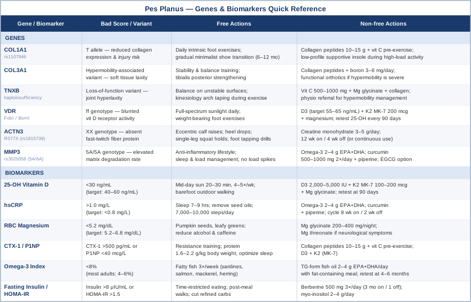
Quick Reference: Genes, Biomarkers, and Actions
The table below summarizes all six genes and six biomarkers covered in this article alongside key action steps at a glance.
The Book That Reframes Everything You Think You Know About Flat Feet
Katy Bowman's Whole Body Barefoot is one of the most practically disruptive books written about foot health. A biomechanist by training, Bowman challenges the foundational assumption behind most orthopedic foot care: that the arch needs external support to function. Her argument, grounded in evolutionary biology and biomechanics research, is that arch supports — when used continuously — progressively weaken the intrinsic foot muscles they were meant to protect, creating dependency and accelerating structural decline. This is not a fringe position: it is consistent with research showing that populations wearing minimal or no footwear have lower rates of pes planus than those in conventional supportive shoes.
The following are ten of the most clinically significant insights from Bowman's framework and the supporting evidence behind each.
1. The Arch Is a Muscle-Tendon System, Not Just a Bone Arch
The medial longitudinal arch is dynamically supported by the tibialis posterior, flexor hallucis longus, and the intrinsic foot muscles — not passively held up by bones alone. When these muscles are chronically unloaded (by arch supports or thick-soled shoes), they atrophy. Bowman frames this as the same principle as cast atrophy: immobilization produces weakness, not recovery.
2. Shoe Shape Is the First Problem
Most conventional shoes taper toward the toe, compressing the toes inward (toe splay is reduced) and preventing the transverse arch from functioning correctly. Bowman argues that restoring toe splay — using wide toe box shoes or toe spreaders — is the single most overlooked intervention in flat foot management. Research has confirmed that toe flexor strength, which requires adequate toe splay space, is significantly correlated with arch height index.
3. Heel Elevation Is a Chronic Hip Flexor and Calf Problem
Conventional shoes with a heel drop (even 10–12 mm in running shoes) chronically shorten the Achilles tendon and gastrocnemius-soleus complex. This altered tension pattern changes ground reaction forces in a way that shifts load medially, directly increasing stress on the spring ligament. Bowman recommends gradual heel drop reduction as a prerequisite to meaningful arch improvement.
4. The Short Foot Exercise Is the Most Evidence-Backed Intervention
The "short foot" or "doming" exercise — contracting the foot to shorten the distance between the ball and the heel without toe curling — activates the intrinsic plantar muscles with high specificity. A 2016 randomized controlled trial published in the Journal of Physical Therapy Science demonstrated that short foot exercise performed 5 days per week for 8 weeks significantly increased arch height index compared to control. Bowman highlights this exercise as foundational.
5. Walking Surface Variability Is Proprioceptive Training
Flat, predictable surfaces (concrete, hardwood floors) reduce proprioceptive input from the plantar fascia and intrinsic foot muscles. Walking on grass, gravel, sand, or uneven terrain forces constant micro-adjustments that strengthen the arch stabilizers. Bowman recommends deliberate "texture walking" for at least 10–15 minutes daily.
6. Sitting Is Not Neutral — It Is Arch Loading by Proxy
Prolonged sitting tightens hip flexors and glutes, which alters pelvis tilt and gait mechanics. An anteriorly tilted pelvis during gait changes load distribution through the foot and increases medial arch stress. Bowman's protocol includes regular standing, ground sitting in various positions, and hip mobility work as part of flat foot correction — not as a peripheral add-on.
7. Foot Mobility Before Foot Strength
Before strengthening weak intrinsic muscles, restricted mobility in the midfoot joints must be addressed. Many people with pes planus have limited talar mobility and restricted first metatarsophalangeal joint extension. Bowman uses specific joint mobilizations and toe extension stretches to restore range of motion before loading the arch in strengthening exercises.
8. The Transition to Minimalist Footwear Must Be Earned, Not Rushed
Bowman is explicit that switching immediately to zero-drop minimalist shoes causes acute overload injuries in deconditioned feet. Her protocol recommends a 6–12 month transition: starting with a 4 mm heel drop and progressing through 2 mm to zero, only advancing when the foot can perform single-leg calf raises and short foot holds without fatigue. This staged protocol substantially reduces injury risk.
9. The Plantar Fascia Is a Proprioceptive Organ, Not Just a Structural Band
The plantar fascia contains mechanoreceptors that send real-time positional information to the nervous system. Bowman argues — consistent with neurological research on plantar sensory input — that padded footwear reduces this signal, impairing balance, gait efficiency, and arch control. Gradually increasing sensory input through thinner soles is framed as neurological rehabilitation as much as structural correction.
10. Whole-Body Alignment Determines Foot Outcome
Bowman's central thesis is that the foot is the downstream consequence of whole-body movement patterns. Tight calves, weak glutes, excessive forward lean, and poor hip extension all funnel abnormal load into the medial arch. No amount of foot-specific exercise corrects a flat foot that is being continuously loaded by a misaligned body above it. This systems-level framing is what distinguishes Bowman's approach from isolated foot strengthening protocols.
Complementary Approaches with Clinical Support
The interventions below have meaningful evidence for pes planus or directly adjacent conditions (plantar fasciitis, subtalar instability, arch-related pain). They are not replacements for the structural work described earlier, but they extend the toolkit meaningfully.
Yoga
Yoga-based foot strengthening protocols directly address two of the central deficits in pes planus: intrinsic foot muscle weakness and reduced ankle-foot proprioception. Poses such as Tadasana (mountain pose), Utkatasana (chair pose), and Virabhadrasana I (warrior I) with conscious arch activation train the neuromuscular system to dynamically support the medial arch under load. The emphasis on barefoot practice adds proprioceptive challenge that standard exercise does not replicate.
A 2015 study in the Journal of Bodywork and Movement Therapies found that yoga-based exercises significantly improved arch height index and foot pain scores in adults with symptomatic flat feet over an 8-week intervention. The protocol involved daily 20-minute sessions focused on intrinsic foot activation and ankle stability. The evidence base is not large, but the risk profile is minimal and the biological mechanism is clear.
For practical application, start with 10–15 minutes of barefoot yoga focused on standing poses and arch activation work daily. Progress to balance-challenging poses (tree pose, eagle pose) as strength improves. Avoid forward folding that stretches the calf into passive range before the intrinsic muscles are strong enough to benefit from the stretch reflex. Beginners with significant arch collapse should work with an instructor experienced in structural correction rather than following a generic online flow.
Tai Chi
Tai chi's slow, deliberate weight shifting and continuous single-leg loading provides a sustained proprioceptive and neuromuscular challenge to the foot and ankle complex. The subtalar joint — whose stability is compromised in most pes planus presentations — is specifically exercised through the rotational weight transfer patterns characteristic of tai chi practice. Balance improvement in this population translates directly to reduced arch loading asymmetry.
A 2012 randomized controlled trial published in the Journal of the American Geriatrics Society found that 16 weeks of tai chi practice significantly improved foot arch structure and dynamic balance in older adults, a population with high rates of acquired flat foot. The tai chi group showed measurable improvement in tibialis posterior activation compared to controls receiving standard balance training.
Begin with a beginner tai chi program 3 times per week, 30–45 minutes per session. Yang-style short form is the most widely researched and accessible entry point. Focus on deliberate heel-to-toe weight transfer and conscious arch engagement during each movement. People with severe ankle instability or significant pain should start with chair-supported versions before progressing to full standing practice.
Massage Therapy
Manual therapy targeting the plantar fascia, tibialis posterior tendon, and calf complex addresses soft tissue restrictions that limit the mechanical efficiency of arch-supporting muscles. Myofascial release of the plantar surface reduces pain and improves tissue extensibility; cross-friction massage of the tibialis posterior tendon reduces adhesion formation that would otherwise impair its arch-lifting function. Calf and Achilles soft tissue work directly addresses the shortened posterior chain that loads the medial arch.
A systematic review of manual therapy for plantar heel pain (which shares significant biomechanical overlap with pes planus) found consistent moderate-quality evidence that soft tissue mobilization combined with exercise produced greater outcomes than exercise alone. In clinical practice, massage therapists experienced in orthopedic or sports soft tissue work offer the most targeted protocols. Self-massage using a lacrosse ball under the plantar surface (2–3 minutes per foot, 3–5 times weekly) provides meaningful benefit as a lower-cost adjunct.
Practically, 4–6 professional sessions over 6–8 weeks targeting the posterior chain and plantar structures, combined with home self-massage, is a reasonable starting protocol. Massage should be followed by strengthening work — soft tissue release without subsequent loading does not produce lasting structural change. Avoid aggressive deep tissue work on an actively inflamed plantar fascia; work the perimeter and address calf restrictions instead until acute inflammation resolves.
Biofeedback
Visual or auditory biofeedback for gait retraining provides real-time data on how weight is distributed across the foot during walking. In pes planus, patients typically overload the medial forefoot and underload the lateral midfoot, creating a loading pattern that progressively stresses the spring ligament. Biofeedback helps individuals learn to consciously shift load toward a more biomechanically neutral distribution — a skill that eventually becomes automatic with sufficient practice.
Research in gait retraining using real-time feedback has shown that even 8–10 treadmill sessions with visual force-plate feedback can produce lasting improvements in foot strike pattern that persist at 1-month follow-up — reported in a 2011 study in Gait & Posture. More recently, wearable insole biofeedback devices have made this approach accessible outside of laboratory and clinical settings.
Clinically, gait biofeedback is most effectively delivered through a physiotherapist or podiatrist with access to a pressure-plate system or instrumented treadmill. Consumer-level options include pressure-sensing insoles (such as those from Moticon or Stridalyzer) that pair with smartphone apps for home gait monitoring. Commit to at least 6–8 weeks of consistent practice for gait pattern changes to transfer into habitual movement.
Low-Level Laser Therapy (Photobiomodulation)
Low-level laser therapy (LLLT) or photobiomodulation delivers near-infrared light into soft tissue at doses that stimulate mitochondrial energy production, reduce inflammatory signaling, and accelerate collagen synthesis without thermal effects. For pes planus-related conditions — particularly plantar fasciitis and tibialis posterior tendinopathy, which frequently co-occur — the anti-inflammatory and tissue repair mechanisms are directly relevant.
Multiple RCTs have evaluated LLLT specifically for plantar fasciitis. A meta-analysis published in Lasers in Medical Science found statistically significant reductions in pain and improvements in function following LLLT compared to placebo, with effects maintained at 3-month follow-up. The evidence is stronger for pain reduction than for structural arch correction, but given the shared tissue pathology, the rationale for use in pes planus is sound.
In practice, clinical LLLT is delivered by physiotherapists, podiatrists, or sports medicine clinics using Class III or Class IV devices. A typical protocol is 6–12 sessions over 3–4 weeks, with 2–3 sessions per week. The treatment itself is painless and takes 5–15 minutes per session. Home devices (red-light panels or targeted handhelds at 630–850 nm wavelengths) are increasingly available and may provide benefit for mild-to-moderate symptoms, though clinical devices are more powerful. Do not use over active infections, directly on cancerous tissue, or over the thyroid gland.
Conclusion
Pes planus is a condition with many biological layers. The arch does not collapse in isolation — it does so against a backdrop of connective tissue genetics, inflammatory load, nutrient status, and movement patterns that no single arch support can address. The six biomarkers covered here — vitamin D, hsCRP, RBC magnesium, collagen turnover markers, omega-3 index, and fasting insulin — provide a concrete starting point for understanding the internal environment your foot is operating in. The six genetic variants — COL1A1, COL3A1, TNXB, VDR, ACTN3, and MMP3 — explain why some people's arches are structurally more vulnerable and what targeted interventions can shift that trajectory.
The next smart step is to pick one or two blood tests you have not yet run and schedule them. If your vitamin D or omega-3 index has never been measured, start there. If you have never done a short foot exercise or a single-leg calf raise, try both today and note how quickly each fatigues. These are data points, not verdicts. Use them to build a more precise intervention rather than a more hopeful guess.