This article contains AI generated content.
· UpdatedPhenylketonuria Arthropathy — 5 Genes And 6 Biomarkers To Track
Introduction
Joint pain and stiffness are not the first things people associate with phenylketonuria. Most conversations about PKU revolve around the diet, phenylalanine levels, and neurological outcomes. But for a meaningful number of people living with PKU, arthropathy — disease affecting the joints and connective tissue — is a real and often unaddressed part of daily life. Joints feel stiff in the morning, movement becomes less fluid over time, and standard rheumatological workups often return inconclusive results because the underlying metabolic driver is not on most clinicians' radar.
The challenge is compounded by how individually PKU presents. Two people with the same diagnosis may have very different genetic profiles, dietary adherence histories, amino acid formula use, and nutritional gaps. One person may have significant connective tissue involvement while another has none. Without examining both what is happening in the body right now — the biomarkers — and the genetic architecture shaping those processes, it becomes extremely difficult to build a rational, targeted approach.
Generic advice — keep your Phe levels controlled — is not wrong, but it is rarely sufficient when joints are involved. Arthropathy in PKU appears to emerge from several converging factors: chronic phenylalanine toxicity affecting connective tissue, micronutrient deficiencies caused by the restrictive dietary protein regime, bone density loss that changes how load is distributed across cartilage, and in some patients, a heightened inflammatory background partly driven by specific genetic variants. Each factor is addressable — but each requires its own measurement and its own intervention logic.
This article offers two of those lenses. The primary section covers six biomarkers that provide a precise, measurable picture of where joint stress is building and what can be done about each one — with and without supplementation. A second section covers five genes that explain much of the individual variation in how PKU arthropathy develops and how severely it manifests. Two further sections draw on Peter Attia's proactive medicine framework and present three evidence-supported complementary approaches. Used together, these frameworks make it possible to move from broad symptom management toward genuinely targeted intervention.
Summary
This article covers six of the most informative biomarkers for PKU arthropathy — blood phenylalanine, the Phe:Tyr ratio, 25-OH vitamin D, bone turnover markers (CTX-I and P1NP), high-sensitivity CRP, and zinc-selenium status — each with measurement methods, cost ranges, and action plans with and without supplementation. A genetics section follows with five key genes (PAH, MTHFR, VDR, IL-6, and COL2A1), explaining what each variant does to joint health and what compensation strategies exist. The article then pulls ten of the most applicable principles from Peter Attia's Outlive, a landmark book on proactive medicine, and closes with three well-matched complementary approaches — mindfulness-based stress reduction, low-level laser therapy, and yoga — each presented with condition-relevant evidence and realistic application protocols. The goal throughout is practical, specific guidance grounded in real evidence, not broad reassurance.
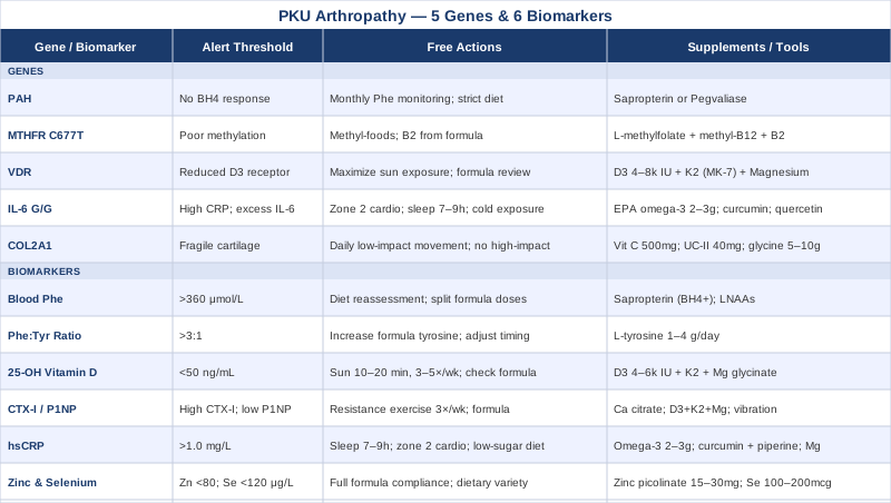
Tracking the right numbers changes what is possible. Here is exactly what to track, and what to do with what you find.
The 6 Biomarkers That Give the Clearest Picture of PKU Arthropathy
Symptoms are lagging indicators — by the time they appear, the underlying process has usually been running for months or years. Biomarkers are leading indicators: they show where physiological stress is accumulating before significant tissue damage occurs. For PKU arthropathy specifically, the following six measurement categories offer the most complete and actionable picture currently available. None requires exotic testing; all can be ordered through standard metabolic or primary care channels.
1. Blood Phenylalanine Level
Blood phenylalanine is the foundational measurement in PKU management, and it is equally foundational when thinking about arthropathy. Chronically elevated Phe does not affect only the brain — it disrupts connective tissue homeostasis throughout the body. At high concentrations, phenylalanine competes with other amino acids for membrane transport and appears to interfere with the synthesis and cross-linking of structural proteins, including collagen. Some researchers also suspect that elevated Phe contributes to increased oxidative stress in synovial tissue, accelerating joint wear that shows up clinically as stiffness and reduced range of motion.
Beyond its direct effects on connective tissue, poorly controlled Phe also acts as a proxy for how well the overall dietary and formula plan is functioning. High Phe generally means more natural protein intake than the metabolic system can handle, which simultaneously means less amino acid formula — and with it, fewer of the micronutrients the formula provides. The arthropathy risk compounds on multiple levels at once.
How to Measure It
Phenylalanine is measured using a dried blood spot (DBS) collected via finger prick, or more accurately through plasma amino acid analysis from a venous blood draw. DBS testing is standard in PKU clinics and many patients perform it at home, mailing samples to specialist metabolic labs. Costs range from $50 to $150 for DBS processing at US metabolic labs. Full plasma amino acid panels typically cost $200 to $500 and offer more detail. For arthropathy management, frequency should be at minimum every 4 to 8 weeks; monthly is preferable when joint symptoms are active.
Most European guidelines and US guidelines from the Genetic Metabolic Dietitians International recommend adult Phe levels below 360 μmol/L (6 mg/dL), with stricter targets of 120-240 μmol/L for those with significant neurological sensitivity. For joint health specifically, there is growing reason to believe that even the upper end of what is considered "acceptable" may not be sufficient to protect connective tissue over time.
If the Score Is High: The Plan Without Supplements
The most direct approach is dietary reassessment. This means recalculating natural protein intake, redistributing phenylalanine-containing foods across smaller and more frequent portions throughout the day, and increasing the proportion of Phe-free amino acid formula in the daily diet. A metabolic dietitian experienced with PKU can calculate tolerable daily Phe loads with precision based on current blood levels.
Beyond dietary changes, protein timing matters considerably. Consuming amino acid formula in three to five divided doses spread across the day — rather than one or two large boluses — maintains a steadier competitive environment at membrane transport sites in joint tissue and reduces peak Phe spikes. Regular moderate physical activity also modestly improves amino acid uptake efficiency, meaning even a consistent walking routine can contribute to lower fasting Phe levels. This is an underappreciated and entirely free tool.
If the Score Is High: The Plan With Supplements or Equipment
For those with BH4-responsive PAH mutations, sapropterin dihydrochloride (Kuvan) remains the most powerful pharmacological option for reducing blood Phe. BH4 responsiveness requires formal testing — a BH4 loading test over 24 to 48 hours — but for responders it can reduce Phe by 30% or more, fundamentally changing dietary flexibility and joint tissue exposure.
Large Neutral Amino Acids (LNAAs) — mixtures containing leucine, isoleucine, valine, tryptophan, tyrosine, and other large neutral amino acids without phenylalanine — compete directly with Phe for intestinal absorption and blood-brain barrier transport. In practice, LNAA supplements are most useful for adults who have difficulty with dietary adherence. They are available as Phe-free formulas under names such as NeoPhe and PheMD Plus. Dosing is typically 0.5 to 1 gram per kilogram of body weight per day spread across meals; they can be taken continuously with no cycling required. Side effects are mild and mostly gastrointestinal at higher doses. Always verify that any supplement is confirmed Phe-free before use.
2. Phenylalanine-to-Tyrosine Ratio
The Phe:Tyr ratio is a more nuanced biomarker than Phe alone. Because phenylalanine hydroxylase normally converts Phe to tyrosine, when PAH function is compromised, tyrosine levels frequently fall at the same time that Phe rises. This is important for arthropathy because tyrosine is not just a neurotransmitter precursor — it is also a critical component of structural proteins and plays a role in collagen synthesis. Deficiency in tyrosine can impair the tissue repair capacity of cartilage and ligaments independently of Phe toxicity.
A Phe:Tyr ratio above 3:1 is generally considered a warning sign of poor metabolic control. Ratios above 6:1 signal significant biochemical dysfunction. More importantly, low tyrosine — even when Phe is borderline acceptable — can create a state where connective tissue repair cannot keep pace with normal wear, accelerating joint degradation through a separate pathway from Phe toxicity itself.
How to Measure It
The ratio is calculated from the same sample as blood Phe — it requires plasma amino acid analysis or a more detailed DBS panel that includes tyrosine. No additional testing cost is incurred if included in the panel. Target ratio for adults: ideally below 3:1; below 2:1 is optimal. Tyrosine itself should ideally sit above 40 μmol/L, with optimal concentrations in the 60 to 90 μmol/L range for adequate connective tissue and neurological function.
If the Ratio Is High: The Plan Without Supplements
If the ratio is elevated primarily because Phe is high, the dietary strategies outlined in the previous section apply directly. If the ratio is elevated because tyrosine is too low while Phe is only marginally elevated, the focus shifts to ensuring adequate tyrosine intake from the amino acid formula. Different PKU formulas contain different amounts of tyrosine, and switching to a formula with a higher tyrosine content may be sufficient. Reviewing the formula composition specifically for tyrosine concentration is a worthwhile clinic conversation that many PKU patients have never initiated.
Timing also matters: consuming tyrosine-containing formula in windows separated from large natural-protein meals can improve its bioavailability by reducing competition at intestinal transport sites.
If the Ratio Is High: The Plan With Supplements or Equipment
Tyrosine is available as a standalone supplement (L-tyrosine), typically at 1 to 4 grams per day in divided doses, taken away from meals for best absorption. Most commercial L-tyrosine supplements are derived from fermentation processes and are Phe-free — but verification with the manufacturer is essential. Side effects are rare at these doses; very high doses above 12 g/day have been associated with headache and nausea in some people. No cycling is required; tyrosine can be supplemented continuously. For BH4-responsive patients already on sapropterin, the increased phenylalanine hydroxylation produced by the medication will naturally boost endogenous tyrosine production, reducing the need for standalone supplementation.
3. 25-OH Vitamin D
Vitamin D deficiency is one of the most consistently documented nutritional concerns in PKU, and it connects directly to arthropathy through multiple mechanisms. The restrictive low-protein PKU diet frequently eliminates the richest natural dietary sources of vitamin D — fatty fish, egg yolks, fortified dairy — and amino acid formulas vary considerably in how much D3 they contain. Some older formulas provide minimal amounts, leaving a significant and often undetected gap.
The connection to joint health is multi-layered. Vitamin D regulates calcium homeostasis, which governs bone mineral density. When bone density declines, joints experience altered biomechanical loading — mechanically softer bone changes how force is transmitted through cartilage at the contact surfaces. Studies in PKU adults have consistently found lower bone mineral density compared to healthy controls, and vitamin D deficiency is a recurring finding in this population. Beyond bone, vitamin D acts as a potent immunomodulator: insufficient levels are associated with elevated systemic inflammation, which directly worsens arthropathy by sustaining the inflammatory environment in synovial tissue.
How to Measure It
25-hydroxyvitamin D (25-OH-D) is measured from a standard venous blood draw and is widely available through primary care, metabolic clinics, and direct-to-consumer lab services. Cost: $30 to $80 in the US. Testing two to three times per year is reasonable for PKU patients, particularly in autumn and winter when sun exposure is lowest and after any major dietary changes. Optimal range for joint and immune health: 50 to 80 ng/mL (125 to 200 nmol/L). The standard "sufficient" threshold of 30 ng/mL is widely considered by functional medicine practitioners and bone specialists as insufficient for individuals managing chronic metabolic conditions.
If the Score Is Low: The Plan Without Supplements
Sun exposure remains the most efficient way to generate vitamin D3 endogenously. For most adults, 10 to 20 minutes of midday sun with arms, legs, and face exposed — without sunscreen during that brief window — on three to five days per week can maintain reasonable levels during spring and summer. However, latitude, skin tone, season, and cloud cover dramatically reduce efficacy, and sun exposure alone rarely corrects significant deficiency in adults. Still, it costs nothing and has no phenylalanine-related implications.
Reviewing the amino acid formula to confirm whether it contains D3 — and in what quantity — is also worthwhile. Many metabolic teams do not specifically track vitamin D status through the lens of joint health; flagging this explicitly at the next clinic appointment often leads to a useful conversation that would not otherwise happen.
If the Score Is Low: The Plan With Supplements or Equipment
Vitamin D3 (cholecalciferol) is the preferred supplemental form and is Phe-free in all standard formulations. For correction of deficiency, doses of 4,000 to 6,000 IU per day for 8 to 12 weeks are commonly used, followed by maintenance doses of 2,000 to 4,000 IU per day. Retesting 8 to 10 weeks after starting a new dose confirms whether the target range has been reached.
Critical co-factors that dramatically improve vitamin D3 outcomes: - Vitamin K2 (MK-7 form): 100 to 200 mcg/day — directs calcium into bone rather than soft tissue and reduces vascular calcification risk associated with higher-dose D3 - Magnesium glycinate: 300 to 400 mg/day — required for the enzymatic activation of vitamin D in both the liver and kidney; magnesium deficiency directly blocks vitamin D from functioning even when blood levels appear adequate - Vitamin A (retinol): high-dose retinol supplements can antagonize vitamin D; standard dietary intake is usually sufficient and no supplemental retinol is needed alongside D3
No cycling is required for D3 at these doses. Side effects below 10,000 IU/day in adults without granulomatous disease are very rare; monitoring every 3 to 6 months prevents accumulation.
4. Bone Turnover Markers: CTX-I and P1NP
Bone remodeling involves a constant cycle of breakdown (resorption) and formation. When resorption chronically outpaces formation, bone mass declines — and research in PKU adults has documented exactly this pattern. What makes bone turnover markers particularly valuable is that they detect this imbalance before it becomes visible on bone density scans (DEXA), often providing a window of a year or more for intervention before structural bone loss becomes measurable.
CTX-I (C-telopeptide of type I collagen) is a resorption marker released into circulation when osteoclasts break down bone matrix. Elevated CTX-I signals excessive bone breakdown. P1NP (procollagen type I N-terminal propeptide) is a formation marker reflecting osteoblast activity and collagen synthesis. Together, the relative values and trends of these two markers provide a precise picture of bone metabolism. For PKU arthropathy, the warning pattern is elevated CTX-I with normal or suppressed P1NP — indicating bone is being broken down faster than it is being replaced.
How to Measure It
Both markers require a venous blood draw. CTX-I should ideally be collected fasting in the morning, as values fluctuate with food intake. P1NP is more stable across the day. Costs in the US: CTX-I approximately $80 to $150; P1NP approximately $100 to $200. These are typically ordered by endocrinologists or metabolic bone specialists; they can be requested directly from a metabolic team when framed in the context of joint and bone health monitoring. Testing every 6 to 12 months provides a useful trend picture, with more frequent testing (every 3 months) when actively treating.
If the Markers Are Unfavorable: The Plan Without Supplements
The most powerful non-pharmacological intervention for bone formation is resistance exercise — specifically, weight-bearing movements that mechanically load the skeleton and stimulate osteoblast activity. Swimming, while joint-friendly, is not optimal for bone density. For PKU patients with joint discomfort, beginning conservatively with body weight squats, step-ups, and light resistance band work at least three times per week is a realistic starting point. The bone-protective effect of resistance exercise is cumulative and dose-dependent.
Adequate protein synthesis through consistent amino acid formula compliance is also essential. Bone matrix is fundamentally protein-based (type I collagen), and the amino acid building blocks required for P1NP production must be present in adequate amounts. This is a direct and often underappreciated connection between formula compliance and bone health.
If the Markers Are Unfavorable: The Plan With Supplements or Equipment
- Calcium: assess formula contribution first; additional calcium citrate (preferred over carbonate for absorption) at 500 mg twice daily if formula intake is insufficient; split doses — calcium absorption declines above 500 mg at a single sitting - Vitamin D3 + K2 + Magnesium: as detailed in the previous section — these directly support P1NP production and suppress excessive CTX-I elevation through multiple complementary pathways - Whole-body vibration therapy: low-magnitude, high-frequency vibration (30 to 50 Hz) has shown modest positive effects on bone density in human studies involving osteoporosis and conditions with limited mobility; sessions of 10 to 20 minutes three to five times per week using devices such as Power Plate or equivalent; useful for patients with joint pain limiting conventional exercise
5. High-Sensitivity C-Reactive Protein
Inflammation is both a driver and a consequence of joint disease. In PKU, chronically elevated phenylalanine appears to activate low-grade systemic inflammation — not the acute, dramatic kind associated with infections, but the chronic, smoldering kind that erodes tissue architecture over years. High-sensitivity CRP (hsCRP) is the most accessible and clinically validated marker of this systemic inflammatory burden.
The distinction between standard CRP and hsCRP is important. Standard CRP only registers when inflammation crosses a significant threshold (above 10 mg/L). High-sensitivity CRP detects concentrations as low as 0.1 mg/L, capturing the chronic low-grade inflammatory state that both rheumatologists and metabolic specialists now recognize as the operating environment of progressive arthropathy. For PKU arthropathy, where the inflammation may be modest but continuous, hsCRP is the correct test.
How to Measure It
hsCRP is a standard blood test available at any laboratory. Cost: $10 to $50 in the US; often included in comprehensive metabolic panels or cardiovascular risk panels. No special preparation is required except avoiding recent intense exercise or intercurrent illness, both of which transiently elevate CRP unrelated to chronic inflammatory status. Testing every 6 to 12 months is reasonable; quarterly if actively trying to reduce inflammation.
Optimal target: below 1.0 mg/L. Values of 1.0 to 3.0 mg/L indicate moderate inflammatory and cardiovascular risk. Above 3.0 mg/L warrants investigation. Lipidologist Thomas Dayspring, known for his rigorous approach to cardiovascular biomarkers, considers below 0.5 mg/L as the genuinely low-risk target.
If the Score Is High: The Plan Without Supplements
Sleep quality and duration are the most underrated tools for reducing hsCRP. Research consistently shows that sleeping fewer than six to seven hours per night significantly raises CRP through multiple neuroimmune pathways. For PKU patients whose neurological involvement may affect sleep architecture, optimizing sleep hygiene — consistent bedtime and wake time, a cool and dark room, limiting blue-light exposure after sunset — is often the highest-yield, zero-cost behavioral intervention available.
Regular moderate aerobic exercise — particularly zone 2 training (a conversational pace sustained for 30 to 45 minutes, four to five times per week) — consistently reduces hsCRP in adults with chronic conditions by improving mitochondrial efficiency and modulating IL-6 signaling. Psychological stress reduction matters independently: chronic stress activates the same HPA and NF-κB inflammatory pathways that elevate CRP. Including adequate polyphenol-rich vegetables and fruits compatible with the low-Phe diet, and minimizing refined sugars and ultra-processed foods, reinforces the dietary anti-inflammatory effect.
If the Score Is High: The Plan With Supplements or Equipment
- Omega-3 fatty acids (EPA + DHA): 2 to 3 grams per day from pharmaceutical-grade fish oil, verified Phe-free; consistently reduces hsCRP in multiple clinical trials; take with food to improve absorption and reduce GI side effects; no cycling required; note mild blood-thinning effect if on anticoagulants - Curcumin with piperine: 500 to 1,000 mg of standardized curcumin extract (95% curcuminoids) with 5 to 10 mg piperine per dose; meta-analyses support its hsCRP-lowering effect through NF-κB suppression; take with meals; continuous use is well-tolerated; occasional mild GI side effects - Magnesium glycinate: 300 to 400 mg at night; anti-inflammatory through multiple pathways and simultaneously improves sleep quality, compounding the hsCRP benefit through two independent mechanisms
6. Zinc and Selenium Status
These two trace minerals are among the most commonly depleted in people living with PKU on restricted diets, and both are directly relevant to joint and connective tissue health. Zinc is essential for collagen synthesis, immune regulation, and cartilage matrix maintenance — it acts as a cofactor for the matrix metalloproteinases that remodel connective tissue. Selenium is critical for the antioxidant selenoprotein system — specifically glutathione peroxidase — that protects joint tissue from the oxidative damage that drives cartilage degradation.
PKU amino acid formulas are designed to compensate for nutrients lost by restricting natural protein, but formulation quality and trace mineral content vary significantly across brands and age-specific products. Someone who is not fully compliant with their formula, or who uses an older formulation, may have meaningful zinc and selenium gaps that never appear on standard PKU follow-up panels. Arthropathy symptoms that persist despite acceptable Phe control are often connected to undetected micronutrient deficiencies rather than Phe itself.
How to Measure It
Zinc is measured from plasma (plasma zinc is the most widely available and clinically meaningful form). Selenium is best measured from plasma or whole blood; whole-blood selenium provides a longer-term picture of status. Costs: plasma zinc approximately $30 to $80; plasma or whole-blood selenium approximately $50 to $120. Both are available through standard labs. Testing annually is a reasonable baseline; twice yearly when supplementing or after significant dietary changes.
Optimal plasma zinc: 80 to 110 μg/dL. Optimal plasma selenium: 120 to 160 μg/L, with some researchers recommending 150 to 200 μg/L for full selenoprotein enzyme saturation.
If the Levels Are Low: The Plan Without Supplements
In PKU, the most sustainable non-supplement approach to zinc and selenium status is ensuring full formula compliance. Most modern metabolic formulas designed for PKU include both minerals in meaningful amounts. Working with a metabolic dietitian to confirm that formula targets are actually being met — and identifying whether the specific formula being used has adequate trace mineral content — is the practical starting point. Formula composition sheets can be reviewed directly with the manufacturer.
Some naturally low-Phe foods also contribute to trace mineral intake: certain fruits, vegetables, and low-protein specialty foods provide small but meaningful amounts of both minerals. The cumulative effect of dietary variety within PKU constraints should not be dismissed.
If the Levels Are Low: The Plan With Supplements or Equipment
- Zinc picolinate or zinc glycinate: 15 to 30 mg elemental zinc per day; take with food (zinc causes nausea on an empty stomach); do not take alongside calcium (they compete for the same transporter); do not exceed 40 mg/day without medical oversight — sustained high-dose zinc depletes copper; if supplementing above 25 mg/day, add copper at 1 to 2 mg/day; 5 days on, 2 days off is a reasonable cycling approach - Selenomethionine: the most bioavailable selenium form; 100 to 200 mcg/day; selenium has a narrow therapeutic window — do not supplement above 400 mcg/day; always verify formula contribution before adding supplemental selenium; retest levels 3 months after starting to confirm adequate but not excessive repletion; no cycling required at 100 to 200 mcg/day
Together, these six biomarkers form a surveillance system for joint health in PKU that exceeds what standard metabolic monitoring provides. Presenting these results at clinic appointments — and explicitly connecting them to arthropathy symptoms — is often the most productive clinical conversation a PKU patient can initiate.
What Your Genes Reveal About Joint Vulnerability in PKU
Biomarkers tell you what is happening. Genes help explain why it is happening — and why two people with similarly controlled PKU can have very different joint outcomes. Genetic factors in PKU arthropathy operate across multiple levels. The PAH gene itself determines the fundamental metabolic architecture. Other genes — several of them well-studied in broader genetics and epigenetics research — shape the inflammatory environment, connective tissue quality, and nutrient metabolism that determine how well any individual's joints withstand the biochemical stresses of living with PKU. Knowing your variants in these five genes does not determine outcomes, but it provides meaningful context for building a smarter, more individualized approach.
1. PAH — Phenylalanine Hydroxylase
The PAH gene, located on chromosome 12q23.2, encodes the enzyme that converts phenylalanine to tyrosine using tetrahydrobiopterin (BH4) as an essential cofactor. Over 1,000 pathogenic variants have been identified in this gene, each producing a different degree of enzyme dysfunction. The clinical spectrum runs from classic PKU (PAH activity below 1% of normal) to mild hyperphenylalaninemia (PAH activity above 10%), with manageable dietary flexibility in milder forms.
The connection to arthropathy is direct: PAH genotype determines the Phe burden the person carries lifelong, which in turn drives cumulative connective tissue stress. But PAH genotype also determines BH4 responsiveness — the distinction between a patient for whom sapropterin is transformative and one for whom it does nothing. Patients with at least one class 3 to 5 severity variant are often BH4-responsive; those with two class 1 to 2 variants typically are not. This single genetic determination has more downstream impact on joint health over a lifetime than almost any other factor.
If the Variant Is Unfavorable: The Plan Without Supplements
Work with a metabolic specialist to confirm both PAH variants through full gene sequencing if not already done — a comprehensive panel rather than targeted variant testing captures the full picture. Without pharmacological support, management remains dietary: the more severe the Phe tolerance dictated by the genotype, the more precisely formula compliance must be maintained. Monthly or more frequent Phe monitoring with prompt dietary adjustment whenever levels drift upward is the essential discipline.
If the Variant Is Unfavorable: The Plan With Supplements or Equipment
BH4 responsiveness testing should be conducted if not yet performed, particularly for patients with at least one mild or moderate PAH variant. For non-responders, pegvaliase (Palynziq) — an enzyme substitution therapy — can dramatically reduce blood Phe levels in adults regardless of genotype, and it has been approved in the US and Europe for adults with uncontrolled PKU. Both sapropterin and pegvaliase require specialist management and ongoing monitoring. The joint health benefit of either agent comes specifically from the sustained Phe reduction they enable over months and years, not from any direct joint effect.
2. MTHFR — Methylenetetrahydrofolate Reductase
The MTHFR gene encodes the enzyme central to folate and methionine metabolism. The most studied variants are C677T (rs1800562) and A1298C (rs1801131). C677T homozygosity reduces MTHFR enzymatic activity by approximately 70% compared to wild type. In the general population, MTHFR variants primarily create concerns around elevated homocysteine and folate pathway inefficiency.
In PKU patients, where dietary variety is already constrained and protein is largely formula-derived, MTHFR variants amplify the nutritional risk. Methylation is foundational to connective tissue maintenance, epigenetic regulation of inflammatory gene expression, and cellular repair in cartilage and synovial tissue. A sluggish methylation cycle means slower recovery from joint microtrauma, a more pro-inflammatory tissue environment, and impaired capacity to regulate the genes that govern joint maintenance — compounding the Phe-related burden from a completely different direction.
If the Variant Is Unfavorable: The Plan Without Supplements
Prioritize dietary folate in naturally methylated forms from vegetables compatible with the low-Phe diet, rather than relying solely on the synthetic folic acid (not methylfolate) that most PKU formulas provide. Limiting enriched flours — common in many low-protein specialty staple foods — reduces unconverted synthetic folic acid accumulation that can paradoxically compete with active methylfolate. Ensuring adequate riboflavin (B2) through the amino acid formula is important: B2 is a direct cofactor for MTHFR function, and MTHFR C677T specifically is classified as riboflavin-responsive.
If the Variant Is Unfavorable: The Plan With Supplements or Equipment
- L-methylfolate (5-MTHF): 400 to 1,000 mcg/day in the active form (brands: Quatrefolic, Metafolin) — this bypasses the MTHFR conversion step entirely; start at the lower dose and increase gradually; continuous use, no cycling required - Methylcobalamin (methyl-B12): 500 to 2,000 mcg/day sublingual — active B12 that works in tandem with methylfolate in the methionine cycle; sublingual absorption bypasses GI variation - Riboflavin (B2): 25 to 200 mg/day; specifically shown in clinical research to improve MTHFR C677T enzyme function; inexpensive, safe, and a high-value intervention for this genotype; no cycling needed
3. VDR — Vitamin D Receptor
The VDR gene encodes the nuclear receptor through which circulating vitamin D exerts its effects on hundreds of genes — including those regulating bone formation, immune modulation, and inflammatory signaling. Four well-studied polymorphisms — FokI (rs2228570), BsmI (rs1544410), TaqI (rs731236), and ApaI (rs7975232) — affect how efficiently the receptor responds to vitamin D at any given circulating level.
In PKU arthropathy, the clinical implication is significant. PKU patients already face elevated vitamin D deficiency risk due to dietary restrictions. VDR variants can mean that even patients who supplement to "adequate" blood levels still experience suboptimal downstream effects on bone formation and immune regulation — because the receptor sensitivity is reduced. Standard thresholds for vitamin D sufficiency may not apply to individuals with unfavorable VDR genotypes, and testing becomes even more important than in the general population.
If the Variant Is Unfavorable: The Plan Without Supplements
Maximize the natural inputs that do not depend on VDR sensitivity at the activation step: sun exposure frequency and duration within reason for skin type and latitude; dietary sources of vitamin D where compatible with the low-Phe diet; avoiding medications known to reduce VDR expression (long-term glucocorticoids being the most significant). These are baseline measures that should be in place regardless.
If the Variant Is Unfavorable: The Plan With Supplements or Equipment
VDR-unfavorable individuals often need higher circulating 25-OH-D targets — 60 to 80 ng/mL rather than the standard 40 to 60 ng/mL — to achieve adequate receptor signaling for bone protection and anti-inflammatory effects. Achieving this typically requires 4,000 to 8,000 IU of D3 daily, always paired with K2 (MK-7, 100 to 200 mcg) and magnesium (300 to 400 mg) as co-factors. Retesting every 3 months during dose adjustment confirms progress. Some clinicians consider activated vitamin D (calcitriol, 1,25-OH-D) in select VDR-unfavorable patients, but this requires close specialist monitoring due to a narrow therapeutic window and hypercalcemia risk.
4. IL-6 — Interleukin-6 Promoter Variant
The IL-6 gene encodes interleukin-6, one of the most studied cytokines in joint and inflammatory disease. The -174 G/C promoter polymorphism (rs1800795) is particularly well-characterized: individuals with the G/G genotype produce higher IL-6 in response to inflammatory stimuli compared to C allele carriers. Higher IL-6 production is associated with elevated CRP, more aggressive inflammatory joint disease in conditions including rheumatoid arthritis, and faster progression of synovial inflammation under otherwise comparable disease burdens.
In PKU, where elevated phenylalanine may already trigger low-grade inflammatory signaling in joint tissue, a G/G IL-6 genotype compounds the risk substantially. These individuals may show disproportionately elevated hsCRP relative to their Phe levels, and their arthropathy symptoms may be more severe than metabolic control alone would predict. Knowing this genotype can explain why conventional PKU management is not resolving joint symptoms and why a more aggressive anti-inflammatory approach is warranted.
If the Variant Is Unfavorable: The Plan Without Supplements
Regular moderate aerobic exercise is the most consistently effective non-pharmacological IL-6 modulator: exercise acutely releases IL-6 for muscle signaling, but chronic exercise training consistently reduces basal systemic IL-6 production. Aim for four to five sessions per week of zone 2 cardio. Sleep, again, is central: IL-6 production is acutely dysregulated by sleep loss. Cold exposure — brief cold showers or cold-water immersion at 10 to 15°C for two to five minutes — has shown meaningful anti-inflammatory effects through IL-6 pathway modulation in human studies, and is accessible and free.
If the Variant Is Unfavorable: The Plan With Supplements or Equipment
- EPA-rich omega-3 oil: EPA specifically (rather than DHA) has the strongest evidence for reducing IL-6 in inflammatory conditions; target 2 to 3 grams EPA per day from a concentrated EPA formula (verify Phe-free); continuous use - Curcumin with piperine: inhibits IL-6 production via NF-κB pathway suppression; 500 to 1,000 mg standardized extract daily with meals; no cycling required; safe for long-term use at this dose - Quercetin with bromelain: 500 to 1,000 mg quercetin daily; broad anti-inflammatory with evidence for IL-6 modulation; bromelain improves bioavailability; generally safe long-term; mild GI side effects in some; take with food
5. COL2A1 — Type II Collagen, Alpha 1
COL2A1 encodes type II collagen, the primary structural protein of articular cartilage. While severe COL2A1 mutations cause recognized skeletal dysplasias such as Stickler syndrome, milder sequence variants in this gene affect cartilage matrix quality more subtly — making cartilage more susceptible to breakdown under mechanical or biochemical stress without producing a clinical syndrome on their own. In PKU, where elevated Phe may reduce the building blocks available for collagen assembly and oxidative stress may accelerate matrix degradation, COL2A1 variants add a third layer of risk operating through cartilage structure rather than inflammation or bone density.
This gene is less commonly tested in standard clinical settings but is accessible through direct-to-consumer genetic services and clinical genetic panels for musculoskeletal conditions. Interpreting variant significance appropriately requires guidance from a genetics counselor familiar with skeletal biology.
If the Variant Is Unfavorable: The Plan Without Supplements
The most important behavioral strategy for COL2A1-unfavorable individuals is maintaining continuous low-to-moderate joint loading through regular movement. Research on cartilage biology consistently shows that cyclical mechanical loading stimulates proteoglycan synthesis and matrix maintenance in articular cartilage, while prolonged immobility accelerates degradation. For people whose cartilage is structurally less resilient, movement is not optional — it is biochemically essential. Daily walking, cycling, swimming, and gentle yoga should be habitual, not occasional. Simultaneously, avoiding sustained high-impact or repetitive joint loading (heavy running on hard surfaces, uncontrolled plyometrics) protects cartilage that has a reduced capacity to compensate for acute microtrauma.
If the Variant Is Unfavorable: The Plan With Supplements or Equipment
- Vitamin C: 500 to 1,000 mg/day; essential hydroxylation cofactor for collagen cross-linking; universally Phe-free; no cycling needed; take with food - Undenatured type II collagen (UC-II): 40 mg/day on an empty stomach; distinct mechanism from hydrolyzed collagen — works via oral tolerance to reduce immune-mediated cartilage degradation; multiple human trials in osteoarthritis and joint health support its benefit for joint comfort and functional markers - Glycine: 5 to 10 g/day; the most abundant amino acid in collagen; verify Phe-free in the specific product; also improves sleep quality at these doses — a secondary joint-health benefit; well-tolerated; continuous use - Boron: 3 to 6 mg/day from food-grade boron supplementation; evidence supports its role in COL2A1 expression and calcium-cartilage metabolism; inexpensive and well-tolerated at these doses
10 Insights From Outlive by Peter Attia That Apply Directly to PKU Arthropathy
The biomarker and genetics framework described throughout this article connects to a broader shift in how thoughtful practitioners are approaching chronic disease prevention. Few people have articulated this shift more rigorously than Peter Attia, MD, in his 2023 book Outlive: The Science and Art of Longevity. While Attia does not write about PKU specifically, the principles he lays out — drawn from decades of clinical practice and a thorough review of the longevity research literature — apply with remarkable precision to PKU arthropathy management. The following ten insights are drawn directly from Outlive and translated to this specific context.
Intercept Disease Years Before It Announces Itself
Attia's core thesis is that medicine reacts too late. By the time a diagnosis of significant joint disease is made, the underlying process has usually been running silently for years. For PKU patients, this means beginning comprehensive biomarker surveillance — vitamin D, bone turnover markers, hsCRP, mineral status — well before joint symptoms emerge. The window between detectable biomarker deviation and clinical disease is the window where intervention is most effective and least expensive.
"Normal" Lab Ranges Are Designed Not to Miss Disease, Not to Optimize Health
A 25-OH-D level of 32 ng/mL clears the standard "sufficient" threshold but may be substantially suboptimal for a PKU patient with VDR variants. A Phe of 600 μmol/L may not trigger a clinical alert but still chronically stresses joint tissue. Attia repeatedly returns to the gap between avoiding disease and performing optimally. For PKU arthropathy, this means being explicitly curious about what level of each biomarker produces the best joint outcomes, not merely what level avoids a red flag.
Bone Density Is a Longevity Marker, Not Just a Fracture Risk
Outlive dedicates significant attention to bone density as a predictor of long-term health and independence — not solely because low bone density causes fractures, but because it reflects the overall metabolic environment of the body. For PKU patients who already carry documented lower bone mineral density compared to age-matched controls, this reframe is important: bone health is a whole-system signal, and tracking CTX-I and P1NP is part of monitoring that signal proactively rather than waiting for a DEXA scan to confirm bone loss.
Zone 2 Cardio Is Among the Highest-Yield Single Interventions for Metabolic Health
Attia emphasizes zone 2 aerobic training — sustained exercise at a pace where conversation remains possible, targeting approximately 60 to 70% of maximum heart rate — for three to four hours per week as a foundational longevity practice. For PKU arthropathy, zone 2 training is particularly relevant because it consistently reduces systemic inflammation (including hsCRP and IL-6), improves mitochondrial density in joint-supporting muscle tissue, and supports metabolic flexibility — all without placing excessive mechanical stress on vulnerable joints.
Muscle Mass Protects Joints — Resistance Training Is Not Optional
Every kilogram of functional muscle acts as a shock absorber, load distributor, and metabolic buffer for adjacent joints. Attia argues that sarcopenia — age-related muscle loss — is among the most significant longevity threats because of its cascading effects on every system, including joint loading mechanics. For PKU patients, where protein restriction challenges may contribute to lower muscle mass, resistance training two to three times per week is among the most direct joint-protective investments available. Beginning conservatively and progressing gradually is appropriate when joints are symptomatic.
Sleep Is the Master Regulator of Inflammation and Tissue Repair
Attia cites extensive research showing that even a single night of poor sleep measurably raises CRP and impairs the cellular repair processes that maintain cartilage and synovial integrity. For PKU patients, many of whom have some degree of neurological involvement that can affect sleep architecture, sleep quality deserves to be treated as a clinical priority equivalent to dietary compliance. Attia recommends seven to nine hours in a cool, dark room with consistent sleep and wake times — a discipline that pays dividends across almost every biomarker covered in this article simultaneously.
Protein Quality and Completeness Determine Tissue Repair Capacity
Outlive is attentive to protein quality and amino acid completeness in a way most mainstream clinical guidance is not. Attia writes about the specific amino acids — particularly leucine — required to stimulate muscle protein synthesis, and about how inadequate protein availability limits tissue repair throughout the body. For PKU patients relying on amino acid formulas, the parallel question is whether the formula provides all essential amino acids — including tyrosine for collagen and neurotransmitter synthesis — in adequate amounts. Reviewing formula composition through this lens is something Attia's framework would explicitly endorse.
Personalized Medicine Means Rejecting Population Averages
One of Outlive's clearest arguments is that applying population-average data to individual patients is systematically imprecise and often wrong. The "average" response to vitamin D supplementation, or the "average" tolerable Phe level, obscures enormous individual variation. Genetic testing — including the five genes described in this article — is one of the tools Attia consistently advocates for moving beyond population averages toward decisions that are actually calibrated to the individual in front of you.
Oral Health and Systemic Inflammation Are More Connected Than Most Clinicians Acknowledge
Attia discusses periodontal disease as an underappreciated driver of systemic inflammation with measurable cardiovascular implications. The same pathway matters for arthropathy: active gum disease raises hsCRP, which sustains the joint inflammatory environment. PKU patients using acidic amino acid supplements may face elevated enamel erosion risk over time. Regular dental care, rinsing with water after formula consumption, and specifically addressing oral inflammatory burden can have non-trivial downstream effects on joint health — through a pathway most PKU teams never mention.
Measure Before and After Every Intervention — Then Adjust
The meta-principle woven through Outlive is that the only way to know whether any intervention is working is to measure relevant markers before the change and again 8 to 12 weeks afterward. Attia applies this rigorously to every patient: no supplements, no exercise programs, no sleep protocols without baseline and follow-up data. Applied specifically to PKU arthropathy, this means establishing baseline hsCRP, vitamin D, bone markers, and Phe:Tyr ratio before making any change — then measuring again afterward to confirm the signal actually moved. Clinical impression is not a substitute for data.
Three Evidence-Supported Approaches for Joint Health in Chronic Metabolic Conditions
The strategies in the previous sections address root causes and underlying physiology. A smaller set of non-pharmacological modalities has meaningful clinical evidence for joint health in chronic conditions and can complement — not replace — those approaches. Three stand out as both practically accessible and well-supported by human data relevant to PKU arthropathy. It should be noted upfront that clinical trials specifically in PKU arthropathy do not yet exist for these modalities; the evidence is drawn from related joint conditions with overlapping mechanisms.
Mindfulness-Based Stress Reduction
Mindfulness-Based Stress Reduction (MBSR) is an 8-week structured program developed by Jon Kabat-Zinn that combines body scanning, breath-focused meditation, and gentle movement practices for approximately 45 minutes per day. Its relevance to PKU arthropathy comes from two directions: first, the well-established bidirectional relationship between psychological stress and systemic inflammation — chronic stress upregulates NF-κB and IL-6 signaling, directly worsening joint inflammation; second, the clinical reality that living with a lifelong metabolic condition creates sustained psychological pressure that conventional medical care rarely addresses.
A widely cited 2014 meta-analysis by Goyal and colleagues in JAMA Internal Medicine (covering 47 randomized controlled trials, over 3,500 participants) found that mindfulness meditation programs produced moderate reductions in pain severity, anxiety, depression, and inflammatory markers compared to control conditions. For chronic inflammatory conditions affecting joints, MBSR consistently outperforms waiting list and education-only comparators in patient-reported outcomes.
Practical application for PKU arthropathy: MBSR is most accessible through structured programs delivered via apps (Insight Timer, Calm's guided MBSR courses) or community health centers. Formal 8-week MBSR programs are offered by hospitals, universities, and mindfulness centers in most cities; costs range from free (scholarship programs) to $300-600 for in-person 8-week courses. Starting with a 10-minute daily body scan practice before progressing to the full protocol is a realistic entry point. Consistency over 6 to 8 weeks is where the measurable effect on inflammatory markers becomes observable.
Low-Level Laser Therapy and Photobiomodulation
Low-level laser therapy (LLLT) — also called photobiomodulation (PBM) — delivers specific wavelengths of red and near-infrared light (typically 630 to 980 nm) to tissue at non-thermal intensities. At the cellular level, this photon energy is absorbed by mitochondrial cytochrome c oxidase, increasing ATP production, reducing oxidative stress in local tissue, and modulating the local inflammatory response in synovial and cartilage cells. The mechanism does not require pharmaceutical intervention and carries an excellent safety profile at appropriate doses.
A 2017 Cochrane systematic review on low-level laser therapy for osteoarthritis found statistically significant reductions in pain and improvements in functional outcomes compared to sham treatment across multiple trials. A separate body of evidence from rheumatoid arthritis trials also supports LLLT for joint pain reduction. While no trial has specifically studied LLLT in PKU arthropathy, the overlapping mechanisms of joint inflammation and oxidative stress make it a reasonable adjunct for individuals with persistent joint symptoms. The evidence is most consistent for knee and hand joint applications.
For practical application in PKU arthropathy: devices with 808 to 850 nm near-infrared wavelength at 100 to 200 mW deliver effective doses. Professional application by a physiotherapist trained in LLLT is the safest starting point (typically $50 to $150 per session; treatment protocols are usually 8 to 12 sessions). Home devices (panels or handheld units from reputable manufacturers such as Joovv or Mito Red Light) are available for ongoing use after an initial clinical assessment; sessions of 10 to 15 minutes on affected joints, four to five times per week, represent a realistic home protocol. Results take 4 to 6 weeks to assess. Contraindications include active malignancy over the treatment area and photosensitizing medications; always disclose all current medications before starting.
Yoga
Yoga — specifically slow-flow, restorative, and chair-adapted styles — is relevant to PKU arthropathy because it directly addresses three of the physical consequences that joint disease creates over time: reduced range of motion, muscle weakness around affected joints, and pain-related movement avoidance. Unlike high-impact activity, yoga provides progressive joint loading, isometric muscle activation, and sustained stretching with a very low injury risk when properly taught. It also integrates breath regulation, which contributes modestly to systemic inflammation reduction through vagal tone improvements.
A 2019 randomized controlled trial published in Musculoskeletal Care found that an 8-week yoga intervention produced significant improvements in pain, stiffness, and physical function in adults with rheumatoid arthritis compared to a control group. Multiple systematic reviews of yoga for osteoarthritis and inflammatory arthropathies have reported consistent benefit for pain outcomes and functional mobility. The evidence base, while not derived from PKU-specific populations, is applicable through shared mechanisms of joint inflammation and physical deconditioning.
Practical application for PKU arthropathy: begin with a restorative or gentle yoga class specifically designed for people with joint limitations; avoid classes labeled as power yoga, hot yoga, or vinyasa flow until joint stability and tolerance have been established. Iyengar yoga — characterized by precise alignment with props and modifications — is particularly well-suited to individuals with joint disease because it provides personalized adaptations for each pose. Two to three sessions per week of 45 to 60 minutes, with a rest day between sessions, is a realistic starting protocol. In-person instruction for the first 4 to 8 sessions ensures safe form; once familiar with modifications, a consistent home practice is sustainable and cost-free.
Conclusion
PKU arthropathy is not a mystery condition — it is a predictable consequence of specific, measurable physiological processes that can be tracked, understood, and in many cases meaningfully modified. The six biomarkers covered in this article — blood phenylalanine, Phe:Tyr ratio, vitamin D, bone turnover markers, hsCRP, and zinc-selenium status — provide a surveillance framework that goes substantially beyond standard PKU monitoring and gives both patients and clinicians the data needed to act specifically rather than generically. The five genetic variants — PAH, MTHFR, VDR, IL-6, and COL2A1 — explain much of the individual variation in joint vulnerability and point toward personalized compensation strategies with clear, actionable steps.
The next smart step is not dramatic. It is requesting a 25-OH vitamin D and hsCRP test at the next clinic visit if those have not been run recently, reviewing amino acid formula composition for tyrosine and trace mineral content, and beginning a consistent moderate movement habit if one does not already exist. Discussing bone turnover markers with a metabolic specialist — framing it explicitly as a joint health question — is a conversation most PKU teams will welcome. Better information makes better decisions possible. That is the only reasonable claim being made here, and it is a reliable one.
Musculoskeletal: Bone Conditions Joint Conditions
Autoimmune: Inflammatory Conditions Connective Tissue Conditions