This article contains AI generated content.
Physeal Arrest Genes and Biomarkers: 4 Genes and 7 Biomarkers to Track
If your child has had a growth plate injury, or you've been told there's a bony bridge forming across a physis, you've probably already noticed that most articles online stop at the basics: what a growth plate is, what Salter-Harris fractures are, and a vague reassurance that "kids heal well." That's true as far as it goes, but it doesn't answer the question that actually keeps parents up at night — is this particular growth plate going to keep growing normally, and if not, how much time do we have to act on it.
Generic advice about "watching for limb length differences" or "following up with your doctor" isn't wrong, it's just too broad to be useful when the situation is a specific bone, a specific fracture pattern, and a specific number of years of growth left. The physis is one of the few tissues in the body where a small structural problem compounds over time — a millimeter of asymmetric growth this year can become a centimeter of discrepancy or a measurable angular deformity a few years later. That makes tracking, not just watching, the more relevant strategy.
This article takes a more mechanistic approach. It looks at the biomarkers — imaging findings, growth measurements, and blood tests — that actually reflect what's happening at the growth plate itself, and at the genes that shape how much growth potential a child has to work with and how their body responds to injury or nutrient support. Neither list is a substitute for an orthopedic surgeon's judgment, but both can make the conversation with that surgeon sharper and more informed.
None of this amounts to a cure for a damaged growth plate — a true bony bridge doesn't reverse itself with any known supplement or lifestyle change. But better information about where growth potential stands, and where it's headed, consistently leads to better-timed decisions: earlier referral, better-chosen surgery, or confident reassurance that nothing needs to be done yet. That's a realistic kind of hope, and it's the one this article is built around.
Summary
Physeal arrest happens when part or all of a growth plate stops functioning normally, usually after a fracture, infection, or other injury to the physis — and the consequences depend entirely on which bone, how much of the plate is affected, and how many years of growth remain. This article walks through the seven biomarkers worth tracking after a physeal injury, from MRI-based bridge mapping to bone turnover markers, with realistic detail on how each is measured, what it costs, and what to do if the number comes back unfavorable — with and without supplements or equipment. It then looks at four genes (NPR2, SHOX, FGFR3, and VDR) that shape a child's baseline growth potential and nutrient sensitivity, drawing on the genomics and biomarker frameworks popularized by researchers like Ali Torkamani and educators like Gary Brecka. A summary of Catherine Shanahan's Deep Nutrition adds a nutrient-and-epigenetics angle that most orthopedic advice skips entirely, and a final section covers which complementary approaches have actual human evidence behind them for pain, recovery, and bone healing — and which don't. Read on for the specifics behind each of these, including the exact tests to ask for and the thresholds that matter.
Understanding what's actually being measured makes the rest of this article far more useful, so let's start with the biomarkers that give the clearest picture of a growth plate's real status.
The Biomarkers That Actually Reflect Growth Plate Status
Most of what gets called "monitoring" after a growth plate injury is really just waiting for a visible problem to show up — a limp, a limb that looks shorter, a knee that starts to angle. By the time those are obvious, the discrepancy has often been building for a year or more. The biomarkers below are the ones that catch the problem earlier, or that quantify it precisely enough to guide a surgical decision rather than a guess. They split into three categories: direct imaging of the physis itself, growth-tracking measurements, and blood markers of the hormonal and mineral environment the growth plate depends on.
MRI Physeal Bar Mapping
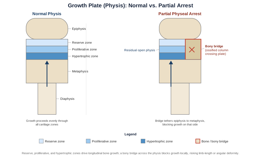
This is the single most important biomarker if a bony bridge is already suspected. A three-dimensional MRI can map exactly what percentage of the growth plate's cross-sectional area has been replaced by bone, and where that bridge sits relative to the plate's center versus its periphery. This matters because the decision to attempt a bar resection (physically removing the bony bridge and interposing fat or another material to keep the plate open) versus opting for a corrective procedure like guided growth or epiphysiodesis hinges almost entirely on bridge size and location. A study using 3D MR reconstruction found bridges ranging from under 1 percent to 47 percent of the physeal volume, with the 3D model closely matching what surgeons actually found during operation, which is why this has become the reference standard for surgical planning (3D MRI assessment of physeal growth arrest).
How to measure it
A dedicated physeal-mapping MRI protocol (not a routine knee or ankle MRI) is done at a hospital or imaging center with pediatric orthopedic experience, typically costing between 1,000 and 3,000 US dollars depending on whether sedation is needed and whether contrast is used. It's usually ordered by a pediatric orthopedic surgeon once a bridge is suspected from X-ray findings or growth arrest lines.If the score is bad: the plan without supplements
A bridge under roughly 25 to 30 percent of a peripheral physis, in a child with substantial growth remaining, is often the best candidate for bar resection. Without any device or supplement, the supporting plan is activity modification to avoid further stress on the plate, physical therapy to maintain joint range of motion around the affected bone, and a repeat MRI in 6 to 12 months to see whether the bridge is stable or expanding.If the score is bad: the plan with supplements or equipment
For bridges that are large, central, or in a child with limited growth remaining, the realistic "equipment" options are surgical: bar resection with fat or Cranioplast interposition, guided growth using a temporary tension-band plate or staple on the healthy side of the plate, or, if a discrepancy has already developed, a limb-lengthening nail (such as a magnetic intramedullary lengthening nail). These carry real surgical risks — infection, regrowth of the bridge, overcorrection, or nerve and vascular injury — and are done on a one-time or staged basis rather than a recurring schedule. Supplements have no established role in bridge resolution itself, though the nutrient support discussed under the bone turnover markers below is relevant to how well the bone remodels afterward.Bone Age and Skeletal Maturity
Bone age — assessed from a hand-and-wrist X-ray using the Greulich-Pyle atlas or the more knee-focused Sauvegrain method — tells you how much growth potential is actually left, independent of the child's chronological age. Two eight-year-olds can have very different amounts of remaining growth depending on skeletal maturity, and this number is what orthopedic surgeons use to time interventions like epiphysiodesis or guided growth so they land at the right moment. A comparison of bone age methods around the knee found that Greulich-Pyle-based estimates, particularly automated scoring, predicted remaining growth more reliably than chronological age alone (comparison of bone age methods in predicting remaining growth).
How to measure it
A single hand-and-wrist or knee X-ray, read by a radiologist or orthopedic surgeon against a standard atlas, costs roughly 50 to 150 dollars. It's typically repeated every 6 to 12 months when growth is actively being monitored.If the score is bad: the plan without supplements
If bone age is significantly advanced or delayed relative to chronological age, the non-supplement plan is straightforward: consistent sleep (growth hormone pulses concentrate in deep sleep), adequate total calorie and protein intake for age, and addressing any chronic illness or inflammation that could be suppressing or accelerating maturation.If the score is bad: the plan with supplements or equipment
A bone age that's markedly out of step with chronological age warrants referral to a pediatric endocrinologist rather than self-directed supplementation. If an underlying growth hormone deficiency or precocious puberty is found, treatment (GH injections, or medication to delay puberty) is prescription-based, given on a daily or monthly schedule depending on the therapy, and requires monitoring for side effects such as joint pain, fluid retention, or, rarely, elevated intracranial pressure.Growth Velocity and Limb Length Tracking
This is the simplest biomarker and arguably the most underused one. Standing height every 3 to 6 months, plus a scanogram (a specialized long-leg X-ray that measures limb segments precisely) when a discrepancy is suspected, catches a slowing growth plate well before it becomes visually obvious. A growth velocity that drops off on one side relative to the other is often the first clue that a physis is underperforming, even before a bridge is visible on standard imaging.
How to measure it
Standing height can be tracked at home with a wall-mounted stadiometer (20 to 40 dollars) or at any pediatric visit. A scanogram, done at a radiology center, costs roughly 100 to 300 dollars and is usually ordered every 6 to 12 months once a discrepancy is suspected.If the score is bad: the plan without supplements
For a mild, stable discrepancy, a shoe lift, gait-focused physical therapy, and continued measurement every 3 to 6 months is often all that's needed, particularly if there's still meaningful growth remaining and the trend isn't worsening.If the score is bad: the plan with supplements or equipment
For a discrepancy that's progressing, the equipment-based options are guided growth (a small plate or staple placed on the longer side's plate to slow it temporarily) or epiphysiodesis (permanently stopping growth on the longer side at the calculated right time), both one-time outpatient procedures. Larger established discrepancies may call for a lengthening nail, which lengthens the shorter bone gradually over weeks with periodic imaging to confirm the new bone is forming correctly. None of these are supplement-driven; nutrient support (protein, calcium, vitamin D — see below) mainly matters for how well the newly formed bone consolidates.Serum IGF-1 and IGFBP-3
Insulin-like growth factor 1 and its main binding protein are downstream of growth hormone and correlate reasonably well with a child's overall growth drive, including skeletal maturity. They're not physeal-arrest-specific, but a child with low IGF-1 relative to age and sex has less hormonal "fuel" behind whatever growth potential remains in an injured plate, which is relevant when deciding how aggressively to intervene. Research on IGF-1, IGFBP-3, and their ratio has shown a real correlation with skeletal maturity stage, supporting their use as a biochemical proxy for growth status (serum IGF-1 and IGFBP-3 as growth maturity indicators).
How to measure it
A blood draw analyzed for IGF-1 and IGFBP-3, typically ordered by a pediatrician or endocrinologist, costs roughly 100 to 250 dollars depending on whether it's bundled with other hormone panels.If the score is bad: the plan without supplements
Low IGF-1 in an otherwise healthy child often responds to addressing the basics first: sufficient total calories and protein (IGF-1 is nutritionally sensitive and drops quickly under-fed states), consistent sleep timing, and resolving any chronic illness, since inflammation suppresses the GH-IGF-1 axis.If the score is bad: the plan with supplements or equipment
If IGF-1 stays low after nutrition and sleep are corrected, the next step is endocrinology referral rather than supplementation — true growth hormone deficiency is treated with prescription GH, given as a daily subcutaneous injection, with IGF-1 rechecked every 3 to 6 months to titrate the dose. Over-treatment risk includes accelerated bone age advancement (which can close growth plates prematurely) and mild insulin resistance, both of which are why this isn't a self-directed intervention.25-Hydroxyvitamin D
Vitamin D governs how efficiently calcium and phosphate are absorbed and delivered to the growth plate's mineralization front. Chronic deficiency during active growth is one of the more correctable contributors to poor bone mineralization and slower healing after a physeal injury, and it's also one of the cheapest things to check and fix. Research on vitamin D's regulation of bone mineralization during growth outlines exactly how deficiency can impair the mineralization process at the growth plate (vitamin D regulation of bone mineralization during growth).
How to measure it
A standard blood test, widely available through any pediatrician or lab, costs roughly 40 to 100 dollars. It's reasonable to check at diagnosis of any physeal injury and again 3 months after starting supplementation.If the score is bad: the plan without supplements
Moderate, sensible sun exposure (roughly 10 to 20 minutes a few times a week, depending on skin tone and latitude) and dietary sources like fatty fish, egg yolks, and fortified milk can meaningfully move a mildly low level, though this is slower and less reliable than supplementation for a true deficiency.If the score is bad: the plan with supplements or equipment
For a confirmed deficiency, pediatric guidance typically supports 1,000 to 2,000 IU of vitamin D3 daily, taken with a fat-containing meal for absorption, rechecked at 3 months. Pairing it with magnesium and vitamin K2 (which helps direct calcium toward bone rather than soft tissue) is a reasonable, low-risk addition. Side effects are rare at these doses but toxicity (hypercalcemia) can occur with very high or poorly monitored dosing, which is why recheck testing matters rather than dosing indefinitely without follow-up.Bone Turnover Markers: P1NP and CTX
P1NP (a marker of new collagen formation) and CTX (a marker of bone breakdown) together describe the overall pace of bone remodeling. In a child recovering from a physeal or fracture injury, an imbalance — resorption outpacing formation for an extended period — can flag delayed healing or nutrient insufficiency worth addressing before it affects the healing bone. Pediatric turnover markers behave differently than in adults and change substantially with age and pubertal stage, so a recent review is useful for interpreting them correctly rather than applying adult reference ranges (bone turnover markers in children: clinical interpretation).
How to measure it
A blood draw for P1NP and CTX together typically runs 80 to 200 dollars and is best interpreted by a pediatric endocrinologist or orthopedic specialist familiar with age-specific reference ranges, since these markers vary enormously across normal childhood development.If the score is bad: the plan without supplements
For a formation-resorption imbalance during fracture or physeal healing, appropriate weight-bearing activity (as cleared by the treating surgeon), adequate dietary protein, and avoiding prolonged unnecessary immobilization all support the formation side of the ledger.If the score is bad: the plan with supplements or equipment
A combined calcium, vitamin D3, and K2 regimen, dosed to age-appropriate levels and rechecked in 3 to 6 months, is the standard low-risk supplement approach. Bisphosphonates or other bone-remodeling medications are reserved for specific, specialist-managed situations (such as certain metabolic bone diseases) and are not a routine response to an isolated turnover marker abnormality, given their side-effect profile and long skeletal half-life in growing bone.Calcium, Phosphate, PTH, and Alkaline Phosphatase Panel
This basic mineral panel screens for the metabolic conditions that can masquerade as or worsen a growth problem — things like renal phosphate wasting, vitamin D-resistant rickets, or parathyroid dysfunction. It's inexpensive, widely available, and worth running at least once in any child with an unexplained growth plate problem, since an undiagnosed metabolic bone disease changes the entire management plan.
How to measure it
This panel is usually included in a standard metabolic blood panel, costing roughly 100 to 250 dollars depending on whether it's ordered as a standalone or bundled test, and can be drawn at any standard lab.If the score is bad: the plan without supplements
Dietary calcium and phosphate (dairy, leafy greens, fish with bones, legumes) alongside sensible sun exposure address mild deficiencies. This isn't sufficient if the abnormality stems from a renal or parathyroid disorder, which requires medical management rather than diet alone.If the score is bad: the plan with supplements or equipment
Persistent abnormalities warrant pediatric endocrinology or nephrology referral before starting supplementation, since treating the wrong mechanism (for example, giving standard vitamin D for a renal phosphate-wasting condition) won't fix the underlying problem and can occasionally worsen it. Once a mechanism is identified, treatment is specific to it — active vitamin D analogs, phosphate supplementation, or other targeted therapy — dosed and monitored by the specialist, with side effects and monitoring schedule dependent on the exact condition.Biomarkers describe what's happening right now; genes describe the baseline potential and sensitivity a child's biology brings into that picture, which is where the next section picks up.
The Genes Behind Growth Plate Potential
It's worth being direct about the evidence here: physeal arrest itself is overwhelmingly a mechanical and traumatic problem — a fracture through the growth plate, an infection, a burn, or a vascular injury — not a genetic disease. No gene test will tell you whether a specific bar has formed after a specific fracture. What genetics does inform is the baseline growth potential and nutrient-pathway sensitivity a child has to work with once an injury has occurred, and how aggressively that might need to be supported. This is the same framing that genomics researchers like Ali Torkamani have pushed in precision-health circles — using genetic and biomarker data together rather than either alone — and that biomarker-focused educators like Gary Brecka have popularized for framing personal health decisions. Applied here, it means genetics is context, not a diagnosis.
NPR2
NPR2 encodes the receptor for C-type natriuretic peptide (CNP), a signal that drives chondrocyte proliferation directly at the growth plate. Loss-of-function variants cause acromesomelic dysplasia (Maroteaux type) and shorter stature, while gain-of-function variants are linked to tall stature — this pathway is a genuine, well-studied driver of how much a growth plate proliferates on its own (CNP-related short and tall stature disorders).
If this pathway runs on the weaker side, the plan without supplements centers on making sure nothing else is additionally suppressing growth: adequate sleep, sufficient caloric and protein intake, and avoiding chronic under-fueling, since these compound with a naturally lower proliferative baseline. The plan with equipment is more specialized: vosoritide, a CNP analog delivered as a daily subcutaneous injection, is FDA-approved for achondroplasia (a different, FGFR3-driven condition) and works by amplifying signaling through this same CNP-NPR2 axis. It is not currently approved or established for isolated NPR2 variants or for post-traumatic physeal arrest, but it illustrates that this pathway is druggable, and it's a reasonable question to raise with a pediatric endocrinologist if growth potential is a specific concern. Side effects include injection-site reactions and transient low blood pressure, and it requires ongoing specialist monitoring, not self-directed use.
SHOX
SHOX is expressed in the hypertrophic chondrocytes of the growth plate and is one of the more clearly established growth genes in humans — deficiency causes Leri-Weill dyschondrosteosis and a share of what's otherwise labeled idiopathic short stature, with well-documented effects on limb growth specifically (SHOX deficiency disorders).
Without supplements, the practical response to a known or suspected SHOX-related growth limitation is the same nutritional and sleep foundation as above, since SHOX-driven short stature isn't caused or worsened by diet, but a poor nutritional foundation still subtracts from whatever growth potential is present. With equipment, growth hormone therapy is actually an approved, well-established treatment specifically for SHOX deficiency, dosed as a daily injection and monitored with growth velocity and bone age checks every 6 months to avoid advancing bone age too quickly, which would close the growth plates prematurely and defeat the purpose. Side effects mirror standard GH therapy: injection-site irritation, occasional joint discomfort, and rare intracranial pressure elevation, all of which are why this is prescribed and monitored by a specialist rather than self-managed.
FGFR3
FGFR3 does the opposite job of NPR2 — it restrains chondrocyte proliferation and differentiation, and a gain-of-function variant (most commonly p.Gly380Arg) constitutively over-activates this brake, causing achondroplasia (achondroplasia gene review).
Without supplements, management is supportive: physical therapy, monitoring for orthopedic and neurological complications (like spinal stenosis) that come with this specific condition, since the core growth restriction is structural rather than nutrient-driven. With equipment, vosoritide again applies here — this time on-label, since achondroplasia is its approved indication — increasing growth velocity by roughly 1 to 2 cm per year in clinical trials through daily injection, with the same monitoring requirements and side-effect profile noted above (injection reactions, blood pressure changes, and required specialist oversight). This is relevant to physeal arrest discussions mainly as context: FGFR3 activity is one of the biological "brakes" that determines how much proliferative reserve a physis has to draw on when recovering from injury.
VDR
The vitamin D receptor gene has several well-studied polymorphisms (FokI, BsmI, TaqI among them) that influence how efficiently a given amount of circulating vitamin D gets converted into a biological effect on bone. The evidence here is more modest than for SHOX or FGFR3 — associations with bone mineral density and calcium absorption efficiency are real but smaller and less consistent across studies, so this should be read as a modifier rather than a determinant.
Without supplements, the practical implication is behavioral: a child whose VDR variants make them a less efficient vitamin D responder benefits disproportionately from consistent, moderate sun exposure and vitamin D-rich foods, since they're starting from a lower baseline efficiency. With supplements, the same vitamin D3 dosing described in the biomarker section above still applies, but the practical takeaway is to lean on the 25-hydroxyvitamin D blood level itself as the target, rather than a fixed dose, since a "normal" dose may leave a less-efficient VDR variant carrier still functionally low. Rechecking levels 3 months after any dose change is the way to confirm the supplement is actually working for that individual, and standard vitamin D3 dosing carries the same low toxicity risk noted earlier, requiring monitoring rather than open-ended increases.
Genes and biomarkers both describe biology already at work — the next section looks at a book that reframes how nutrient signaling shapes that biology in the first place, which is a perspective most orthopedic guidance leaves out entirely.
What Deep Nutrition Gets Right About Growth Plates
Catherine Shanahan's Deep Nutrition: Why Your Genes Need Traditional Food isn't a growth-plate or orthopedics book, but it spends more time on nutrient-driven skeletal development than almost anything written for a general audience, and its central argument challenges a common assumption in pediatric care: that genetic growth potential is fixed and diet is a minor variable around the edges. Shanahan, a physician, argues instead that nutrient signaling during pregnancy and childhood actively shapes how growth-plate genes get expressed — which makes it a useful complement to the more mechanistic sections above.
Genes need instructions, not just raw material
The book's core claim is that DNA is a set of possibilities, not a fixed blueprint, and that specific nutrients act as the signals that tell growth-plate genes which possibility to express. A diet missing those signals doesn't just under-fuel growth, it can under-instruct it.The four pillars of traditional diets
Shanahan organizes ancestral eating patterns around four recurring elements: meat cooked on the bone, organ meats, fermented and sprouted foods, and fresh (unprocessed, unoxidized) foods — arguing that each supplies growth-relevant nutrients that are largely absent from a modern processed diet.Bone broth and cartilage matrix
Broth made from bones and connective tissue supplies glycine, proline, and collagen-building blocks that overlap substantially with what growth-plate cartilage matrix is made of — a specific, practical link between an old kitchen habit and the biology discussed in the bone turnover section above.Fat-soluble vitamins direct calcium traffic
Vitamins A, D, and K2 work together to determine whether calcium gets deposited in bone or in soft tissue — echoing the same vitamin D and K2 pairing discussed under the biomarker plans above, but framing it as a coordinated system rather than a single nutrient to fix in isolation."Genetic amnesia" across generations
One of the book's more provocative claims is that generations of nutrient-poor eating can progressively narrow a population's expressed growth potential — including facial and skeletal proportions — even though the underlying DNA sequence hasn't changed. This is presented as an epigenetic effect, and it's the part of the book most directly relevant to how growth-plate genes get switched on or off by environment.Prenatal and early-childhood windows matter most
Shanahan places heavy emphasis on nutrient status during pregnancy and the first years of life as the period with the most lasting influence on skeletal development, arguing that catch-up later in childhood is possible but partial rather than complete.Vegetable oils and inflammatory signaling
The book argues that oxidized vegetable oils promote a low-grade inflammatory state that interferes with normal growth signaling — a claim that's more debated in mainstream nutrition science than the fat-soluble vitamin points above, and worth treating as a hypothesis rather than settled fact.Facial structure as a visible growth-plate story
Because craniofacial bones grow via their own growth-plate-like sutures, Shanahan uses jaw and dental arch development as a visible, easy-to-photograph proxy for the same nutrient-driven growth processes happening less visibly in the long bones — a useful mental model even though it's not a direct measurement of limb physes.Real fat and cholesterol are growth substrates, not just calories
The book pushes back on reflexive fat restriction in growing children, arguing that dietary fat and cholesterol are structural building blocks for developing tissue, not simply an energy source to be minimized.Diet as a modifiable input, not a replacement for medical care
To Shanahan's credit, the book doesn't claim nutrition can undo a structural injury like a bony bridge — its argument is about baseline potential and resilience, which is exactly how it should be read alongside the biomarker and gene sections above: as a modifier of the environment those genes and growth plates are operating in, not a substitute for orthopedic management of an actual arrest.Nutrition and genetics describe the internal environment; the next section turns to what can realistically help from outside that environment — supportive therapies with actual human evidence behind them.
Complementary Approaches Worth Considering
None of the following changes the structural reality of a bony bridge across a growth plate. What they can realistically help with is pain, anxiety around procedures and recovery, and — in a couple of cases — the mechanics of bone healing itself. The evidence quality varies a lot across these, and it's worth being honest about which ones are well-supported versus which are plausible but thin.
Photobiomodulation (Low-Level Laser Therapy)
Photobiomodulation uses low-intensity laser light to stimulate cellular activity in healing tissue, and it has a genuine, if still limited, human evidence base specifically in bone consolidation — which makes it more directly relevant to physeal and fracture recovery than most complementary options on this list.
A human trial applying low-level diode laser during the bone consolidation period after mandibular distraction osteogenesis found improved bone density and shortened consolidation time on the treated side compared to the control side within the same patients (low-level laser therapy in distraction osteogenesis bone consolidation).
Realistically, this is something to discuss with the treating orthopedic surgeon rather than attempt independently — it requires a clinical-grade device and appropriate dosing parameters, and the evidence, while genuine, comes from small trials and a different bone-lengthening context (mandibular distraction) rather than physeal arrest directly, so expectations should stay modest.
Guided Imagery
Guided imagery is a structured relaxation technique using directed mental visualization, most useful here for the anxiety and pain that come with imaging appointments, injections, cast changes, or surgical recovery in children with a physeal injury.
A randomized controlled trial comparing guided imagery to virtual reality for pediatric procedural pain and distress found comparable effectiveness between the two, with guided imagery showing a particular advantage in patients with higher baseline anxiety (guided imagery and virtual reality for pediatric procedural pain).
It's low-risk and easy to apply practically: a short, age-appropriate recorded script or a trained child-life specialist before an injection, cast change, or MRI, with no equipment required beyond a phone or tablet for audio.
Music Therapy
Music-based interventions before or during pediatric surgery have one of the stronger evidence bases on this list, specifically for reducing perioperative pain and anxiety — relevant for children facing bar resection, guided growth surgery, or lengthening procedures.
A systematic review and meta-analysis of randomized controlled trials on perioperative music interventions in pediatric surgery found a statistically significant reduction in postoperative pain and anxiety across the pooled studies (perioperative music interventions in pediatric surgery).
This is easy and low-cost to apply: familiar, calming music played before anesthesia induction and in the recovery room, ideally chosen with the child rather than for them, with no known downside beyond simply not being the right fit for every child's preferences.
Mindfulness Meditation
Mindfulness-based approaches are more relevant to the chronic pain and prolonged recovery some children face after limb-lengthening or repeated surgical procedures than to the acute injury itself.
A pilot study of mindfulness meditation for pediatric chronic pain found no significant reduction in reported pain intensity, but did find modest functional improvement and a notable reduction in parental worry, with the intervention rated as feasible and acceptable by participating families (pilot study of mindfulness meditation for pediatric chronic pain).
Given that mixed but genuinely reported result, this is worth trying as a low-risk adjunct for a child managing a long recovery (particularly during external fixator lengthening, which can take months), rather than as a primary pain-management strategy on its own.
Massage Therapy
Massage therapy is commonly suggested for postoperative orthopedic recovery, but the direct pediatric evidence is thin and, in the study available, not clearly positive.
A pilot study of massage therapy in postoperative rehabilitation for children and adolescents after lower-limb orthopedic surgery found no significant reduction in heart rate (used as a stress proxy) in the massage group compared to controls, in a very small sample (massage therapy in pediatric postoperative orthopedic rehabilitation).
Given the limited and inconclusive evidence, massage is reasonable to offer for general comfort and parent-child bonding during recovery, but it shouldn't be expected to produce a measurable physiological effect, and it's not a substitute for prescribed physical therapy around a healing physis.
Bringing It Together
Physeal arrest is fundamentally a mechanical problem — most often the result of a Salter-Harris fracture, with the higher-grade patterns carrying the greatest risk of premature closure (Salter-Harris fracture classification) — and no supplement, gene test, or complementary therapy changes that underlying structural reality. What does change is how early a problem is caught and how well-informed the decisions around it are. MRI bridge mapping and bone age tell you what's structurally happening and how much time is left to act; growth velocity, IGF-1, vitamin D, and the mineral and turnover panels tell you whether the hormonal and nutrient environment is helping or hindering whatever growth potential remains; and genetics adds useful context about baseline potential without pretending to predict or prevent an injury-driven bridge. The complementary approaches covered here are genuinely useful for pain and recovery, and in the case of photobiomodulation, plausibly for bone consolidation itself, but they sit alongside orthopedic management, not in place of it.
If there's one practical takeaway, it's this: don't wait for a visible limp or an obvious limb-length difference to start tracking. A baseline MRI or bone age reading soon after a significant physeal injury, followed by growth velocity checks every few months and a basic blood panel if anything looks off, gives an orthopedic surgeon real data to work with instead of a guess. Bring these specific tests to your next appointment, ask directly whether a bridge has been mapped and how much growth remains, and let that conversation — not general reassurance — set the schedule for what gets checked next.
Musculoskeletal: Bone Conditions Sports Injuries