This article contains AI generated content.
· UpdatedPityriasis Rosea Genes and Biomarkers: 5 Genes and 6 Biomarkers to Track
Introduction
If you have been diagnosed with pityriasis rosea, the conversation with your dermatologist probably ended with some version of the same advice: keep the skin moisturized, avoid hot showers, and wait. Most cases resolve within six to twelve weeks. For many people, that is true. But for those who experience recurrent episodes, widespread or intensely itchy rashes, or a prolonged course that disrupts daily life, the standard script leaves a frustrating gap between what is happening and what can actually be done about it.
Pityriasis rosea is not simply a skin irritation. It is a dermatological syndrome driven, in most cases, by the reactivation of Human Herpesvirus 6B (HHV-6B) and Human Herpesvirus 7 (HHV-7) — two viruses that most people carry silently for life after childhood exposure. The question is not why you were exposed to these viruses (almost everyone has been), but why your immune system permits reactivation while others' does not. That selective vulnerability is biological, measurable, and in many respects modifiable.
Not everyone with HHV-6 or HHV-7 latency develops pityriasis rosea. Immune control, inflammatory signaling, vitamin D status, antiviral cytokine output, and herpesvirus-specific antibody responses all vary significantly from person to person — and much of that variation has a genetic foundation. Understanding your immune landscape can help explain why episodes happen, why they recur, how severe they tend to be, and what targeted interventions are most likely to work for your specific biology.
This article explores that landscape through two lenses. The primary section maps six biomarkers that reflect your immune readiness, inflammatory baseline, and viral vulnerability — each with guidance on how to test and what to do when results fall outside the optimal range. A second section then examines five gene variants that shape susceptibility at a deeper level, providing the mechanistic explanation for why those biomarkers trend the way they do. Beyond those two frameworks, you will find a synthesis of what current research on viral reactivation and skin immunity reveals — and four evidence-supported complementary approaches worth knowing about. The goal throughout is not to promise a cure but to replace vague advice with precise, actionable information.
Summary
Pityriasis rosea is a viral-immune event, and who develops it, how severely, and how often depends on a specific set of biological variables that most dermatology visits never measure. This article breaks down six key biomarkers — HHV-6/HHV-7 antibody titers, high-sensitivity CRP, vitamin D, lymphocyte-to-monocyte ratio, IL-4, and interferon-gamma — each with clear guidance on how to test, what out-of-range results mean, and what to do about them both with and without supplements. It then explores five relevant gene variants — TLR3, TNF-α, IL4, CCR5, and HLA-B — explaining how each one shapes your immune susceptibility and what practical compensatory strategies look like. After that, ten research-backed insights on viral reactivation biology provide a framework for long-term immune management, followed by four evidence-supported complementary approaches — including light therapy, mindfulness, microbiome support, and photobiomodulation — that have real clinical data behind them for this type of condition. If your dermatologist told you to wait it out, this is the map they didn't hand you.
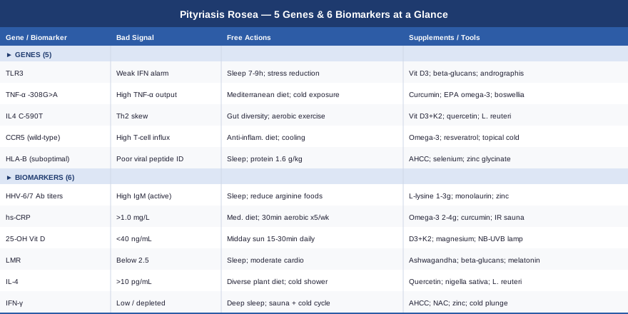
6 Biomarkers Worth Tracking If You Have Pityriasis Rosea
Standard clinical care for pityriasis rosea focuses almost entirely on symptom management: what the rash looks like and how uncomfortable it is. But the immune story unfolding beneath those lesions is measurable in blood and can be tracked over time. For people with recurrent episodes or a prolonged course, these six biomarkers provide a practical monitoring framework that connects your individual biology to actionable interventions.
Biomarker 1: HHV-6 and HHV-7 Antibody Titers
Why it matters: The most consistent finding in pityriasis rosea research is its association with the reactivation of Human Herpesvirus 6B and Human Herpesvirus 7. These viruses, belonging to the betaherpesvirus subfamily, establish latent infection in early childhood — in T-lymphocytes and salivary gland tissue — and remain dormant indefinitely in most people. In a subset, immune disruption (stress, illness, sleep deprivation, immune suppression) allows reactivation, and that reactivation triggers the cytokine cascade that produces the characteristic PR rash. Multiple studies, including Drago et al. (Dermatology, 1997), detected HHV-7 DNA in plasma during acute PR episodes and found elevated IgM antibodies indicating active viral replication. This is not incidental — it is the most probable mechanism in most cases.
Measuring IgG and IgM antibodies to both HHV-6 and HHV-7 provides a window into whether your immune system has recently controlled a reactivation event (elevated IgG, normal IgM) or is currently dealing with one (rising IgM). For people with recurrent PR, serial testing during and between episodes can reveal a clear reactivation cycle that correlates with known stressors.
How to Measure It
HHV-6 and HHV-7 IgG/IgM antibody panels are available through major reference labs including LabCorp and Quest. Cost: $80–$200 depending on the panel. PCR testing for HHV-6B DNA directly in plasma or saliva is more informative during an active episode and costs $150–$300 through specialty labs or through the HHV-6 Foundation's recommended diagnostic centers. An infectious disease specialist or immunologist can order these; some direct-to-consumer labs also offer them.
If the Marker Is Elevated: The Plan Without Supplements
Active reactivation (elevated IgM) should prompt a structured review of your recent immune stressors: sleep quality, psychosocial stress load, any concurrent illness, and recent physical overtraining. Prioritizing 7–9 hours of sleep is the single highest-impact step, since NK cell activity — the immune arm most responsible for suppressing herpesviruses — is tightly coupled to deep sleep duration. High-intensity exercise should be moderated during active episodes; intense exertion transiently suppresses NK cell function. Reducing arginine in the diet during vulnerable periods is also worthwhile: herpesviruses depend on arginine for replication, and minimizing intake from peanuts, chocolate, and excess nuts reduces this substrate.
If the Marker Is Elevated: The Plan With Supplements or Equipment
L-Lysine: 1,000–3,000 mg/day during active or high-risk periods. Lysine competes with arginine for herpesvirus replication machinery. Cycle: use during outbreaks or periods of immune stress, then discontinue. Side effects at sustained high doses include gastrointestinal discomfort. Monolaurin: 600–1,800 mg/day has antiviral properties against enveloped viruses including herpesviruses. Evidence is primarily in vitro, but safety profile is favorable. Zinc glycinate or picolinate: 25–40 mg/day supports NK cell function. Pair with 1–2 mg copper daily for long-term use to prevent copper depletion. Red light therapy (photobiomodulation, 630–850 nm): 10–20 minutes per session daily. Supports mitochondrial function in immune cells and may modulate inflammatory signaling, though PR-specific evidence is currently limited.
Biomarker 2: High-Sensitivity C-Reactive Protein (hs-CRP)
Why it matters: hs-CRP is the most widely available and clinically validated marker of systemic inflammation. While pityriasis rosea is often framed as a localized skin condition, the immune activation driving it is systemic — and many people with recurrent or severe PR have a chronically elevated inflammatory baseline even between episodes. This elevated baseline acts as a permissive environment for HHV reactivation by suppressing the antiviral immune tone that keeps latent viruses dormant.
Peter Attia, who regularly uses hs-CRP as one of his primary clinical monitoring tools in longevity medicine, emphasizes that it responds reliably to lifestyle interventions and that its prognostic value extends far beyond cardiovascular risk — including immune regulation. For people managing PR recurrence, baseline hs-CRP is one of the most actionable single markers to track because its trajectory reflects multiple system-level changes simultaneously.
How to Measure It
hs-CRP is a standard blood test costing $15–$40, available at virtually any lab and often covered by insurance. Optimal target: below 0.5 mg/L for people with immune-related conditions. Levels of 1.0–3.0 mg/L indicate mild but meaningful chronic inflammation; above 3.0 mg/L is considered elevated and warrants further investigation. Retest every 3–6 months when tracking the effect of interventions.
If the Score Is Elevated: The Plan Without Supplements
The Mediterranean dietary pattern — rich in oily fish, extra-virgin olive oil, leafy vegetables, legumes, and whole grains — has the strongest evidence base for reducing hs-CRP. Eliminating ultra-processed foods, refined carbohydrates, and industrial seed oils removes the primary dietary drivers of inflammatory signaling. Thirty minutes of moderate-intensity aerobic exercise at least five days per week is one of the most reliable hs-CRP–lowering interventions in the clinical literature. Improving sleep quality and treating any underlying sleep apnea also produce significant CRP reductions — disrupted sleep itself is a powerful inflammatory stimulus.
If the Score Is Elevated: The Plan With Supplements or Equipment
Omega-3 fatty acids (EPA + DHA): 2–4 g combined EPA+DHA per day. Multiple meta-analyses confirm CRP reduction. Take with meals to reduce gastrointestinal effects. Maintenance dose of 2 g/day is appropriate long-term; higher doses should be reassessed every three months. Curcumin (bioavailable form): 500–1,000 mg/day as phospholipid complex or nanoparticle formulation. Inhibits NF-κB and directly reduces CRP. Cycle 8 weeks on, 4 weeks off. At high doses, has mild blood-thinning properties — relevant if on anticoagulants. Infrared sauna: 2–3 sessions per week at 150–180°F for 15–20 minutes has emerging evidence for reducing circulating hs-CRP and supporting immune resilience. Avoid using during active, widespread skin flares.
Biomarker 3: 25-OH Vitamin D
Why it matters: Vitamin D is better understood as an immune hormone than as a nutrient. The vitamin D receptor (VDR) is expressed on nearly every immune cell type — T-cells, NK cells, dendritic cells, and macrophages — and vitamin D signaling directly regulates antiviral interferon production, inflammatory cytokine balance, and the differentiation of immune cell subtypes critical to herpesvirus control. Vitamin D deficiency (below 30 ng/mL) is associated with reduced NK cell activity, impaired interferon-alpha and interferon-beta responses, and heightened inflammatory cytokine production — all of which create a more permissive environment for HHV reactivation.
For people with recurrent pityriasis rosea, vitamin D is one of the highest-priority biomarkers because deficiency is common (affecting over 40% of adults in Western countries), testing is inexpensive, and correction is safe and well-studied. Multiple dermatology clinicians now recognize low vitamin D as a modifiable factor in recurrent viral skin conditions.
How to Measure It
A 25-OH vitamin D blood test costs $30–$60 and is frequently covered by insurance. Optimal range for immune function: 40–70 ng/mL (100–175 nmol/L). Most labs flag deficiency at below 20 ng/mL, but for immune-related conditions, functional medicine clinicians typically target 50–65 ng/mL. Retest every six months if actively supplementing.
If the Score Is Low: The Plan Without Supplements
Midday sun exposure (arms and legs exposed, 15–30 minutes at solar noon) produces 1,000–5,000 IU of vitamin D depending on skin tone, latitude, and season — far more efficiently than dietary sources. This approach is ideal where practical. Dietary contributions (fatty fish, egg yolks, fortified dairy) are meaningful but modest, and are unlikely to correct established deficiency on their own.
If the Score Is Low: The Plan With Supplements or Equipment
Vitamin D3: 2,000–5,000 IU/day for maintenance; 5,000–10,000 IU/day for 8–12 weeks to correct deficiency, then retest and reduce. Always pair with Vitamin K2 (MK-7 form): 100–200 mcg/day to direct calcium appropriately to bones rather than arterial walls. At sustained doses above 10,000 IU/day without blood level monitoring, vitamin D toxicity is possible — track serum levels. Magnesium glycinate or malate: 200–400 mg/day supports the enzymatic conversion of D3 to its bioactive form (1,25-OH vitamin D); deficiency in magnesium renders vitamin D supplementation less effective. Take in the evening; well-tolerated long-term. A narrowband UVB home lamp (311 nm) used for 10–15 minutes three times per week is an effective option for those with limited outdoor sun access. Dose should be calibrated carefully by skin type to avoid burns. Do not use during an active, inflamed PR episode.
Biomarker 4: Lymphocyte-to-Monocyte Ratio (LMR)
Why it matters: The lymphocyte-to-monocyte ratio is a simple calculation derived from a standard complete blood count with differential, and it captures the balance between adaptive immune capacity (lymphocytes) and innate inflammatory monocyte activity. A low LMR — typically below 2.5 — signals that the inflammatory, non-specific innate immune arm is dominant over the adaptive, virus-specific arm. This pattern appears during acute viral illness, after significant physiological stress, and in states of chronic systemic inflammation.
During an active pityriasis rosea episode, LMR commonly falls as monocyte counts rise in response to viral reactivation. Tracking LMR between episodes reveals whether your immune system has fully recovered its adaptive balance or remains in a sub-optimal inflammatory state that creates ongoing vulnerability. This is rarely discussed in dermatology literature, but it is highly informative and costs nothing beyond the CBC itself.
How to Measure It
A complete blood count with differential is a standard test costing $10–$30 at most labs. Divide the absolute lymphocyte count by the absolute monocyte count. Target: above 3.5 indicates good immune balance. Below 2.5 is worth addressing. Repeat at three-month intervals when monitoring recovery from recurrent PR.
If the Ratio Is Low: The Plan Without Supplements
The primary drivers of low LMR are chronic stress (which elevates cortisol and downstream monocyte production), insufficient or disrupted sleep, and sedentary lifestyle. Sleep restoration — specifically targeting 7–9 hours of high-quality sleep with consistent timing — is the most powerful single lever for normalizing immune cell distribution. Moderate aerobic exercise (not exhaustive) raises circulating lymphocytes and NK cell counts. Reducing chronic stressors decreases the cortisol signal that sustains monocyte elevation.
If the Ratio Is Low: The Plan With Supplements or Equipment
Ashwagandha (KSM-66 extract): 300–600 mg/day has documented effects on reducing cortisol and modulating immune cell ratios. Cycle: 8–12 weeks on, 4 weeks off. Occasional GI discomfort reported; theoretical thyroid effects at very high sustained doses. Beta-glucans (from oats or yeast): 250–500 mg/day support NK cell and macrophage maturation without amplifying pro-inflammatory signaling. Generally safe for long-term use. Low-dose melatonin: 0.3–1 mg taken 30 minutes before sleep supports sleep architecture and has direct immune-modulating properties, including lymphocyte support. Avoid higher doses (5–10 mg) unless specifically guided — they can alter circadian signaling and suppress endogenous melatonin production over time.
Biomarker 5: Interleukin-4 (IL-4) and Th2 Cytokine Profile
Why it matters: Pityriasis rosea involves a mixed immune response, with research indicating that the acute inflammatory phase is characterized in part by Th2-skewed cytokine activity — including elevated IL-4 and IL-13. These cytokines promote mast cell activation, eosinophil recruitment, and IgE-mediated inflammatory responses, which likely explain the intense pruritus (itching) that a significant minority of PR patients experience — an itch pattern more characteristic of atopic conditions than of typical viral rashes.
Persistent Th2 skewing between episodes may indicate incomplete immune recalibration and could reflect underlying genetic variants (see Gene 3: IL4 below). Tracking IL-4 or its surrogate markers can help determine whether your immune system's inflammatory tone has normalized or whether it remains in a state of ongoing Th2 dominance that predisposes you to recurrent flares.
How to Measure It
Direct IL-4 measurement requires a specialty cytokine panel, available through labs such as Cyrex Arrays or Vibrant America, costing $150–$400 depending on panel size. A more accessible and affordable surrogate is the eosinophil count from a standard CBC — elevated eosinophils (above 300 cells/µL) are a reliable marker of Th2 immune dominance. Reference range for serum IL-4 is typically below 10 pg/mL in most clinical assays, but laboratory-specific ranges apply.
If the Score Is Elevated: The Plan Without Supplements
Dietary patterns most strongly associated with Th2 reduction are whole-food, minimally processed diets with high fiber diversity. Reducing common dietary allergens (if concurrent atopy is present) and supporting gut barrier integrity through fermented foods and prebiotic fiber can meaningfully shift Th1/Th2 balance via gut-immune cross-talk. Brief cold exposure (cold showers or cold plunge) transiently amplifies Th1 signaling and provides a counterbalancing effect on Th2 dominance.
If the Score Is Elevated: The Plan With Supplements or Equipment
Vitamin D3 (as above): Directly suppresses IL-4 gene expression via the vitamin D receptor. Target blood level 50–65 ng/mL. Quercetin phytosome: 500 mg twice daily. Inhibits mast cell degranulation and IL-4 release at the cellular level. Cycle 8 weeks on, 4 weeks off. Well-tolerated. Nigella sativa (black seed oil): 2–3 mL/day or 500 mg standardized capsule twice daily. Published human evidence shows reduction in Th2 cytokine levels and eosinophil counts in inflammatory conditions. Use for 8–12 week cycles. Probiotics (L. rhamnosus GG or L. reuteri DSM 17938): specific strains with published evidence for modulating Th1/Th2 balance via the gut–immune axis. Minimum 8 weeks of consistent use to assess effect.
Biomarker 6: Interferon-Gamma (IFN-γ)
Why it matters: Interferon-gamma is the central antiviral cytokine of the Th1 immune response. It activates macrophages, coordinates cytotoxic T-cell activity, enhances antigen presentation, and directly suppresses herpesvirus replication in infected cells. Low IFN-γ output is associated with reduced viral clearance and is a plausible contributing factor in the incomplete immune control that allows HHV-6B and HHV-7 to periodically reactivate.
In the acute phase of pityriasis rosea, IFN-γ levels are often elevated — reflecting the immune system's attempt to contain the reactivation event. Between episodes, chronically low IFN-γ may signal an undertrained or depleted antiviral immune response. Measuring this marker gives a direct read of the antiviral arm's functional capacity.
How to Measure It
Serum IFN-γ is measurable through specialty immunology labs and academic medical centers, costing $100–$250. Stimulated whole-blood assays provide a better measure of functional immune capacity than a single serum snapshot. A more affordable proxy is the NK cell count (CD56+ lymphocytes) from a lymphocyte subset panel — NK cells are the primary IFN-γ–producing antiviral cells and their count reflects the same antiviral arm. Lymphocyte subset panels typically cost $100–$200.
If the Level Is Low: The Plan Without Supplements
Slow-wave (deep) sleep is the primary physiological event during which IFN-γ production peaks — making sleep quality the highest-leverage intervention for restoring antiviral cytokine output. Adequate dietary protein (1.6–2.0 g/kg body weight daily) supplies the amino acid substrate for immune cell synthesis. Alternating heat and cold exposure (sauna followed by cold plunge, or cold showers) has the most consistent evidence for acutely stimulating NK cell activity and IFN-γ output. Moderate aerobic exercise (not exhaustive intensity) raises NK cell count durably over time.
If the Level Is Low: The Plan With Supplements or Equipment
Medicinal mushrooms (AHCC, Reishi, Shiitake extract): 500–1,000 mg/day of standardized beta-glucan–containing extracts. Randomized controlled trial evidence exists for increasing NK cell activity and IFN-γ production (AHCC particularly). Use with a one-month break every three months. Zinc glycinate: 25–40 mg/day directly supports IFN-γ receptor signaling. N-acetylcysteine (NAC): 600–1,200 mg/day supports glutathione production, which protects NK cells from oxidative damage during immune activation. Take separated from zinc to avoid interaction. Cold plunge or cold shower: Water temperature 10–15°C, 3–5 minutes, three sessions per week. Multiple controlled studies confirm NK cell activation following cold water immersion. Avoid during active PR episodes when skin barrier is compromised.
With six measurable signals in hand, you now have a monitoring framework built around the actual biology of pityriasis rosea. What those markers reveal about your immune architecture also has a genetic foundation — and understanding that foundation helps explain why these patterns emerge and which interventions are most likely to move them in the right direction.
5 Key Genes That Shape Your Pityriasis Rosea Susceptibility
Genetics does not determine fate, but it does establish a biological baseline. Certain gene variants — particularly those governing antiviral signaling, inflammatory amplification, and immune cell trafficking — create environments that are more or less permissive to herpesvirus reactivation and inflammatory skin responses. Knowing which variants are relevant to your immune profile can explain patterns that otherwise seem random, and it points toward the interventions most likely to compensate effectively.
Gene 1: TLR3 (Toll-Like Receptor 3)
What it does: TLR3 encodes a pattern-recognition receptor that detects double-stranded RNA — the molecular signature produced during active replication of DNA viruses including herpesviruses. When TLR3 detects viral RNA, it initiates an interferon signaling cascade that suppresses viral spread and activates antiviral immune cells. The TLR3 rs3775291 variant (L412F) is a well-characterized loss-of-function single nucleotide polymorphism that impairs this signaling pathway, meaning carriers mount a delayed or reduced interferon response to viral replication events.
The connection to herpesvirus susceptibility is not theoretical. Research has linked TLR3 loss-of-function variants to increased susceptibility to herpes simplex encephalitis — one of the most striking demonstrations that this specific receptor is critical for herpesvirus immune control in the central nervous system and likely in peripheral tissue as well. For HHV-6B and HHV-7 reactivation driving PR, a suboptimal TLR3 response means the initial interferon alarm signal arrives late, giving the virus more opportunity to replicate before containment.
If the Gene Is Unfavorable: The Plan Without Supplements
Consistent sleep is the most powerful non-supplement intervention, because TLR3-mediated interferon production is regulated by circadian immune rhythms — the signaling pathway is enhanced during the nocturnal rest phase. Chronic sleep deprivation specifically impairs interferon-based antiviral signaling. Stress reduction is equally important: cortisol directly suppresses TLR3 expression, creating a compounding vulnerability in people who are both stress-exposed and carry this variant. Maintaining gut barrier health is also relevant — a significant portion of TLR3 activity occurs in gut epithelial cells, where it monitors luminal microbial and viral signals.
If the Gene Is Unfavorable: The Plan With Supplements or Equipment
Vitamin D3: Upregulates TLR signaling pathways through the vitamin D receptor — a direct mechanism for partially compensating reduced TLR3 activity. Target blood level 50–70 ng/mL. Beta-glucans (yeast-derived): Activate innate immune cells through TLR2 and Dectin-1 pathways independently of TLR3, providing a compensatory antiviral alerting signal. 250–500 mg/day, suitable for long-term use. Andrographis paniculata (standardized extract): 400–600 mg/day during high-risk periods (illness season, stress periods, seasonal change). Supports innate interferon induction via TLR-independent pathways. Use in 2–3 week cycles with breaks; potential interactions with blood-thinning medications. Near-infrared light (810–850 nm): 10–15 minutes per day applied to the chest region has emerging evidence for supporting innate immune cell function via mitochondrial activation in immune cells.
Gene 2: TNF (Tumor Necrosis Factor-Alpha, -308G>A Polymorphism)
What it does: The TNF gene encodes tumor necrosis factor-alpha — one of the most potent pro-inflammatory cytokines in the immune system. The -308G>A promoter polymorphism (rs1800629) increases TNF-α gene transcription, meaning that people carrying the A allele produce substantially more TNF-α in response to immune triggers. In the context of pityriasis rosea, where the HHV reactivation event triggers an inflammatory cascade, higher TNF-α production amplifies the dermal inflammatory infiltrate — potentially explaining why some individuals experience more widespread lesions, more severe itching, or a more prolonged course than others exposed to the same viral trigger.
This polymorphism is also associated with elevated susceptibility to inflammatory skin conditions including psoriasis and atopic dermatitis, making it relevant context if you have a personal or family history of overlapping immune skin conditions alongside PR.
If the Gene Is Unfavorable: The Plan Without Supplements
Anti-inflammatory dietary patterns (Mediterranean) directly reduce TNF-α secretion from adipose tissue and immune cells. Intermittent fasting using a 16:8 time-restricted eating window activates AMPK and autophagy pathways that downregulate TNF-α gene expression. Cold exposure — 2–5 minute cold showers or cold plunge — acutely reduces circulating TNF-α post-session through catecholamine-mediated anti-inflammatory signaling. Tobacco and excess alcohol are powerful TNF-α amplifiers and should be eliminated. Regular moderate aerobic exercise (not exhaustive) suppresses baseline TNF-α over weeks to months.
If the Gene Is Unfavorable: The Plan With Supplements or Equipment
Curcumin (phospholipid complex or nanoparticle form): 500–1,000 mg/day. Among the most studied natural TNF-α inhibitors — inhibits NF-κB, the transcription factor that drives TNF-α expression. Cycle 8 weeks on, 4 weeks off. Mild blood-thinning effect at high doses — discuss with a physician if taking anticoagulants. EPA-dominant omega-3: EPA specifically inhibits TNF-α via competitive eicosanoid pathways. 2–4 g EPA/day during inflammatory periods; 1–2 g/day for maintenance. Boswellia serrata (AKBA standardized extract): 100–250 mg/day. Inhibits both TNF-α and the 5-LOX inflammatory pathway. Well-tolerated long-term. Infrared sauna: 3 sessions per week with documented effects on reducing circulating TNF-α in patients with inflammatory conditions.
Gene 3: IL4 (Interleukin-4, C-590T Promoter Variant)
What it does: The IL4 gene encodes interleukin-4, the master driver of Th2 immune polarization. The C-590T promoter variant (rs2243250) increases IL-4 transcription, leading to stronger Th2 immune responses. In the skin, elevated IL-4 promotes mast cell activation, eosinophil infiltration, and IgE-driven inflammation — the same immune fingerprint associated with atopic dermatitis and allergic skin responses. People with pityriasis rosea who carry this variant may experience more intense pruritus and are more likely to have concurrent or subsequent atopic conditions.
Importantly, this variant also means that the Th1/Th2 immune balance is tilted by genetic default — not just by environmental triggers. The biomarker most directly connected to this gene is IL-4 itself (Biomarker 5 above), and eosinophil count from a standard CBC serves as the most accessible monitoring tool.
If the Gene Is Unfavorable: The Plan Without Supplements
Gut microbiome diversity is the strongest non-supplement lever for Th2 recalibration. Higher microbial diversity — cultivated through dietary plant diversity (30+ different plant foods per week), daily fermented foods, and avoidance of unnecessary antibiotics — is consistently associated with reduced atopic disease risk and better Th1/Th2 balance across the scientific literature. Regular aerobic exercise and brief cold exposure both amplify Th1 signaling, providing a physiological counterweight to the IL4 variant's Th2-promoting effect.
If the Gene Is Unfavorable: The Plan With Supplements or Equipment
Vitamin D3 + K2: Directly suppresses IL-4 gene expression via the VDR. Target 50–65 ng/mL blood level. Quercetin phytosome: 500 mg twice daily, inhibits IL-4 release from mast cells at the molecular level. Cycle 8 weeks, 4 weeks off. Lactobacillus reuteri DSM 17938: One of the best-studied probiotic strains for shifting Th2 toward Th1 balance via gut-immune cross-talk. 100–500 million CFU daily, minimum 8 weeks for assessment. Red light therapy (630–660 nm): Applied to active skin areas for 10 minutes per session once or twice daily. Reduces local inflammatory cytokine production and may counteract IL-4–driven skin inflammation in active lesions.
Gene 4: CCR5 (C-C Chemokine Receptor 5)
What it does: CCR5 is a chemokine receptor expressed on T-cells and macrophages that guides these immune cells to sites of infection and inflammation. In pityriasis rosea, CCR5-expressing T-cells migrate into the dermis in response to HHV-7 reactivation, forming the perivascular inflammatory infiltrate that characterizes active PR lesions. The CCR5-Δ32 deletion variant (rs333) produces a non-functional truncated receptor, reducing T-cell trafficking to inflamed tissue.
Homozygous CCR5-Δ32 carriers are the same individuals known to be naturally resistant to CCR5-tropic HIV — a well-documented genetic protective effect. In heterozygous form, the deletion reduces CCR5 surface expression by approximately 50%, potentially resulting in a less severe dermal infiltrate during PR episodes. Wild-type CCR5 (no deletion) — the most common status — means normal or higher CCR5 expression, which allows vigorous T-cell migration to inflamed skin. This is neither universally good nor bad, but it does mean that reducing the chemokine signals driving CCR5-mediated migration (primarily CCL5/RANTES) is a relevant strategy during active episodes.
If the Gene Shows Elevated CCR5 Expression: The Plan Without Supplements
Reducing systemic inflammatory load decreases CCL5/RANTES signaling that drives CCR5-mediated T-cell migration. The same anti-inflammatory dietary pattern, moderate exercise, and stress reduction measures recommended throughout this article collectively reduce CCL5 expression. During active PR flares, cooling compresses applied to lesion areas reduce local vasodilation and transiently decrease T-cell infiltration, providing symptomatic relief alongside a physiological benefit.
If the Gene Shows Elevated CCR5 Expression: The Plan With Supplements or Equipment
EPA-dominant omega-3: Reduces CCL5 chemokine expression. 2–4 g EPA+DHA per day during active periods. Resveratrol (trans-resveratrol, micronized form): 250–500 mg/day inhibits NF-κB — the master transcriptional regulator of CCL5 and other pro-migratory chemokines. Take with meals for better absorption. Cycle 8 weeks on, 4 weeks off. Potential estrogen-modulating effects at high doses — relevant to discuss with a physician in certain contexts. Topical cooling therapy: Gel cooling packs or dedicated cold therapy devices applied to lesion areas for 10–15 minutes twice daily during active episodes. Reduces local vasodilation, decreases inflammatory cell infiltration, and provides direct symptomatic relief for pruritus without any systemic effect.
Gene 5: HLA-B (Human Leukocyte Antigen-B)
What it does: HLA-B is part of the MHC class I system — the molecular machinery that presents viral peptides on the surface of infected cells for recognition and elimination by cytotoxic T-cells (CD8+ T-cells). Different HLA-B alleles present viral peptides with different efficiency and binding affinity. If your specific HLA-B type presents HHV-6B or HHV-7 peptides suboptimally, your cytotoxic T-cells will have a harder time identifying and killing infected cells, leaving reactivating virus to replicate for longer before containment.
HLA associations in pityriasis rosea are still being characterized — the research is less advanced than for conditions like psoriasis (HLA-Cw6) or drug hypersensitivity reactions (HLA-B*57:01). But given the fundamental role of HLA class I in herpesvirus recognition across the herpesvirus family, this gene family is clearly relevant to understanding individual susceptibility. Full HLA typing is now accessible through commercial genetic testing platforms and provides the most complete picture of your immune recognition landscape.
If the HLA Type Is Suboptimal: The Plan Without Supplements
Your HLA type does not change, but the efficiency of the cytotoxic T-cell response operating through that HLA type very much does. Preserving CD8+ T-cell number and function requires adequate protein intake (1.6–2.0 g/kg body weight daily), consistent high-quality sleep, zinc, and vitamin D — the same foundational pillars that support broader antiviral immune function. Avoiding unnecessary immunosuppressive medications (systemic steroids, certain antihistamines in excess) is particularly important for people whose viral recognition is already less efficient at the HLA level.
If the HLA Type Is Suboptimal: The Plan With Supplements or Equipment
AHCC (Active Hexose Correlated Compound): 3 g/day taken on an empty stomach. Multiple human clinical trials have demonstrated increases in NK cell and CD8+ T-cell activity — providing a compensatory boost to antiviral cytotoxic immunity regardless of HLA efficiency. Use during high-risk periods; 4-week breaks every 3 months. Selenium (selenomethionine): 100–200 mcg/day. Directly supports cytotoxic T-cell function and antiviral competence via selenoprotein-dependent antioxidant defense in immune cells. Long-term use above 400 mcg/day risks selenosis — stay within recommended range. Zinc glycinate: 25–40 mg/day. Supports MHC class I antigen presentation machinery and cytotoxic T-cell differentiation. Medically supervised narrowband UVB therapy: Activates skin-resident T-cells and Langerhans cells, the local antigen-presenting sentinels of the skin immune system, which operate downstream of — and partially independently of — systemic HLA-mediated recognition.
The genetic picture adds a layer of explanatory depth that biomarkers alone cannot provide. Understanding why your inflammatory signaling trends the way it does, why your antiviral response may be slower to engage, or why your immune system produces more intense skin inflammation in response to the same viral trigger as someone else — these insights transform the intervention recommendations from generic advice into individualized strategy. The next section draws on one of the most accessible and evidence-grounded sources for putting all of this biology into practical context.
What Research on Viral Reactivation and Skin Immunity Reveals: 10 Insights That Change How You Manage PR
The synthesis of neuroscience, immunology, and practical medicine that Andrew Huberman and his collaborators have assembled over hundreds of podcast episodes provides a uniquely useful framework for understanding conditions like pityriasis rosea — conditions where the immune system, the nervous system, and the skin intersect in complex ways. No single episode addresses PR directly, but the principles of viral latency, NK cell biology, stress-immune coupling, and sleep-immune architecture apply with remarkable precision to the HHV-driven dermatological syndrome that PR represents. Here are the ten most directly applicable insights.
1. Herpesviruses Are Permanent Residents — Management Is the Goal, Not Eradication
HHV-6 and HHV-7, like all herpesviruses, establish lifelong latent infection in host cells following primary exposure. The immune system does not eliminate these viruses; it establishes a dynamic containment relationship. Understanding this shifts the entire strategic frame: the goal is not to clear an infection but to maintain the immune conditions under which the virus remains suppressed. Every intervention in this article — from vitamin D to cold exposure to stress reduction — is best understood as a tool for strengthening that containment relationship, not treating a temporary illness.
2. Sleep Is the Most Powerful Single Antiviral Intervention Available
Cytotoxic T-cells and NK cells — the two immune populations most responsible for controlling herpesvirus reactivation — reach peak activity and peak IFN-γ production during slow-wave sleep. Controlled sleep restriction studies show that even six hours per night across two weeks reduces NK cell cytotoxic activity by up to 70%. For someone carrying TLR3 or HLA-B variants that already reduce viral recognition efficiency, layering chronic sleep restriction on top creates compounding vulnerability. Sleep is not a lifestyle preference — it is an antiviral intervention with a measurable dose-response.
3. Cortisol Is the Virus's Ally
Chronic psychosocial stress increases cortisol, and cortisol directly suppresses the antiviral immune arm — specifically NK cell cytotoxicity, CD8+ T-cell function, and IFN-γ production. Beyond the direct immunosuppressive effect, catecholamines released during chronic stress have been shown to directly activate latent herpesvirus replication machinery. Even anticipatory stress — worry about upcoming events without actual exposure to the stressor — produces measurable suppression of antiviral immunity. Managing stress is therefore not psychological self-care adjacent to treatment; it is a mechanistically direct antiviral action.
4. Cold and Heat Exposure Train NK Cell Function
The research on cold water immersion and sauna as tools for NK cell activation is consistent and growing. Brief cold water exposure at 10–15°C for 3–5 minutes produces a catecholamine surge that transiently activates NK cells and increases circulating lymphocyte count. Heat exposure at 80–90°C for 15–20 minutes generates a heat shock protein response that supports immune surveillance and NK cell maturation over time. Alternating both creates a hormetic training effect on the antiviral immune arm. Three sessions per week — whether through dedicated cold plunge and sauna access or consistent cold showers — represents one of the most evidence-grounded non-pharmacological immune resilience practices available.
5. Exercise Dose Matters — Too Much Is Immunosuppressive
Moderate-intensity aerobic exercise at 65–75% of maximum heart rate for 30–45 minutes consistently raises NK cell count and CD8+ T-cell activity for hours post-session and improves immune resilience over weeks to months. But exhaustive high-intensity exercise — particularly daily high-intensity interval training or endurance sports above 90 minutes at high intensity — creates a transient post-exercise open window of immune suppression during which NK cell activity drops and herpesvirus reactivation becomes more likely. For people managing recurrent PR, this is a critical nuance: the training strategy should favor volume at moderate intensity over maximum intensity.
6. The Gut-Immune Axis Is a Genuine Therapeutic Target
Approximately 70% of immune cells reside in or immediately adjacent to the gut-associated lymphoid tissue. Gut microbial diversity and the short-chain fatty acid (SCFA) output of fermentative bacteria directly regulate systemic immune tone — including NK cell training, Th1/Th2 balance, and dendritic cell maturation. For PR patients with a Th2-skewed immune profile (elevated IL-4, high eosinophils), improving gut microbiome health through dietary fiber diversity and fermented foods is a mechanistically grounded intervention with systemic immune consequences — not a vague wellness recommendation.
7. Vitamin D Functions as an Immune Hormone, Not a Supplement
The vitamin D receptor is expressed on virtually every immune cell type. Deficiency does not merely cause bone loss — it fundamentally impairs the innate antiviral response by reducing TLR3 and TLR4 signaling, decreasing NK cell cytotoxicity, and suppressing the interferon responses that contain herpesvirus replication. The distinction between vitamin D as a "supplement" and vitamin D as a "hormone precursor with broad immune regulatory function" matters practically: deficiency should be corrected with the same urgency as any other clinically significant immune impairment, not treated as an optional wellness optimization.
8. Nasal Breathing and Nitric Oxide Are Antiviral Frontlines
The nasal passages produce nitric oxide — a molecule with direct antiviral properties, including activity against enveloped viruses such as herpesviruses. Nasal breathing maintains this production; mouth breathing bypasses it. The upper respiratory epithelium also hosts a microbiome and innate immune apparatus that serves as a primary surveillance site for viral pathogens. Optimizing this frontline through nasal breathing, managing allergic rhinitis that forces mouth breathing, and maintaining upper respiratory health through appropriate humidity and air quality is a meaningful antiviral maintenance practice.
9. Sunlight Has Direct Immune Effects Beyond Vitamin D
Beyond driving vitamin D synthesis, UV-A and near-infrared components of sunlight have direct immunomodulatory effects in skin tissue: activating Langerhans cells (the skin's resident antigen-presenting cells), driving nitric oxide release from skin stores, and calibrating circadian immune rhythms through melanopsin-mediated light signaling. This broader mechanism explains why NB-UVB phototherapy produces faster PR resolution beyond what vitamin D alone would predict — and provides a biological basis for moderate, non-burning sun exposure as an immune maintenance practice between episodes.
10. Gene Expression Is Modifiable — Epigenetic Context Determines Phenotypic Outcome
Perhaps the most practically important insight for anyone reading their genetic variants: genes are not fate. They are default settings. Epigenetic modifications — driven by sleep quality, dietary patterns, stress, exercise, sun exposure, cold, and social environment — directly regulate the transcription rate of genes including TNF-α, IL4, TLR3, and others covered in this article. A person carrying the TNF -308A allele who sleeps eight hours, exercises moderately, eats an anti-inflammatory diet, and manages stress will likely express less TNF-α than someone with the same allele living under chronic stress and sleep deprivation. The variant loads the gun; epigenetic context pulls — or holds — the trigger.
Complementary Approaches With Meaningful Clinical Evidence
The following four modalities have human clinical evidence relevant to pityriasis rosea or its underlying mechanisms. None is a standalone treatment, and none replaces appropriate medical evaluation. For people managing recurrent or severe PR, they represent evidence-informed adjuncts that address different dimensions of the condition.
Light Therapy (Narrowband UVB)
Narrowband ultraviolet B phototherapy at 311 nm is the most studied light-based intervention for pityriasis rosea, and its mechanism is directly relevant to the biology described throughout this article. NB-UVB suppresses inflammatory cytokine production in keratinocytes, inhibits T-cell activation in the dermis, stimulates local vitamin D synthesis in skin tissue, and may directly inhibit herpesvirus replication within keratinocytes. For patients with widespread or intensely pruritic PR, dermatologists in Europe and North America have incorporated NB-UVB courses (5–10 sessions) into standard management, particularly when the rash persists beyond four weeks.
Randomized controlled evidence supports UVB phototherapy for accelerating PR resolution. A controlled trial by Leenutaphong and Jiamton demonstrated significantly faster lesion clearance with UVB versus visible light control treatment. The evidence base is smaller than for psoriasis or eczema but mechanistically aligned and clinically consistent. This approach is among the most evidence-grounded complementary interventions for PR specifically, not just for inflammatory skin conditions generally.
For practical use, NB-UVB sessions in a dermatology office typically cost $30–$100 per session, with 5–10 sessions recommended per course. Home units are available for $500–$1,200 and are used for ongoing management of phototherapy-responsive conditions. Do not begin light therapy during the acute or vesicular phase without medical guidance — an initial flare before improvement can occur with phototherapy, and skin phototype, current medications, and UV sensitivity must be assessed first. Standard moderate sun exposure (15–25 minutes of midday sun, avoiding burn) provides a lower-dose analog and is a reasonable first step between episodes.
Mindfulness Meditation and MBSR
Mindfulness-based stress reduction (MBSR) — the structured 8-week program developed by Jon Kabat-Zinn — is the most rigorously studied behavioral intervention for modulating the stress–immune axis. Since HHV reactivation is demonstrably stress-triggered and cortisol is a direct suppressor of antiviral NK cell and IFN-γ activity, reducing the neuroendocrine stress response through MBSR is a mechanistically grounded intervention for PR recurrence, not merely a general wellness recommendation.
A landmark randomized study published in Psychosomatic Medicine (Davidson et al., 2003) demonstrated that an 8-week MBSR program produced measurable increases in antibody titers to influenza vaccine compared to a waitlist control group — confirming that mindfulness-induced immune changes are objectively measurable, not simply self-reported. Additional trials have documented reductions in salivary cortisol, IL-6, and TNF-α following MBSR programs, directly addressing the inflammatory and stress-hormone pathways most relevant to PR biology.
For practical use, the Palouse Mindfulness program offers a complete, free online 8-week MBSR course modeled on Kabat-Zinn's protocol. Even 10–15 minutes of daily breath-focused meditation — practiced consistently — has been shown to reduce cortisol area under the curve across the waking day. The evidence consistently favors daily shorter practice over occasional longer sessions. For someone who notices stress as a consistent PR trigger, committing to an 8-week MBSR course is one of the highest-leverage behavioral interventions available.
Microbiome-Directed Therapies
The gut–skin axis is increasingly supported by clinical and mechanistic research as a genuine bidirectional communication system. Gut microbial composition directly influences systemic immune tone, inflammatory cytokine profiles, and Th1/Th2 balance via multiple pathways: SCFA production that feeds intestinal epithelial cells, regulatory T-cell induction, and microbial metabolite signaling to skin-resident immune cells. Dysbiosis patterns — particularly reduced Lactobacillus and Bifidobacterium species — are consistently associated with Th2 skewing and heightened skin inflammatory reactivity.
While PR-specific probiotic RCTs do not yet exist, a systematic review of probiotic interventions in inflammatory skin conditions found consistent reductions in IL-4, hs-CRP, and eosinophil markers with specific strains (particularly L. rhamnosus GG and L. reuteri DSM 17938) over 8–12 weeks. The shared Th2-dominant and inflammatory mechanism between atopic dermatitis and the pruritic subset of PR makes these findings translatable in principle, even in the absence of PR-specific trials.
For practical application, build microbiome support through diet first: 30+ different plant foods per week (for fiber diversity), daily fermented foods (kefir, kimchi, sauerkraut), and minimizing antibiotic use. When supplementing, choose clinically studied strains at doses above 10 billion CFU per day, for at least 8 weeks before assessing response. Track hs-CRP and eosinophil count before and after to measure whether microbiome intervention is producing measurable immune-level change.
Low-Level Laser Therapy and Photobiomodulation
Low-level laser therapy (LLLT), also called photobiomodulation, uses non-thermal red and near-infrared light (630–850 nm) to stimulate cellular energy production, reduce oxidative stress in immune cells, and inhibit inflammatory cytokine release including TNF-α and IL-1β. The primary molecular mechanism involves activation of cytochrome c oxidase in the mitochondrial electron transport chain, increasing ATP production and reducing reactive oxygen species — downstream of which, inflammatory NF-κB signaling is reduced.
Clinical evidence for LLLT in inflammatory skin conditions includes published studies showing reductions in inflammatory markers and lesion size with 630 nm red light protocols. PR-specific LLLT evidence is preclinical and anecdotal at present — the condition has not been the primary subject of controlled photobiomodulation trials. However, given the anti-inflammatory mechanism (directly relevant to the TNF-α and IL-4 pathways discussed in this article), the absence of meaningful side effects at appropriate doses, and the accessibility of consumer-grade devices, it represents a reasonable adjunctive approach for people seeking to address active lesions alongside their primary management strategy.
Consumer red light panels delivering at least 50 mW/cm² at 630–660 nm (for surface skin effects) and 810–850 nm (for deeper anti-inflammatory effects) are available from $100–$600. A practical protocol for stable PR lesions: device held 10–15 cm from skin, 10 minutes per area, once or twice daily. Do not apply over actively inflamed, weeping, or vesicular skin — wait until the rash has stabilized before initiating. Red light therapy is entirely distinct from UVB phototherapy and does not carry the same UV precautions or requirement for medical supervision.
Conclusion
Pityriasis rosea is not an unexplained mystery, and the standard advice to simply wait it out is not the whole picture. It is a measurable biological event — herpesvirus reactivation in individuals whose immune readiness, inflammatory baseline, and genetic architecture create permissive conditions — and many of those conditions are testable and modifiable. The six biomarkers covered here give you a monitoring framework built around the actual biology of the condition. The five genes explain why those biomarkers trend the way they do. The complementary approaches offer evidence-grounded adjuncts for the dimensions of PR that remain underaddressed in standard care.
The most practical next step is to choose one or two biomarkers most relevant to your pattern — hs-CRP and 25-OH vitamin D are the highest-yield and lowest-cost first tests for most people — and use those results to focus your initial interventions. If you have a history of recurrent episodes or significant pruritus, adding HHV antibody titers and a CBC with differential gives you a much richer picture. From there, a functional medicine physician, dermatologist with an interest in phototherapy, or integrative clinician can help translate those results into a personalized plan. Better information does not guarantee perfect outcomes, but it consistently improves the quality of decisions — which, over time, is what changes trajectories.
Skin: Inflammatory Skin Conditions Infectious Skin Conditions
Autoimmune: Inflammatory Conditions
Infectious: Viral Infections