This article contains AI generated content.
· UpdatedPoliomyelitis Genes And Biomarkers — 6 Genes And 7 Biomarkers To Track
Introduction
If you or someone close to you has lived with the effects of poliomyelitis — or received a diagnosis of post-polio syndrome decades after the original infection — you already know that standard advice rarely fits. "Rest more," "pace yourself," "stay active but not too active" sounds reasonable until the fatigue becomes unpredictable, or the muscle weakness that seemed stable quietly begins to advance again. Generic guidance was not designed for a condition this specific.
The core challenge with poliomyelitis sequelae is that they sit at the intersection of virology, immunology, and neuromuscular biology. The poliovirus itself is long gone in survivors, yet the damage it caused to anterior horn motor neurons continues to ripple outward for decades. Post-polio syndrome affects an estimated 25 to 40 percent of polio survivors, typically emerging 15 to 40 years after the original infection. Without specific markers to follow, it is nearly impossible to know what is actually happening inside the body or what interventions are genuinely helping.
This is where precision tracking changes the conversation. Instead of guessing whether a lifestyle change or supplement protocol is working, looking at measurable biological signals gives you objective ground to stand on. And separately, understanding your individual genetic profile can reveal how your immune system was predisposed to respond to the poliovirus, how robustly your neurons are capable of regenerating, and how aggressively your body produces chronic inflammation — the three levers most relevant to post-polio progression.
This article covers two complementary approaches in depth. The first identifies seven key biomarkers you can track through regular blood testing to monitor neurological integrity, muscle health, and systemic inflammation. The second explores six genes whose variants meaningfully shape both susceptibility to severe poliovirus infection and long-term recovery trajectory. Together, they offer a more precise, evidence-informed picture of what your body is dealing with — and what targeted actions are most worth pursuing.
7 Biomarkers to Track for Poliomyelitis and Post-Polio Syndrome
Post-polio syndrome does not follow a single predictable pattern. Some survivors face primarily fatigue and cognitive fog. Others experience new muscle weakness, respiratory difficulties, or chronic pain. This variability makes biomarker tracking particularly valuable: it converts subjective, fluctuating symptoms into measurable data points that can be monitored over time and compared against specific interventions.
The seven biomarkers below were chosen for their direct relevance to the biological processes most disrupted by poliovirus infection — motor neuron damage, chronic neuroinflammation, impaired neuromuscular repair, and neuroendocrine dysregulation. Most are available through standard blood panels; a few require more specialized laboratories.
1. High-Sensitivity C-Reactive Protein (hsCRP) — The Inflammation Sentinel
Why it matters: CRP is produced by the liver in response to tissue damage and infection, and the high-sensitivity version (hsCRP) detects low-grade chronic inflammation that a standard CRP test misses entirely. In post-polio syndrome, persistent neuroinflammation — driven by ongoing microglial activation in the spinal cord — is one of the primary mechanisms behind progressive motor neuron loss. Studies from Scandinavian post-polio research centers have documented elevated inflammatory cytokines and acute-phase reactants in PPS patients compared to healthy controls, suggesting inflammation is not simply residual but actively contributing to deterioration over time.
Optimal range: Under 1.0 mg/L is considered low cardiovascular and inflammatory risk. Between 1.0 and 3.0 mg/L is moderate. Above 3.0 mg/L indicates significant inflammatory burden.
How to measure it: A standard venous blood draw. Widely available and typically costs $20 to $60 through direct-to-consumer labs or via a primary care physician. Test in a fasted, resting state for the most reproducible result.
If the score is bad — the plan without supplements: The most evidence-supported free intervention for lowering hsCRP is consistent moderate aerobic activity — but in PPS, overexertion is a genuine clinical risk. A pacing protocol is essential: low-intensity activity such as swimming or water aerobics for 20 to 30 minutes, three times per week, never approaching fatigue. An anti-inflammatory dietary pattern — reducing refined carbohydrates, seed oils, and ultra-processed foods while increasing oily fish, colorful vegetables, legumes, and olive oil — has a strong and consistent evidence base. Sleep quality is equally important; prioritize 7 to 9 hours and address any sleep-disordered breathing, which is significantly more common in PPS than in the general population.
If the score is bad — the plan with supplements or equipment: Omega-3 fatty acids (EPA + DHA): 2 to 4 grams per day of combined EPA and DHA from fish oil reduces hsCRP across multiple randomized trials. Continuous use is generally well tolerated. Watch for blood-thinning effects if on anticoagulants or aspirin therapy.
Curcumin with piperine: 500 to 1000 mg curcumin standardized extract plus 5 to 10 mg piperine, taken with a fat-containing meal. Anti-inflammatory evidence is robust in metabolic and inflammatory conditions. Use for 8 to 12 weeks, then retest. Minimal side effects at standard doses.
Magnesium glycinate: 200 to 400 mg at night. Magnesium deficiency independently elevates CRP. Well tolerated; the glycinate form avoids the laxative effect of magnesium oxide and may improve sleep quality as a secondary benefit.
2. Creatine Kinase (CK) — The Muscle Integrity Marker
Why it matters: Creatine kinase is an enzyme released by muscle cells when they are damaged or stressed beyond their capacity. In post-polio syndrome, surviving motor neurons attempt to compensate for lost neurons by sprouting new axon collaterals to reinnervate orphaned muscle fibers. This process — while adaptive and essential — places mechanical overload on already fragile muscles. Chronically elevated CK signals ongoing muscle fiber breakdown and indicates that the body's compensatory mechanisms are being pushed past their sustainable limit. In PPS, monitoring CK over time is one of the clearest ways to calibrate whether activity levels are too high before symptoms worsen.
Optimal range: For sedentary to moderately active adults, normal CK is typically 30 to 200 U/L. In PPS, even persistent elevations in the 200 to 500 U/L range deserve clinical attention.
How to measure it: Standard blood panel, often included in or ordered alongside a basic metabolic panel. Cost: $15 to $40. Measure 48 to 72 hours after any unusual physical activity to get a baseline picture rather than an acute spike.
If the score is bad — the plan without supplements: Activity modification is the cornerstone. The energy conservation model in PPS management recommends keeping any given physical task under 50% of maximum effort. Rest intervals between exertion periods are non-negotiable. Physical therapy with a clinician experienced in neuromuscular conditions — ideally a physiatrist — can identify which specific movements are creating excessive muscle stress. Using mobility aids, orthoses, or adaptive equipment to reduce mechanical overload on affected limbs has documented benefit in reducing ongoing muscle fiber damage.
If the score is bad — the plan with supplements or equipment: Creatine monohydrate: 3 to 5 grams per day has evidence for supporting muscle repair and reducing exercise-induced CK elevation in neuromuscular conditions. Continuous use is appropriate. A loading phase (20g per day for 5 to 7 days) is optional; it may cause temporary water retention. Generally well tolerated; mild gastrointestinal discomfort in a small minority.
CoQ10 (ubiquinol form): 100 to 300 mg per day. Supports mitochondrial function in muscle cells and reduces oxidative stress that contributes to myofibril damage. The ubiquinol form is significantly better absorbed than ubiquinone in older adults. Continuous use; reassess at 3 months.
Surface EMG biofeedback equipment: Used in rehabilitation settings to quantify muscle recruitment patterns in real time, allowing identification of overuse before symptoms escalate. Portable units are available from $150 to $400 for home monitoring of specific muscle groups.
3. IGF-1 (Insulin-Like Growth Factor 1) — The Nerve and Muscle Regeneration Signal
Why it matters: IGF-1 is the primary mediator of growth hormone's anabolic effects and plays a central role in motor neuron survival and peripheral nerve regeneration. Research has established that IGF-1 promotes the survival of injured and stressed motor neurons through PI3K-Akt signaling pathways — precisely the type of neuroprotection that post-polio survivors need. As IGF-1 declines with normal aging (which all adults experience), PPS survivors face an accelerated erosion of the compensatory motor units they depend on. Restoring and maintaining IGF-1 within a functional range is therefore not just a fitness or anti-aging concern — it has direct neurological relevance.
Optimal range: Generally 150 to 300 ng/mL for adults aged 30 to 60, though optimal values are age-dependent. Many PPS patients, particularly older women, fall below 120 ng/mL.
How to measure it: Serum IGF-1 blood test. Cost: $50 to $100. Some direct-to-consumer labs offer it; a physician referral is often required. Test in a fasted morning state for consistency.
If the score is bad — the plan without supplements: IGF-1 is powerfully regulated by sleep quality — specifically deep (slow-wave) sleep, during which the majority of growth hormone is secreted. Prioritizing 7 to 9 hours of uninterrupted sleep is the most accessible and evidence-supported free lever. Adequate dietary protein (1.2 to 1.6 grams per kilogram of body weight) provides substrate for IGF-1-mediated protein synthesis. Light resistance work on unaffected muscle groups stimulates IGF-1 production; in PPS, this must be approached with strict energy-conservation principles under professional guidance.
If the score is bad — the plan with supplements or equipment: Zinc: 15 to 30 mg elemental zinc per day. Zinc is a required cofactor for growth hormone receptor signaling and downstream IGF-1 production. Use for 8 to 12 weeks, then reassess. Take with food to reduce nausea; balance with 1 to 2 mg copper daily to prevent copper depletion with extended use.
Vitamin D (if concurrently deficient): The IGF-1 and vitamin D signaling pathways interact significantly. Correcting concurrent vitamin D deficiency can independently support IGF-1 production and sensitivity.
Passive heat therapy (sauna or warm bath): Regular sauna use — approximately 20 minutes at 80°C, three to four times per week — is associated with elevated growth hormone and IGF-1 in healthy adults. For PPS patients with heat sensitivity (a common feature due to impaired thermoregulation), keep temperature moderate and sessions short. Monitor tolerance carefully and never use post-exertion.
4. 25-OH Vitamin D — Immune Regulation and Neuromuscular Function
Why it matters: Vitamin D deficiency has reached epidemic proportions in populations with limited sun exposure, but in neuromuscular conditions its consequences are particularly serious. Vitamin D receptors are present on both muscle cells and motor neurons. Deficiency is independently associated with muscle weakness, pain, and impaired neuromuscular transmission — all symptoms that can mirror or significantly worsen post-polio syndrome. Additionally, vitamin D plays a regulatory role in immune function, and its deficiency is associated with higher inflammatory cytokine production — including IL-6 and TNF-α, the two cytokines most implicated in post-polio neuroinflammation.
Optimal range: Most functional medicine researchers and physicians, including Peter Attia, target 40 to 60 ng/mL (100 to 150 nmol/L). Clinical deficiency is typically defined as below 20 ng/mL, but values between 20 and 30 ng/mL are suboptimal for neuromuscular function.
How to measure it: Standard serum 25-OH vitamin D test. Cost: $30 to $70, widely available. For the most clinically relevant reading, test in late winter when levels are at their seasonal low point.
If the score is bad — the plan without supplements: Midday sun exposure — UVB radiation is generated between approximately 10am and 2pm — to arms and legs for 15 to 30 minutes daily, without sunscreen on the exposed areas, is the most effective natural approach. Fatty fish (salmon, mackerel, sardines, herring) and egg yolks provide modest dietary vitamin D but cannot realistically correct a significant deficiency alone. For PPS patients with limited mobility, even seated outdoor sun exposure helps, though supplementation is typically necessary to reach optimal ranges.
If the score is bad — the plan with supplements or equipment: Vitamin D3 combined with K2: 2000 to 5000 IU of D3 daily, always paired with 100 to 200 mcg of menaquinone-7 (MK-7) K2 to direct calcium appropriately and prevent arterial calcification. Retest at 3 months and adjust dose to reach target range. Toxicity risk is very low under 10,000 IU per day but retesting is still essential. Take with the largest fat-containing meal of the day.
UV-B lamp: For those with severely limited mobility or in northern latitudes during winter, a medical-grade UVB lamp is a practical and effective alternative. Follow manufacturer guidelines for distance and duration (typically 5 to 15 minutes). No significant side effects at appropriate dosing.
5. Neurofilament Light Chain (NfL) — The Neuronal Damage Marker
Why it matters: Neurofilament light chain is a structural protein released into the bloodstream when neurons are damaged or undergo cell death. It has emerged as one of the most sensitive and specific biomarkers of active neurodegeneration across conditions including ALS, multiple sclerosis, and traumatic brain injury. For post-polio syndrome specifically, serum NfL may be the most direct available window into whether ongoing motor neuron attrition is occurring — and whether an intervention is actually slowing that process. While NfL is not yet standard of care for PPS monitoring, its clinical use is expanding and it is increasingly accessible through commercial specialized labs.
Optimal range: Normal values are age-dependent. For adults under 50, under 10 pg/mL is typical. Values rise modestly with healthy aging. Elevated values above 20 to 30 pg/mL in adults over 50 deserve neurological evaluation.
How to measure it: Plasma NfL is available through specialized labs using Simoa (single molecule array) technology. Cost: approximately $100 to $300. Not universally reimbursed, but increasingly offered through functional neurology and specialized neurological clinics. Some academic medical centers include it in PPS research protocols.
If the score is bad — the plan without supplements: Neuroprotection starts with removing avoidable stressors from motor neurons. Rigorous sleep optimization is the highest-yield intervention — sleep is when glymphatic clearance of neuronal debris occurs and when the nervous system undergoes primary maintenance. Avoidance of excessive heat reduces metabolic demand on fragile neurons. Careful pain management matters because chronic uncontrolled pain elevates glucocorticoids that accelerate neuronal attrition. Avoiding neurotoxic substances — including alcohol and certain medications known to have neurotoxic effects — is particularly important when NfL is elevated.
If the score is bad — the plan with supplements or equipment: Lion's Mane mushroom (Hericium erinaceus): 500 to 1000 mg of standardized extract per day. Contains hericenones and erinacines that stimulate nerve growth factor (NGF) production, a complementary neurotrophic pathway. Human evidence is still early but several small trials show cognitive and neurological benefits. Cycle: 8 to 12 weeks on, then reassess. Generally well tolerated; rare allergic reactions in mushroom-sensitive individuals.
Methylcobalamin (active B12): 1000 to 5000 mcg sublingually or via intramuscular injection. B12 is essential for myelin synthesis and maintenance. Deficiency — common in older adults and those on metformin or proton-pump inhibitors — directly drives NfL elevation. Continuous supplementation is safe and inexpensive.
Phosphatidylserine: 100 to 300 mg per day. Supports neuronal membrane structural integrity and phospholipid turnover. Evidence is stronger in cognitive contexts but mechanistically relevant to general neuronal membrane health. Continuous use; minimal documented side effects.
6. Serum Interleukin-6 (IL-6) — The Chronic Inflammation Driver
Why it matters: While hsCRP reflects the overall inflammatory signal, IL-6 is a specific pro-inflammatory cytokine that both drives and indicates the chronic neuroinflammatory state characteristic of post-polio syndrome. Research from Scandinavian post-polio research centers has documented elevated serum IL-6 in PPS patients, and IL-6 signaling is directly linked to motor neuron vulnerability through JAK-STAT pathways. Chronically elevated IL-6 also drives muscle catabolism — accelerating the muscle wasting that already compounds functional deficits in PPS. Tracking IL-6 separately from CRP helps identify whether inflammatory burden is cytokine-driven, which responds to different targeted interventions.
Optimal range: Under 2 pg/mL. Levels above 5 pg/mL indicate meaningful chronic inflammatory activity. Values above 10 pg/mL warrant investigation for concurrent immune-mediated conditions.
How to measure it: Requires a specialized cytokine panel, typically ordered by immunologists, rheumatologists, or functional medicine physicians. Cost: $80 to $200. Not universally available through standard labs; quest and labcorp both offer it.
If the score is bad — the plan without supplements: Time-restricted eating in a 12 to 16-hour window has strong evidence for reducing IL-6 independent of weight loss, driven by fasting-induced autophagy and insulin pathway modulation. A Mediterranean dietary pattern — specifically high in extra-virgin olive oil, colorful vegetables, legumes, and oily fish — is associated with meaningful IL-6 reduction in longitudinal studies. Stress management is critical: cortisol and IL-6 form a bidirectional amplification loop, so sustained psychological stress perpetuates cytokine elevation. Consistent, gentle movement (distinct from intense exertion) supports IL-6 clearance through myokine signaling.
If the score is bad — the plan with supplements or equipment: EPA-rich omega-3s: Eicosapentaenoic acid (EPA) at 1.5 to 2 grams per day has more consistent evidence for IL-6 reduction than DHA. Use an EPA-dominant formula for a minimum of 12 weeks before reassessing the marker.
Quercetin with vitamin C: 500 to 1000 mg quercetin combined with 500 mg vitamin C, taken with food. Quercetin is a plant flavonoid with specific NF-kB and IL-6 inhibitory activity demonstrated in human trials. Cycle: 8 weeks on, 2 weeks off. Minor gastrointestinal sensitivity in some individuals.
Photobiomodulation (red and near-infrared light): Emerging evidence suggests low-level light therapy in the 630 to 850 nm wavelength range reduces inflammatory cytokines including IL-6 through mitochondrial signaling. Home panel devices are available from $200 to $600. Use for 10 to 20 minutes per day to affected areas. No significant documented side effects at appropriate exposure levels.
7. Free Testosterone and DHEA-S — The Muscle and Energy Axis
Why it matters: Post-polio syndrome effectively accelerates the neuromuscular aging process, and the hormonal decline that accompanies normal aging — particularly falling free testosterone and DHEA — further compounds muscle wasting and fatigue. Free testosterone (not just total) and DHEA-S are key indicators of anabolic reserve and adrenal steroidogenic capacity. In PPS patients, low testosterone is associated with greater muscle weakness, more severe fatigue, and reduced quality of life. This applies to both men and women, though reference ranges differ significantly. Tracking these markers adds a critical layer that purely neurological testing misses entirely.
Optimal range: Free testosterone — men: 15 to 25 pg/mL; women: 1.5 to 3.5 pg/mL. DHEA-S — men: 200 to 400 mcg/dL (age-adjusted downward from age 30 onward); women: 100 to 250 mcg/dL.
How to measure it: Serum free testosterone and DHEA-S. Free testosterone requires either equilibrium dialysis (gold standard) or calculated from total testosterone and SHBG; the latter is widely available. DHEA-S is a standard serum test. A full hormonal panel costs $50 to $150.
If the score is bad — the plan without supplements: Sleep is the most powerful and accessible lever: testosterone pulses primarily during REM sleep, and poor or disrupted sleep is the most common driver of low free testosterone in non-pathological cases. Even moderate resistance exercise — adapted carefully for PPS constraints — stimulates testosterone production and reduces sex hormone binding globulin (SHBG), freeing more testosterone for biological use. Managing visceral adiposity matters: excess abdominal fat drives aromatization of testosterone to estrogen, suppressing free testosterone. Cortisol is directly antagonistic to testosterone and DHEA; sustained stress management has measurable hormonal benefits.
If the score is bad — the plan with supplements or equipment: Micronized DHEA: 25 to 50 mg per morning for men; 10 to 25 mg for women. DHEA is a precursor to both testosterone and estrogen. Monitor with bloodwork at 8 weeks. Cycle: 12 weeks on, 4 weeks off. Side effects can include acne, oily skin, and — in women at higher doses — mild androgenic effects. Not appropriate without baseline hormone testing by a knowledgeable clinician.
Ashwagandha (KSM-66 extract): 300 to 600 mg per day. Multiple randomized controlled trials show 10 to 15 percent increases in testosterone alongside meaningful cortisol reduction. Cycle: 8 to 12 weeks on. Generally well tolerated; mild sedation in some individuals, which can be leveraged by taking it at night.
Zinc and magnesium (ZMA formula): Both are cofactors in testosterone synthesis and are commonly depleted in people with high physiological stress. Standard ZMA dosing (30 mg zinc, 450 mg magnesium, 10 mg B6) taken at night is reasonable when deficiency is suspected. Continuous use is appropriate; retest minerals at 3 months.
With all seven biomarkers covered, the following summary table brings together both the biomarkers above and the genetic variants discussed in the next section — giving a consolidated view of where to look and what to do about it.
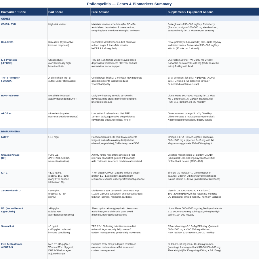
Understanding what your blood says is one layer of the picture. Understanding what your DNA contributes brings a second, often overlooked dimension — why some people were more vulnerable to severe poliovirus infection, and why some survivors deteriorate more rapidly in the post-polio phase.
What Genetics Research Suggests About Poliomyelitis and Recovery
Your genetic profile does not determine your outcome in post-polio syndrome, but it shapes the terrain considerably. How aggressively your immune system responded to the poliovirus, how well your nervous system can regenerate damaged circuits, and how intensely your body sustains chronic inflammation are all meaningfully influenced by heritable variants. The six genes below represent the most studied and clinically meaningful variants in the context of poliovirus susceptibility and post-polio neuromuscular integrity.
Gene 1: CD155 (PVR) — The Poliovirus Entry Gate
What it does: CD155 — also designated the poliovirus receptor (PVR) — is the cell-surface protein that the poliovirus binds to gain entry into human cells. It is expressed at highest levels in the anterior horn of the spinal cord and on motor neurons — precisely the tissues most devastated by poliovirus infection. Variants in the CD155 gene that increase receptor expression or binding affinity may have contributed to higher initial viral loads in some individuals, potentially explaining differential severity at the time of original infection. Beyond its role as a viral entry point, CD155 is also a ligand for natural killer cell receptors (TIGIT and CD226), meaning PVR polymorphisms influence ongoing immune surveillance of damaged tissue — relevant to both initial recovery and chronic post-polio inflammatory dynamics.
If the gene is bad — the plan without supplements: Since CD155 governed viral entry — a historical event for PPS survivors — the current focus is on minimizing ongoing immune stressors that can reactivate neuroinflammation. Maintaining current vaccine schedules is important, particularly for influenza and COVID-19, both of which can independently worsen neuromuscular conditions and trigger post-viral inflammatory cascades. Avoiding known triggers of viral reactivation (sleep deprivation, extreme physical overload, high psychological stress) is practical and evidence-informed. Rigorous sleep hygiene reduces microglial overactivation driven by immune stress.
If the gene is bad — the plan with supplements or equipment: Beta-glucans (from oat bran or Saccharomyces cerevisiae yeast): 250 to 500 mg per day. Beta-glucans modulate innate immune function via macrophage and natural killer cell receptors directly relevant to CD155 signaling pathways. Continuous use is well tolerated, with no significant side effects documented at standard doses.
Elderberry extract (Sambucus nigra): 300 to 500 mg standardized extract during high-risk periods such as autumn and winter. Supports NK cell activity and has antiviral immune-priming properties. Seasonal cycling: 8 to 12 weeks maximum per season. Avoid in autoimmune conditions without medical guidance.
Gene 2: HLA-DRB1 — The Immune Response Architect
What it does: Human leukocyte antigen genes are the foundation of the adaptive immune system — they govern how the body presents fragments of viral proteins to T cells and determines the strength and character of the resulting immune response. Specific HLA-DRB1 variants are associated with more or less effective T-cell responses to poliovirus antigens. Some HLA-DRB1 alleles linked to exaggerated inflammatory responses may have driven a more immunologically destructive course during acute poliovirus infection, and they may continue to sustain chronic low-grade neuroinflammation in PPS through persistent T-cell activation against motor neuron antigens. HLA variants have been investigated in post-polio syndrome and in multiple other post-infectious neurological conditions.
If the gene is bad — the plan without supplements: HLA-DRB1 risk variants that promote hyperactive immune responses make anti-inflammatory lifestyle interventions particularly high-value. A consistently Mediterranean dietary pattern — rather than periodic clean eating — is most effective for sustained cytokine modulation. Eliminating or substantially reducing dietary triggers of immune activation (refined sugar, trans fats, and — in those with detectable immunological reactivity — gluten or casein) reduces the background demand on an already reactive immune system. Systematic monitoring of hsCRP and IL-6 is especially important for those with high-risk HLA variants, given the chronic inflammatory tendency.
If the gene is bad — the plan with supplements or equipment: Palmitoylethanolamide (PEA): 600 to 1200 mg per day in divided doses. PEA is an endogenous anti-inflammatory compound with well-documented activity on microglial activation — the neuroinflammatory process most relevant to HLA-driven post-polio progression. A growing human evidence base supports its use in neuropathic pain and neuroinflammatory conditions. Continuous use is appropriate; tolerability is excellent.
Resveratrol: 250 to 500 mg per day with a fat-containing meal for absorption. Inhibits NF-kB — the master inflammatory transcription factor upstream of HLA-driven cytokine release. Cycle: 12 weeks on, 4 weeks off. Generally well tolerated; avoid at pharmacological doses during pregnancy.
Gene 3: IL-6 Promoter Polymorphism (-174G/C) — The Inflammation Amplifier
What it does: The IL-6 gene promoter polymorphism at position -174 (rs1800795) is one of the most widely studied functional genetic variants in inflammatory medicine. The C allele is associated with higher baseline IL-6 transcription, while the G allele produces less. Individuals carrying the CC genotype tend to have constitutionally elevated IL-6 levels and mount a more intense cytokine response to any inflammatory stimulus. In the context of post-polio syndrome, this variant may explain why some survivors experience far more severe fatigue and progressive weakness than others — their immune systems are biologically predisposed to maintain a higher inflammatory set-point. This is not a life sentence, but it means the threshold for intervention is lower and the evidence-based anti-inflammatory strategies discussed above become even more critical.
If the gene is bad — the plan without supplements: Time-restricted eating within a 12 to 16-hour fasting window produces meaningful IL-6 reduction through autophagy and insulin pathway regulation — and this benefit appears consistent even in constitutionally inflammatory individuals. Avoiding chronic sleep deprivation is critical: even a single night of inadequate sleep significantly elevates IL-6 through HPA axis dysregulation. Any evidence-based stress management practice — mindfulness, cognitive behavioral therapy, structured social support — reduces IL-6 through the cortisol-IL-6 amplification loop.
If the gene is bad — the plan with supplements or equipment: Quercetin plus vitamin C: 500 mg quercetin and 500 mg vitamin C, twice daily with food. This combination has documented IL-6 inhibitory activity in human trials and targets the specific NF-kB-mediated pathway through which the -174C variant drives overproduction. Cycle: 8 weeks on, 2 weeks off.
Boswellia serrata extract: 200 to 400 mg standardized to 65% boswellic acids, twice daily with food. Boswellic acids specifically inhibit 5-lipoxygenase and have direct IL-6 reducing activity documented in human clinical trials. Take with food to minimize the mild gastric irritation that can occur in some individuals.
Gene 4: TNF-α Promoter Polymorphism (-308G/A) — The Neuroinflammation Risk Gene
What it does: The tumor necrosis factor alpha gene polymorphism at position -308 (rs1800629) is among the most studied functional variants in neuroinflammatory disease. The A allele — present in roughly 20 to 30 percent of European ancestry individuals — is associated with significantly higher TNF-α production in response to immune stimulation. TNF-α at chronically elevated levels drives neuronal apoptosis, impairs myelin integrity, and accelerates muscle catabolism. In post-polio syndrome, high TNF-α activity may underlie the overlap with symptoms seen in other chronic inflammatory conditions — including the fatigue, diffuse pain, and cognitive difficulties that are frequently reported but poorly explained within a purely neurological framework.
If the gene is bad — the plan without supplements: Cold exposure — finishing daily showers with 2 to 3 minutes of cold water — has documented acute anti-inflammatory effects including TNF-α modulation through catecholamine release. Regular low-to-moderate aerobic exercise reduces TNF-α expression chronically through anti-inflammatory myokine production; the critical caveat in PPS being never to exercise to the point of fatigue, which paradoxically increases TNF-α through oxidative stress pathways. Reducing visceral adiposity is a high-priority free intervention, as adipose tissue is one of the primary sources of chronic TNF-α overproduction.
If the gene is bad — the plan with supplements or equipment: EPA-dominant fish oil: 3 to 4 grams combined omega-3 with an EPA to DHA ratio of at least 2:1. EPA specifically suppresses the arachidonic acid pathway through which TNF-α drives systemic inflammation. Continuous use; reassess inflammatory biomarkers at 3 months.
Glycine: 3 to 5 grams dissolved in water, taken before bed. Glycine has specific TNF-α inhibitory effects at the macrophage level through glycine-gated chloride channels, and independently improves sleep quality through glycine receptor activation in the brain. Continuous use is safe and the compound is among the most affordable available.
Gene 5: BDNF Val66Met — The Neuroplasticity Bottleneck
What it does: Brain-derived neurotrophic factor (BDNF) is the central nervous system's primary signal for growth, repair, and synaptic maintenance. The BDNF Val66Met polymorphism (rs6265) is one of the most clinically significant neurological variants identified in human genetics. Carriers of the Met allele — approximately 30 percent of the population — have significantly reduced activity-dependent secretion of BDNF. In practical terms, this means the nervous system releases far less repair signal in response to exercise, learning, and other stimulating inputs. This is particularly consequential in post-polio syndrome, where the capacity to maintain compensatory motor unit reinnervation and axonal sprouting is central to preserving function over time. Met allele carriers with PPS may face accelerated decline if BDNF-stimulating inputs are not deliberately amplified.
If the gene is bad — the plan without supplements: Exercise is the most evidence-supported BDNF stimulator available — even in Met allele carriers — but it must be carefully dosed in PPS. Low-intensity aerobic activity (15 to 20 minutes of non-fatiguing movement) done consistently and daily stimulates BDNF more reliably than infrequent intense sessions. Novel learning tasks — a new language, a musical instrument, complex cognitive games — are independently documented BDNF stimulators that carry no physical overload risk. Morning bright light exposure for serotonin production (which precedes BDNF release) and brief cold exposure are low-cost amplifiers relevant to Met allele carriers.
If the gene is bad — the plan with supplements or equipment: Lion's Mane mushroom: 500 to 1000 mg of standardized extract daily. Stimulates nerve growth factor (NGF) production — a complementary neurotrophic pathway to BDNF that is unaffected by the Val66Met polymorphism. Cycle: 8 to 12 weeks on, then assess response. Well tolerated in most individuals.
Magnesium L-threonate: 1.5 to 2 grams per day (providing approximately 140 to 200 mg elemental magnesium). This specific form was developed to cross the blood-brain barrier efficiently and has evidence for increasing synaptic density — directly relevant to neuroplasticity compensation in Met allele carriers. Takes 4 to 6 weeks for neurological effects to accumulate; continuous use is appropriate.
Transcranial photobiomodulation: Delivering red and near-infrared light (810 to 850 nm) transcranially is an emerging area with preliminary human evidence for increasing BDNF and supporting neurological recovery. Home devices in the $300 to $800 range are available. Use 10 to 20 minutes per day to the frontal and temporal regions. Avoid during fever or over active malignancy.
Gene 6: APOE ε4 — The Neurodegeneration Risk Amplifier
What it does: The APOE gene encodes apolipoprotein E, a lipoprotein involved in lipid transport throughout the nervous system and in the clearance of amyloid proteins and neuronal debris. The ε4 variant — carried by approximately 25 percent of the general population — is the strongest known genetic risk factor for Alzheimer's disease, but its relevance extends to any condition involving motor neuron repair. APOE ε4 carriers have impaired clearance of neuronal debris and lipid metabolites following injury, potentially resulting in poorer motor neuron recovery after poliovirus-mediated damage and greater accumulation of neuroinflammatory byproducts over time. Some evidence from post-viral neurological conditions suggests ε4 carriers experience more rapid cognitive decline and neurological deterioration than non-carriers when additional neurological stressors accumulate.
If the gene is bad — the plan without supplements: APOE ε4-specific lifestyle modifications are unusually well-researched thanks to the Alzheimer's research field. A dietary pattern low in saturated fat and refined carbohydrates and higher in monounsaturated fats (olive oil primarily) and long-chain omega-3s appears to specifically benefit ε4 carriers' neurological lipid metabolism. Time-restricted eating — 16 to 18 hours of fasting per day — has compelling evidence for reducing neurological Alzheimer's risk in ε4 carriers through autophagy-mediated clearance of neuronal debris; the same mechanism is directly relevant to post-polio motor neuron health. Aggressive sleep defense is non-negotiable for ε4 carriers: sleep is when glymphatic clearance of neuronal debris occurs, and even modest sleep disruption in ε4 carriers is associated with accelerated neurological accumulation of damaging proteins.
If the gene is bad — the plan with supplements or equipment: DHA-dominant omega-3: 1 to 2 grams DHA per day (different from the EPA-first recommendation for the other genes). For ε4 carriers, DHA specifically supports neuronal membrane phospholipid composition and reduces amyloid precursor protein cleavage through mechanisms tied to APOE lipid transport function. Continuous use; monitor lipid panels at 6 months.
Low-dose lithium orotate: 5 mg per day. Low-dose lithium has a growing neuroprotective evidence base and specifically promotes autophagy — the cellular debris clearance process that is particularly compromised in APOE ε4 carriers. This is fundamentally different from pharmaceutical lithium doses used in psychiatric conditions. Discuss with a physician before starting; not appropriate with impaired renal function.
Ketone supplementation or dietary ketosis: APOE ε4 carriers may have reduced neuronal glucose utilization efficiency; ketones provide an alternative metabolic fuel that bypasses this impairment. Exogenous ketone esters or salts (beta-hydroxybutyrate) can raise ketones acutely. A modified Mediterranean-ketogenic dietary pattern is a sustainable longer-term option. Significant dietary commitment is required; the effects on neurological health in ε4 carriers are supported by mechanistic evidence and early clinical data.
A Book That May Reframe How You Think About Post-Polio Recovery
For anyone managing post-polio syndrome with the persistent sense that conventional medicine has relatively little to offer, The Polio Paradox by Richard L. Bruno is essential reading. Bruno — a psychologist who himself survived polio and directed the Post-Polio Institute in New Jersey — spent decades documenting why PPS symptoms follow the patterns they do and why the mainstream "push through it" approach actively accelerates the condition. The book draws from decades of clinical observation and research across thousands of PPS patients and challenges multiple assumptions that both patients and physicians carry unchallenged.
10 Ideas From The Polio Paradox That Challenge Standard Thinking
1. Polio survivors disproportionately developed overachieving personalities as adaptive coping. Bruno documents how the drive to prove normalcy — socially praised and culturally rewarded — is biologically catastrophic for a neuromuscular system already operating at its functional ceiling. Recognizing this pattern in yourself is the beginning of a different approach.
2. Overuse is the primary driver of new weakness — not disuse. The compensatory motor units that survivors depend on are chronically overburdened. Every time exertion reaches fatigue, the biological cost falls on neurons that cannot afford it. The "no pain, no gain" framework is categorically wrong for this population.
3. Post-polio fatigue is partly a brain phenomenon, not just muscular. Bruno proposes that poliovirus damaged dopamine-producing neurons in the reticular activating system of the brainstem — meaning the fatigue of PPS has a genuine neurochemical component that willpower cannot override. This reframes fatigue as a physiological signal that must be respected.
4. The 80 percent rule is a clinical threshold, not a metaphor. Bruno's protocol caps any physical or cognitive effort at 80 percent of maximum, never reaching the point of fatigue. This is operationally defined: if you feel tired after an activity, you exceeded the threshold. The rule applies to mental effort as well as physical.
5. Cognitive fatigue depletes the same neurological resources as physical exertion. Long workdays, sustained concentration, and emotional stress are not neurologically free. They draw from the same depleted reserve as physical activity, and must be accounted for in daily energy budgeting.
6. Sleep is a primary therapeutic intervention, not a passive restoration. Consistent 8 to 9 hours — not occasional long nights — is described by Bruno as the most evidence-supported intervention for slowing PPS functional decline. Addressing sleep-disordered breathing (very common in PPS due to respiratory muscle weakness) is part of this.
7. Physical therapy has a narrow and specific role. Exercise in PPS is not about building strength in affected muscle groups — it is about maintaining range of motion, preventing secondary deconditioning in unaffected muscles, and redistributing mechanical load. Strength training targeted at already-overloaded motor units is directly counterproductive.
8. Heat is a legitimate medical hazard, not just discomfort. Heat dramatically reduces conduction efficiency in already-damaged peripheral nerves. Symptom worsening in warm weather is physiologically real and not psychological. Cooling strategies — cooling vests, climate control, cold fluids — are legitimate management tools.
9. Social support is measurably neuroprotective. Bruno's clinical data showed PPS patients with strong social support networks had consistently slower functional decline. This operates through real biological mechanisms: social connection suppresses cortisol, reduces inflammatory cytokines, and supports sleep quality.
10. Most physicians are inadequately prepared to manage PPS. Bruno is direct: general neurologists, internists, and even many rehabilitation specialists have limited training in post-polio syndrome and frequently offer advice — "stay active," "push yourself" — that accelerates deterioration. Finding a physiatrist or neurologist with demonstrated PPS expertise is one of the highest-return clinical investments available.
Complementary Approaches With Evidence for Post-Polio Conditions
Biomarker data and genetic insights are tools for understanding what is happening and why. The approaches below translate that understanding into practical interventions — drawn from the modalities with the most meaningful clinical evidence for the core challenges of post-polio syndrome: fatigue, neuromuscular deconditioning, autonomic dysregulation, and chronic neuroinflammation.
Adapted Yoga for Fatigue Regulation and Autonomic Balance
Adapted yoga removes postures requiring maximal muscular effort and replaces them with supported, breath-centered movements designed to activate the parasympathetic nervous system. For PPS, the therapeutic value is not muscular strengthening — it is nervous system regulation. Chronic fatigue in post-polio syndrome has a strong autonomic dysregulation component, and yoga's effects on heart rate variability (a direct measure of autonomic balance) are well-documented across chronic condition populations. Gentle movement coordinated with slow diaphragmatic breathing is one of the most accessible tools for shifting an overactive sympathetic baseline.
A randomized trial published in Topics in Stroke Rehabilitation (Bastille and Gill-Body, 2004) examining yoga-based programs in individuals with chronic neurological conditions found improvements in balance, fatigue perception, and quality of life scores. While direct evidence in PPS is limited, the neurobiological mechanisms are condition-appropriate. Evidence from closely related conditions — multiple sclerosis, Parkinson's disease, chronic fatigue syndrome — consistently supports adapted yoga for autonomic regulation and fatigue reduction.
In practice: begin exclusively with chair yoga or supine yoga — all postures performed while seated in a chair or lying down, with blocks, bolsters, and straps for support. Sessions should not exceed 30 minutes and should never approach fatigue. Emphasis belongs on coordinated slow breathing (5-second inhale, 5-second exhale) throughout every movement. One to two sessions per week is the appropriate starting cadence, with any increase made only when there is zero post-exertional symptom worsening in the 48 hours following each session.
Biofeedback for Muscle Monitoring and Activity Pacing
Biofeedback provides real-time monitoring of physiological signals — heart rate variability, surface EMG muscle activity, skin conductance — to help individuals learn to regulate functions they cannot otherwise observe. For PPS patients, surface electromyography (EMG) biofeedback is particularly relevant: it delivers direct visual feedback on muscle recruitment patterns, enabling patients and rehabilitation clinicians to identify which muscles are chronically overworking and to consciously practice redistributing effort across more mechanically advantaged pathways. This is distinct from general exercise and addresses the compensatory muscle overuse pattern that drives ongoing CK elevation and progressive motor neuron stress.
A review in NeuroRehabilitation examining EMG biofeedback across neuromuscular rehabilitation contexts, including lower motor neuron pathologies with patterns comparable to PPS, found consistent improvements in motor recruitment efficiency and reduced subjective effort during activities of daily living. The mechanistic rationale in PPS — where the problem is inefficient motor unit recruitment, not simply weakness — is well aligned with what biofeedback addresses.
Practically: EMG biofeedback for PPS is best initiated in a physiatry or specialized neuromuscular physical therapy clinic where a clinician can select the appropriate muscle groups and establish baseline patterns. After initial calibration, portable surface EMG biofeedback units ($150 to $400) can be used at home during daily activities to maintain awareness of overuse in real time. Sessions of 20 to 30 minutes, two to three times weekly, focused on everyday movements rather than exercise-for-its-own-sake, represent an appropriate starting protocol.
Mindfulness-Based Stress Reduction (MBSR) for Fatigue and Inflammatory Burden
MBSR, developed by Jon Kabat-Zinn at the University of Massachusetts, is an 8-week structured program combining mindfulness meditation, body scan practices, and gentle movement. Its relevance to PPS is multilayered: it measurably reduces cortisol (which is catabolic to neuromuscular tissue), lowers IL-6 and CRP (the inflammatory biomarkers most consistently elevated in PPS), and addresses the depression and anxiety that accompany chronic neuromuscular conditions at high rates. The cortisol-IL-6 amplification loop, which can maintain a high inflammatory set-point independent of physical factors, is specifically disrupted by mindfulness practice through prefrontal cortex regulation of the HPA axis.
A widely cited 2014 meta-analysis in JAMA Internal Medicine by Goyal et al. found MBSR produced significant, clinically meaningful improvements in anxiety, depression, pain, and stress-related biomarkers across chronic condition populations. For post-polio syndrome, where sustained psychological stress perpetuates the same neuroinflammatory burden as physical overload, MBSR addresses a mechanism that is largely invisible in conventional neurological management protocols.
The standard MBSR format involves weekly 2.5-hour group sessions over 8 weeks with daily 45-minute home practice. For PPS patients, this can be completed in adapted form — chair-based body scans, shorter home sessions of 20 to 30 minutes — without losing the core neurobiological benefit. Fully online MBSR programs are now widely available and make this accessible regardless of mobility status or geographic location.
Breathing-Based Therapy for Respiratory Support and Autonomic Recovery
Respiratory muscle involvement in poliomyelitis is frequently underrecognized. Many survivors have subclinical respiratory muscle weakness that worsens quietly over time as the combined effects of aging and PPS accumulate. Sleep-disordered breathing is significantly more prevalent in PPS than in the general population, and it directly undermines the sleep-based repair processes that are clinically critical in this condition. Separately from frank respiratory weakness, many PPS patients exhibit autonomic dysregulation characterized by elevated sympathetic tone — a pattern that paced breathing techniques are specifically equipped to address.
Slow-paced breathing at approximately 0.1 Hz (about 6 breath cycles per minute) maximally stimulates the baroreflex and increases heart rate variability — a direct measure of autonomic balance. This technique has been studied in chronic fatigue and autonomic dysregulation conditions with consistent beneficial findings. A systematic review in Applied Psychophysiology and Biofeedback documented that resonance-frequency breathing (the physiological term for 6 breaths per minute) reliably and significantly increases HRV across populations with autonomic imbalance.
In practical application: practice 10 minutes of slow-paced breathing twice daily — a 5-second nasal inhale followed by a 5-second nasal exhale, with no breath-holding. A breathing pacer app or a device such as Resperate can guide the rate precisely. Separately and with equal urgency: any PPS patient with suspected sleep-disordered breathing — snoring, witnessed apneas, morning headaches, excessive daytime sleepiness — should be evaluated with a formal sleep study. Treating obstructive or central sleep apnea in this population has outsized benefits given how comprehensively post-polio recovery depends on restorative sleep.
Low-Level Laser Therapy (Photobiomodulation) for Nerve and Muscle Repair
Low-level laser therapy — clinically termed photobiomodulation (PBM) — uses red and near-infrared wavelengths (typically 630 to 850 nm) to stimulate mitochondrial cytochrome c oxidase, increasing cellular ATP production, reducing oxidative stress, and accelerating tissue repair. Its relevance to post-polio syndrome rests on evidence that PBM promotes peripheral nerve regeneration and reduces neuropathic pain — two areas of direct clinical relevance in PPS. Pre-clinical models of motor neuron injury have shown PBM increases axonal sprouting and accelerates myelin repair, consistent with the biological mechanisms that post-polio motor neurons need to maintain compensatory reinnervation.
A systematic review published in Lasers in Medical Science documented PBM's benefits for peripheral neuropathy and neuromuscular rehabilitation, including conditions with lower motor neuron pathology comparable to PPS. Direct evidence in post-polio syndrome specifically is limited to case reports and small series, but the mechanistic alignment is strong and the safety profile is excellent — no significant adverse effects have been documented at appropriate dosing parameters.
For practical use: target affected limb muscle groups and the lumbosacral or cervical spinal segments corresponding to the original poliomyelitis level. Devices using combined 630 nm red and 850 nm near-infrared LED panels are available for home use from approximately $200 to $600. Use for 10 to 20 minutes per session, five days per week, at the recommended distance specified for the device's irradiance level. Avoid direct eye exposure. Contraindicated over active malignancy. For individuals with access to clinical-grade laser systems through a physiotherapy practice, therapeutic dosing is more precise and may produce stronger effects.
Conclusion
Poliomyelitis and post-polio syndrome occupy a quiet corner of medicine — the acute disease largely eradicated, and PPS still underfunded and poorly understood by most clinicians. But the biology is not mysterious, and it is measurable. Chronic neuroinflammation, declining motor neuron integrity, hormonal insufficiency, and genetic vulnerabilities that amplify these processes all contribute to the post-polio trajectory — and virtually all of them can be tracked and acted upon.
The clearest next step is to establish a baseline. Start with the most accessible biomarkers: hsCRP, 25-OH vitamin D, IGF-1, and creatine kinase. If those reveal a meaningful signal, add the more specialized tests — NfL, serum IL-6, free testosterone — to deepen the picture. If genetic testing is available to you, understanding your BDNF Val66Met, APOE, and IL-6 promoter variants can refine your approach substantially and help you prioritize which interventions matter most for your specific biology.
None of this replaces working with a physician who genuinely understands post-polio syndrome — ideally a physiatrist or a neurologist with demonstrated neuromuscular experience. But arriving at that conversation with actual data, a clear picture of your inflammatory status, and an informed understanding of your genetic predispositions gives both you and your clinician something concrete to work with. Informed, precise action is always a better starting point than hope and generic advice.
Musculoskeletal: Muscle Conditions
Neurological: Nerve Conditions Spinal Cord Conditions
Autoimmune: Inflammatory Conditions
Infectious: Viral Infections