This article contains AI generated content.
Polyethylene Wear Syndrome: 5 Genes and 7 Biomarkers to Track
Introduction
If you have a hip or knee replacement and you've started hearing terms like "osteolysis," "wear debris," or "aseptic loosening" from your surgeon, you already know the frustrating part: the standard advice stops at "we'll keep an eye on it with X-rays every year." That's true, and imaging matters, but it tells you almost nothing about what's happening at the cellular level around your implant right now, months or years before a scan would ever show bone loss.
Generic joint-health advice — "stay active," "maintain a healthy weight," "take your vitamin D" — isn't wrong, but it's built for the general population, not for someone whose body is actively processing microscopic polyethylene particles shed from a bearing surface. Two people with the same implant, the same activity level, and the same age can have very different biological responses to that wear debris, because the reaction is driven by inflammatory signaling and bone remodeling pathways that vary meaningfully from person to person.
This article takes a more specific approach. Instead of repeating broad wellness advice, it focuses on the actual biomarkers that reflect what your immune system and bone cells are doing in response to wear particles, and on the genetic variants that help explain why that response differs between individuals. Both angles point toward the same goal: catching a problem while it's still biochemical, not yet structural.
None of this replaces your orthopedic follow-up, and there is no supplement or lab panel that reverses established osteolysis. But better information, tracked consistently, gives you and your surgeon a real head start — the kind that turns a five-year warning into a fifteen-year implant.
Summary
Polyethylene wear syndrome doesn't announce itself with pain until bone loss is already underway, which is exactly why lab tracking matters more here than in almost any other orthopedic condition. This article walks through the seven biomarkers — spanning systemic inflammation, bone turnover, vitamin D status, and synovial fluid analysis — that together can flag a problem years before it shows up on an X-ray, along with realistic cost ranges and both non-supplement and supplement-based ways to correct an abnormal result. It then turns to genetics: five specific genes involved in inflammatory signaling and bone remodeling that help explain why some people develop aggressive osteolysis and others don't, with practical steps for each variant. A dedicated section distills the most relevant, joint- and bone-focused lessons from Peter Attia's Outlive, and a final section reviews the complementary approaches — from tai chi to photobiomodulation — that have actual clinical evidence in joint replacement patients, rather than vague wellness claims. Read on to see exactly which numbers to ask your doctor for at your next visit, and which ones are worth tracking on your own.
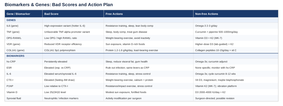
7 Biomarkers That Reveal What's Happening Around Your Implant
Polyethylene wear syndrome — the constellation of chronic inflammation, macrophage activation, and progressive bone resorption (osteolysis) triggered by microscopic wear particles shed from a plastic bearing surface — develops silently. By the time it's visible on a standard X-ray, meaningful bone has often already been lost around the implant. The biomarkers below are the closest thing available to a early-warning system, because they track the biological process itself rather than its structural end result.
A study that followed hip replacement patients for years before their osteolysis diagnosis found that a combination of just two markers — a bone resorption product and an inflammatory cytokine — could identify at-risk patients with very high accuracy, in some cases years ahead of any radiographic sign of bone loss (Landgraeber et al., biomarker discovery study). That's the core logic behind tracking these numbers at all: they move before the imaging does.
High-sensitivity C-reactive protein (hs-CRP)
Why it matters: hs-CRP is a general marker of systemic inflammation produced by the liver in response to circulating cytokines, including the same IL-6 signaling that drives osteoclast activity around a wearing implant. It won't tell you specifically what's happening in your joint, but a persistently elevated hs-CRP in someone with a stable, non-infected implant is a signal that a broader inflammatory process is active and worth investigating alongside more specific markers.
How to measure it: A simple venous blood draw, widely available through primary care or direct-to-consumer lab services. Cost typically runs $10 to $30 out of pocket, or is bundled into routine annual bloodwork covered by insurance. Results in one to two days.
If it's elevated — the plan without supplements: Rule out an unrelated infection or dental source first, since hs-CRP is nonspecific. Beyond that, prioritize sleep consistency (inflammation rises sharply with chronic sleep restriction), reduce visceral fat through diet quality rather than aggressive calorie restriction, and address gum disease, which is an underappreciated chronic driver of systemic hs-CRP. Recheck every 3 months rather than reacting to a single value, since hs-CRP has real day-to-day variability.
If it's elevated — the plan with supplements or equipment: Omega-3 fatty acids (2-3 g/day EPA/DHA combined) have the most consistent evidence for modestly lowering hs-CRP; give it 8-12 weeks before reassessing. Curcumin with piperine (500-1000 mg/day) is a reasonable adjunct with a good safety profile, though evidence in this specific population is limited. Avoid stacking multiple anti-inflammatory supplements at once — it makes it impossible to know what's working, and some (high-dose fish oil in particular) carry a mild bleeding risk that matters if you're anywhere near another surgery.
Erythrocyte sedimentation rate (ESR)
Why it matters: ESR is an older, less specific inflammatory marker, but it's still used clinically alongside CRP because the two don't always move together, and discordance between them can itself be informative. In joint replacement follow-up, ESR is also part of the standard workup to distinguish aseptic wear-related inflammation from periprosthetic joint infection, which needs a completely different treatment path.
How to measure it: Standard blood test, usually ordered together with CRP. Cost is low, generally $10-25, and often included in a basic metabolic or inflammatory panel.
If it's elevated — the plan without supplements: The most important step is diagnostic, not lifestyle: an elevated ESR with joint symptoms should prompt your surgeon to check for infection (via synovial fluid analysis, discussed below) before anything else. If infection is ruled out, the same anti-inflammatory lifestyle levers as hs-CRP apply — sleep, body composition, and periodontal health.
If it's elevated — the plan with supplements or equipment: There's no strong supplement literature targeting ESR specifically; treat it as a marker to watch alongside hs-CRP rather than a target in itself.
Interleukin-6 (IL-6)
Why it matters: IL-6 is one of the central cytokines produced by macrophages that have engulfed polyethylene wear particles, and it directly promotes osteoclast formation through the RANKL pathway. It is one of the two markers that, combined with a bone resorption marker, showed the strongest predictive power for future osteolysis in the biomarker discovery study cited above (Landgraeber et al.), and it's also central to the broader mechanistic picture of wear-debris-driven bone loss (comparative role of IL-1, IL-6, and TNF-α in osteolysis models).
How to measure it: Serum IL-6 is measured via specialized immunoassay, less routinely available than CRP. Expect $40-90 through specialty or research-oriented labs; it's not always part of standard insurance-covered panels, so check first. Synovial fluid IL-6, drawn during a joint aspiration, is more specific but requires a procedural visit.
If it's elevated — the plan without supplements: Since IL-6 rises with visceral adiposity, poor sleep, and unmanaged psychological stress independent of any joint issue, address those systemic contributors first. Resistance training performed consistently (not sporadically) has been shown to lower baseline IL-6 over months, likely through improved body composition and myokine signaling.
If it's elevated — the plan with supplements or equipment: Omega-3s again have the best supporting data here, at similar doses to the hs-CRP protocol. Some clinicians add low-dose curcumin as an adjunct. Cycle any anti-inflammatory supplement regimen (e.g., 8-12 weeks on, reassess, consider a break) rather than staying on it indefinitely without rechecking labs — chronic suppression of inflammatory signaling isn't automatically beneficial, and you want to know if the underlying process is actually improving.
C-terminal telopeptide of type I collagen (CTX-I)
Why it matters: CTX-I is a direct breakdown product of bone collagen, released when osteoclasts resorb bone. It is the most specific of the biomarkers here for the actual process of periprosthetic bone loss, and it was the single strongest predictor of future osteolysis in the study referenced above, distinguishing affected patients years before diagnosis (biomarker discovery study).
How to measure it: Blood test, ideally drawn fasting in the morning since CTX-I has a strong diurnal rhythm. Cost ranges $50-100 through most commercial labs; it's a standard marker in osteoporosis management, so many labs run it routinely.
If it's elevated — the plan without supplements: Weight-bearing exercise is the single most evidence-backed lever for bone turnover, but it needs to be appropriate for your implant — ask your surgeon or physical therapist which impact level is safe. Ensure adequate dietary calcium (roughly 1000-1200 mg/day from food where possible) and protein intake (around 1.2-1.6 g/kg body weight), since inadequate protein intake independently worsens bone turnover markers.
If it's elevated — the plan with supplements or equipment: Vitamin D3 (see below) and magnesium glycinate (200-400 mg/day) support the mineralization side of the equation. If CTX-I is markedly and persistently elevated, this is a conversation for your surgeon about whether a bone-protective medication (such as a bisphosphonate) is warranted — that decision sits outside supplement territory and needs clinical judgment, not self-directed dosing.
Procollagen type I N-terminal propeptide (P1NP)
Why it matters: P1NP is the mirror image of CTX-I — a marker of bone formation rather than resorption. Tracking both together tells you whether bone remodeling is balanced or whether resorption is outpacing formation, which is the actual mechanism behind periprosthetic osteolysis and implant loosening.
How to measure it: Blood test, similarly $50-100, often ordered as a pair with CTX-I in osteoporosis or bone-health panels.
If it's low relative to CTX-I — the plan without supplements: Resistance and impact-appropriate weight-bearing exercise stimulate osteoblast activity, not just slow resorption. Adequate sleep and stress management matter here too, since cortisol elevation suppresses bone formation markers.
If it's low relative to CTX-I — the plan with supplements or equipment: Vitamin K2 (MK-7 form, 90-180 mcg/day) supports proper mineralization once formation is stimulated. Whole-body vibration platforms have some evidence for supporting bone formation markers in older adults, though evidence specific to periprosthetic bone is limited — treat this as a reasonable adjunct, not a primary strategy.
25-hydroxyvitamin D
Why it matters: Vitamin D status directly affects calcium absorption and bone mineralization, and low levels are common in exactly the population most likely to have a joint replacement — older adults with reduced sun exposure and mobility. It's also linked to the VDR gene discussed in the genetics section below, since the receptor and the ligand only work as a pair.
How to measure it: Inexpensive and widely available blood test, typically $20-50, often covered by insurance given how routinely it's checked.
If it's low — the plan without supplements: Modest, regular sun exposure (10-20 minutes, several times weekly, skin-type and latitude dependent) and dietary sources like fatty fish and fortified foods help but rarely correct a significant deficiency on their own.
If it's low — the plan with supplements or equipment: Vitamin D3, typically 2000-4000 IU/day for correction, paired with vitamin K2 to direct calcium toward bone rather than soft tissue. Recheck levels after 8-12 weeks rather than guessing at dose. Very high doses (above 10,000 IU/day sustained) carry a real risk of hypercalcemia, so this is a supplement worth lab-guided dosing rather than a flat daily habit.
Synovial fluid analysis (cell count, differential, and alpha-defensin)
Why it matters: This is the one test on this list that requires a procedure rather than a blood draw, but it's arguably the most clinically decisive. Its primary purpose is distinguishing aseptic wear-related inflammation from periprosthetic joint infection — the two look similar on standard bloodwork but require completely different treatment. Elevated synovial white cell counts with a neutrophil-predominant differential point toward infection; elevated macrophage and wear-particle content without infection markers points toward mechanical wear.
How to measure it: Joint aspiration performed by an orthopedic surgeon or radiologist, typically $200-600 depending on whether imaging guidance is used, plus lab analysis fees. This isn't something to pursue proactively without symptoms — it's indicated when pain, swelling, or rising inflammatory markers raise a real question about what's going on.
If results suggest wear-driven inflammation — the plan without supplements: This confirms the biological process is active; the response is largely the exercise, sleep, and body composition measures described above, combined with closer imaging follow-up as directed by your surgeon.
If results suggest wear-driven inflammation — the plan with supplements or equipment: Same anti-inflammatory adjuncts as above (omega-3s, curcumin), understanding that none of them address the mechanical source of the particles. At this stage, the more consequential decisions — activity modification, or in advanced cases, revision surgery — belong to your surgeon.
Taken together, these seven markers form a layered picture: hs-CRP and ESR flag general inflammation, IL-6 points more specifically at the wear-debris response, CTX-I and P1NP show whether bone remodeling is tipping toward loss, vitamin D shows whether your body has the raw material to remineralize, and synovial analysis settles the infection-versus-wear question when something looks off. Tracking two or three of these consistently — rather than ordering all seven repeatedly — is usually the most practical approach for most patients.
5 Genes That Help Explain Individual Risk
Biomarkers tell you what's happening now; genetics helps explain why the same wear debris load produces a mild reaction in one person and aggressive osteolysis in another. Researchers including Ali Torkamani, whose work on genomic risk prediction has repeatedly shown that common inflammatory and bone-metabolism variants explain meaningful differences in disease susceptibility, and clinicians like Gary Brecka who popularized practical, actionable genetic panels, both emphasize the same principle: a gene variant is not a diagnosis, it's a probability shift you can act on. The evidence base for genetics in periprosthetic osteolysis specifically is smaller than for biomarkers, and much of it comes from candidate-gene association studies rather than large genome-wide studies — worth keeping in mind as you read.
IL6 (interleukin-6 gene)
Certain promoter variants in IL6 are associated with higher baseline production of this cytokine, the same one tracked as a blood biomarker above, and cytokine profiling work has repeatedly implicated IL-6 as a central driver of the osteoclast activation that follows wear-particle exposure (comparative cytokine roles in aseptic loosening).
If the gene is unfavorable — the plan without supplements: Since a high-expression IL6 variant essentially means your inflammatory "volume knob" runs hotter than average, the lifestyle levers that lower IL-6 matter more for you than for someone without the variant: consistent resistance training, adequate sleep duration, and body composition management. None of these are unique to this gene, but they carry more weight when you're carrying a higher baseline.
If the gene is unfavorable — the plan with supplements or equipment: Omega-3 fatty acids (2-3 g/day) again have the best supporting data for blunting IL-6 production; expect to reassess via blood IL-6 or hs-CRP after 8-12 weeks rather than assuming benefit. This is not a variant that responds to a single dramatic intervention — it responds to sustained, boring consistency.
TNF (tumor necrosis factor-alpha gene)
TNF-α is, alongside IL-6, one of the core cytokines implicated in wear-particle-driven osteolysis, and functional promoter variants have been studied in relation to aseptic loosening risk in several candidate-gene studies (genetics and mechanisms of periprosthetic osteolysis review). Evidence here is early and mixed across populations — treat it as a plausible contributor, not a settled finding.
If the gene is unfavorable — the plan without supplements: Same foundational levers as IL6: sleep, body composition, and consistent (not sporadic) physical activity. Reducing overall systemic inflammatory load — including treating periodontal disease and managing any autoimmune conditions — matters more here than any single targeted intervention.
If the gene is unfavorable — the plan with supplements or equipment: Curcumin (500-1000 mg/day with piperine) has some mechanistic evidence for modulating TNF-α signaling specifically. Cycle it — 8-12 weeks on, reassess with hs-CRP, then decide whether to continue — rather than treating it as a permanent daily supplement. Avoid combining with other anti-inflammatory agents you're already taking (including prescription NSAIDs) without checking with your physician, given overlapping effects and GI risk.
TNFRSF11B / TNFSF11 (the OPG-RANKL axis)
This pair of genes encodes osteoprotegerin (OPG) and its counterpart RANKL, which together set the balance between bone formation and resorption. Wear debris pushes this balance toward RANKL-driven resorption, and elevated serum RANKL relative to OPG has been documented specifically in patients with periprosthetic osteolysis (serum OPG and RANKL as markers of periprosthetic osteolysis). Polymorphisms affecting OPG expression have been studied in general osteoporosis populations, with less direct evidence yet in the implant-specific context.
If the gene is unfavorable — the plan without supplements: Mechanical loading is the most direct non-supplement lever on this axis — appropriate, surgeon-approved weight-bearing activity shifts remodeling toward formation. Avoiding prolonged immobility after any setback (illness, minor injury) is more important for you than for someone with a more favorable OPG/RANKL balance.
If the gene is unfavorable — the plan with supplements or equipment: Vitamin K2 (MK-7, 90-180 mcg/day) and adequate vitamin D3 support the formation side of the equation once mechanical loading is in place. There is no supplement that directly shifts the OPG/RANKL ratio in a clinically meaningful, evidence-backed way — be skeptical of products marketed with that specific claim.
VDR (vitamin D receptor gene)
Common VDR polymorphisms (FokI, BsmI, TaqI, ApaI) affect how effectively vitamin D signaling translates into bone mineral density and turnover, with some studies showing meaningful associations with fracture risk and response to bone-protective therapy (VDR polymorphisms and response to antiresorptive therapy, systematic review of VDR polymorphisms and osteoporosis).
If the gene is unfavorable — the plan without supplements: Regular, modest sun exposure and vitamin D-rich foods matter, but a less efficient receptor variant often means diet and sun alone won't fully compensate — this is one case where checking the actual blood biomarker (25-hydroxyvitamin D, above) matters more than usual, since you may need a higher intake to hit the same functional target.
If the gene is unfavorable — the plan with supplements or equipment: Vitamin D3 dosed to a lab-confirmed target (often needing the higher end of the 2000-4000 IU/day range, or more under medical supervision) paired with K2. Retest levels every 8-12 weeks during correction, then every 6 months for maintenance — don't assume a standard dose is hitting target when receptor efficiency is reduced.
COL1A1 (collagen type I alpha 1 gene)
The Sp1 promoter polymorphism in COL1A1 affects collagen production and has a well-documented, if modest, association with lower bone mineral density and increased fracture risk, particularly vertebral fracture, across multiple studies (GENOMOS study on COL1A1 polymorphism, BMD, and fracture risk). Since collagen is the structural scaffold bone mineral is built onto, this variant affects the baseline quality of the bone stock around an implant, independent of the inflammatory pathways above.
If the gene is unfavorable — the plan without supplements: Protein intake sufficiency (1.2-1.6 g/kg body weight daily) is more important for you specifically, since collagen synthesis requires adequate amino acid substrate even when the gene itself limits output efficiency. Weight-bearing exercise remains the strongest non-supplement lever for overall bone strength.
If the gene is unfavorable — the plan with supplements or equipment: Collagen peptide supplementation (10-15 g/day) combined with vitamin C has some supporting evidence for collagen synthesis support, though direct trials in COL1A1-variant carriers specifically are limited. Standard bone-support nutrients — vitamin D3, K2, and magnesium — remain the backbone of any plan here as well.
What Outlive Gets Right About Protecting Aging Joints
Peter Attia's Outlive isn't written about implants or wear debris specifically, but its central argument — that the decisions determining your physical capability at 80 are made decades earlier, and that most doctors don't measure the things that actually predict decline — maps unusually well onto periprosthetic bone health. Here are the ten ideas from the book most relevant to someone managing polyethylene wear syndrome.
1. Medicine 3.0 means acting before the problem is visible
Attia's framing of most conventional medicine as reactive ("Medicine 2.0" — waiting for disease to appear) versus proactive risk management is exactly the logic behind tracking CTX-I and IL-6 before osteolysis shows up on imaging. The whole premise of biomarker tracking in this article is Medicine 3.0 applied to a specific mechanical-biological problem.2. Muscle mass predicts survival more than most people realize
Attia cites data showing older adults in the lowest muscle mass quartile face dramatically higher mortality than those in the highest. For implant patients, muscle mass isn't just a longevity marker — it's the tissue that stabilizes the joint and reduces the fall risk that can catastrophically damage a prosthesis.3. Strength training matters as much as cardio, maybe more
The book argues strength training deserves equal billing with aerobic exercise because it's the primary driver of the muscle mass described above. For someone with a joint replacement, surgeon-approved resistance training is also one of the few interventions that meaningfully influences bone formation markers like P1NP.4. VO2 max is one of the strongest predictors of healthspan
Attia treats cardiorespiratory fitness as a near-vital-sign-level metric. While it doesn't directly measure bone or joint health, it's a proxy for overall physiological resilience, which matters when your body is managing a chronic low-grade inflammatory load from wear particles.5. Annual DEXA scans should be standard, not exceptional
Attia recommends routine DEXA scans for body composition and bone density tracking well before typical osteoporosis screening age. For implant patients, this dovetails directly with tracking CTX-I and P1NP — DEXA shows the cumulative structural result, the blood markers show the process in real time.6. Chronic inflammation ("inflammaging") is a root driver of tissue decline
The book's discussion of low-grade systemic inflammation as a common thread across cardiovascular disease, cognitive decline, and frailty applies just as directly to periprosthetic bone loss, which is fundamentally an inflammatory process triggered by particulate debris.7. Sleep is a non-negotiable lever, not an optional one
Attia is emphatic that inadequate sleep undermines nearly every other intervention, including inflammatory control and tissue repair. This lines up directly with the sleep guidance repeated throughout the biomarker sections above.8. Protein intake needs are higher than most guidelines suggest, especially with age
The book pushes back on conservative protein recommendations, arguing older adults need more to preserve muscle and support tissue repair — consistent with the 1.2-1.6 g/kg range referenced for bone and collagen support above.9. Emotional and mental health directly affect physical outcomes
Attia devotes real attention to the idea that healthspan isn't purely physical — chronic stress elevates cortisol, which suppresses bone formation and raises inflammatory markers, tying psychological state directly back to the biomarkers tracked in this article.10. The "marginal decade" is worth fighting for now, not later
The book's closing argument — that the last decade of life is disproportionately shaped by choices made 20-30 years earlier — is a useful reframe for implant patients: protecting bone stock and controlling inflammation today is what determines whether a joint replacement lasts one revision surgery or none at all.Complementary Approaches Worth Considering
Nothing below replaces surgical follow-up, biomarker tracking, or a bone-protective medication when one is indicated. But several complementary approaches have real, condition-relevant clinical evidence in joint replacement patients, and they're worth understanding rather than dismissing outright or over-relying on without evidence.
Tai chi
Tai chi is a slow, low-impact movement practice combining weight shifting, balance work, and mindful breathing, and it's relevant here for two distinct reasons: it improves proprioception and balance (reducing fall risk around a prosthesis) and it has documented benefit for knee osteoarthritis-related pain and function, a population that substantially overlaps with joint replacement candidates and patients managing the contralateral joint.
A randomized comparative-effectiveness trial found tai chi performed at least as well as standard physical therapy for knee osteoarthritis symptoms over a 52-week follow-up period, with participants doing two sessions weekly for 12 weeks (comparative effectiveness of tai chi versus physical therapy for knee osteoarthritis).
Realistically, this means starting with a instructor experienced in working with joint replacement or arthritis patients, beginning with seated or supported forms if balance is a concern, and treating it as a complement to — not a replacement for — surgeon-directed physical therapy protocols after surgery.
Mindfulness meditation and MBSR
Mindfulness-Based Stress Reduction combines meditation, gentle movement, and body awareness training, and its relevance here is less about the joint itself and more about breaking the loop between chronic pain, stress, and the cortisol elevation that worsens bone formation markers described earlier in this article.
The strongest evidence comes from a large randomized trial in chronic low back pain, not joint replacement specifically, which found MBSR produced meaningfully greater improvement in pain and function than usual care, with effects persisting at one year (MBSR versus CBT versus usual care for chronic low back pain, JAMA). Evidence specific to periprosthetic patients is more limited, so this should be read as reasonable extrapolation rather than condition-specific proof.
A practical starting point is an 8-week structured MBSR course (widely available in person or online), practiced alongside — not instead of — the physical rehabilitation your surgical team prescribes.
Guided imagery
Guided imagery uses structured mental rehearsal and relaxation scripts, typically delivered by audio, and is one of the few complementary approaches with a trial specifically in total knee arthroplasty patients rather than borrowed from an unrelated condition.
A randomized, placebo-controlled pilot study found guided imagery improved postoperative outcomes in total knee replacement patients compared with a non-therapeutic control condition (guided imagery for total knee replacement, randomized placebo-controlled pilot study). As a pilot study, the sample size is modest, so treat the finding as promising rather than definitive.
Practically, this looks like a short (10-20 minute) daily audio-guided session in the weeks before and after surgery, which requires no equipment beyond headphones and costs nothing beyond an app or a set of recordings from your surgical center.
Massage therapy
Massage therapy applied to the surrounding musculature can reduce post-surgical muscle guarding and improve range of motion during rehabilitation, which matters for polyethylene wear syndrome indirectly — better function and mobility support the mechanical loading needed for healthy bone remodeling described in the biomarker sections above.
A systematic review and meta-analysis of eleven randomized trials in total knee arthroplasty patients found massage therapy produced meaningful pain reduction and improved range of motion in the first three weeks after surgery, compared with standard care alone (massage for rehabilitation after total knee arthroplasty, systematic review and meta-analysis).
In practice, this means scheduling massage sessions with a therapist experienced in post-surgical orthopedic care, typically starting once your surgeon clears soft-tissue work, and treating it as an adjunct to formal physical therapy rather than a substitute for it.
Low-level laser therapy / photobiomodulation
Photobiomodulation uses specific wavelengths of low-level laser or LED light applied to the skin over a joint, with proposed effects on local inflammation and pain signaling. Its relevance here is primarily for pain and stiffness management in the arthritic or post-surgical joint, rather than any direct effect on wear debris or osteolysis itself.
A systematic review and network meta-analysis comparing wavelengths found photobiomodulation superior to sham treatment for reducing knee osteoarthritis pain, though effects on function and stiffness were less consistent (network meta-analysis of optimal wavelength for low-level light therapy in knee osteoarthritis). Overall certainty of evidence across this literature is generally rated low to moderate, so it's best treated as a reasonable adjunct rather than a primary pain strategy.
A realistic approach is a course of sessions (often 6-12 over several weeks) through a physical therapy clinic with appropriate equipment, used alongside — not in place of — the exercise and rehabilitation protocols your care team has prescribed.
Conclusion
Polyethylene wear syndrome is a slow, biologically driven process, which means the biggest advantage available to you isn't a single treatment — it's earlier, better information. Tracking hs-CRP, ESR, IL-6, CTX-I, P1NP, vitamin D, and, when clinically indicated, synovial fluid analysis gives you a real window into whether bone remodeling around your implant is balanced or tipping toward resorption, often well before any change would show up on an X-ray. Understanding variants in genes like IL6, TNF, the OPG-RANKL axis, VDR, and COL1A1 helps explain why your particular risk profile might differ from someone else's with an identical implant, and points toward specific, proportionate responses rather than generic advice. None of this — supplements, exercise, complementary therapies, or genetic insight — substitutes for your surgeon's clinical judgment, especially if imaging or symptoms suggest active bone loss.
The most useful next step is a concrete one: bring this list of biomarkers to your next follow-up appointment, ask which ones your surgeon or primary care physician is willing to order, and establish a baseline you can track over time rather than reacting to a single abnormal number in isolation.
Musculoskeletal: Bone Conditions Joint Conditions
Autoimmune: Inflammatory Conditions