This article contains AI generated content.
· UpdatedPolymyalgia Rheumatica — 6 Genes And 7 Biomarkers To Track
Introduction
You wake up in the morning and your shoulders feel like they have been encased in concrete overnight. Rolling over to get out of bed requires a mental rehearsal before the physical act. By the time you have shuffled to the bathroom, your hips and upper arms have joined the protest. This is not the normal stiffness of middle age, and deep down you already know that. The fatigue that accompanies it is not the tired-but-functional feeling of a bad night's sleep — it is a heaviness that sits behind your eyes and drags at your limbs for hours after you finally get moving. If this sounds familiar, you are not imagining it, and you are not alone. Polymyalgia rheumatica, or PMR, affects roughly one in every hundred adults over the age of fifty, making it one of the most common inflammatory conditions in that age group, yet it remains widely misunderstood even by many who are living with it.
The problem with most of the advice that circulates about PMR is that it stops at the diagnosis and the standard treatment. You are told that corticosteroids — typically prednisolone — will bring the inflammation down, and that with time and careful tapering, you may be able to come off them. That is true as far as it goes. But it leaves out an enormous amount of practical information that affects how quickly you respond to treatment, how likely you are to relapse when you taper, what is actually driving the fire inside your joints and blood vessels, and what you can do every single day to influence your trajectory. Generic advice about "reducing inflammation" or "eating well" does not address the specific molecular mechanisms at work in your body, and it does not give you anything concrete to measure or act on.
This article takes a different approach. It is organized around the two layers of information that are most actionable for someone managing PMR: biomarkers you can measure through standard blood tests and track over time, and the genetic variants that influence how susceptible you are to this condition and how aggressively it tends to behave. For each biomarker, you will find what it actually measures, what counts as a concerning value, and two sets of concrete intervention strategies — one built entirely from lifestyle changes, and one that includes evidence-based supplements and tools. For each gene, you will find the same structure: what the variant means and what to do about it whether you choose to use supplements or not.
The goal is not to replace your rheumatologist or to suggest that lifestyle alone can substitute for appropriate medical treatment. The goal is to give you a working map of your own biology so that your appointments are more productive, your choices between visits are more informed, and your sense of agency over your own health is restored. People who understand their inflammation markers and track them consistently tend to catch relapses earlier, taper more successfully, and make lifestyle adjustments that genuinely move the numbers. That is not optimism — it is what the data on self-management in inflammatory conditions consistently shows.
7 Biomarkers That Reveal What Is Happening in Polymyalgia Rheumatica
Blood tests are not just diagnostic tools for the moment of initial workup. For a condition like PMR, they are a living record of how active your disease is, how well your interventions are working, and whether a relapse is brewing before it becomes clinically obvious. The seven biomarkers below each tell a different part of the story. Some are familiar; others are less often discussed in patient conversations but carry real clinical weight.
1. CRP (C-Reactive Protein / hs-CRP)
C-reactive protein is produced by the liver in direct response to signals from interleukin-6, the master cytokine of PMR's inflammatory cascade. When IL-6 rises — which it does reliably in active PMR — the liver accelerates CRP synthesis within hours. This makes CRP one of the fastest-responding markers of disease activity available through standard blood work. The high-sensitivity version of the test, known as hs-CRP, is capable of detecting very low-level chronic inflammation that standard CRP would miss, making it particularly useful for monitoring remission and early relapse.
In active, untreated PMR, CRP is elevated in over ninety percent of patients, often dramatically so. A level of 40 to 80 mg/L at diagnosis is common, and levels above 100 mg/L are not unusual in severe cases. What makes CRP especially useful as a monitoring tool is its responsiveness: it typically falls within one to two weeks of starting corticosteroid therapy, and it tends to start creeping back up before clinical symptoms return during a taper. A patient who tracks CRP monthly while reducing their prednisolone dose has an early warning system that clinical symptoms alone cannot provide.
The clinical significance goes beyond diagnosis and monitoring. Chronically elevated hs-CRP, even in the range of 3 to 10 mg/L, is independently associated with cardiovascular risk — a relevant concern because PMR patients who remain on corticosteroids for extended periods already carry elevated metabolic and cardiovascular risk from the treatment itself. Keeping hs-CRP as close to the normal range as possible through lifestyle and adjunctive strategies is therefore relevant not just for disease control but for long-term vascular health.
How to Measure It
The test is called hs-CRP (high-sensitivity C-reactive protein) or simply CRP depending on the laboratory. Most primary care providers and rheumatologists include it in standard PMR monitoring panels. Out-of-pocket cost through direct-to-consumer labs ranges from approximately $15 to $35 USD. Normal: hs-CRP below 1 mg/L indicates low cardiovascular and inflammatory risk. Values between 1 and 3 mg/L represent moderate elevation. Values above 3 mg/L are elevated; in active PMR, levels above 10 mg/L are common and values above 40 mg/L indicate high disease activity.
If the Score Is Bad — The Plan Without Supplements
Diet is the most evidence-supported lever for hs-CRP reduction outside of medication. A Mediterranean-style diet — olive oil as the primary fat, fatty fish three or more times per week, abundant leafy vegetables, legumes, and a sharp reduction in refined carbohydrates and seed oils — has been shown in multiple randomized trials to reduce hs-CRP by 20 to 35 percent over 12 weeks. The mechanism is partly through omega-3 fatty acids and polyphenols that suppress NF-κB signaling, which drives CRP production. Eliminating ultra-processed foods — particularly those high in refined sugar and industrial trans fats — removes a consistent driver of low-grade hepatic inflammation.
Sleep architecture has a direct effect on CRP. A single night of sleep below six hours raises morning hs-CRP measurably, and chronic short sleep is associated with hs-CRP elevations comparable to smoking. For PMR patients, who are already dealing with pain-disrupted sleep, prioritizing sleep hygiene is genuinely therapeutic: consistent sleep and wake times, blackout curtains, room temperature around 65 to 67 degrees Fahrenheit, and avoidance of screens for 45 minutes before bed are each individually validated. Even a structured 20-minute nap before 3pm can blunt some of the inflammatory cost of a poor night.
Aerobic exercise at moderate intensity — roughly 30 minutes five times per week — consistently reduces hs-CRP in inflammatory conditions including rheumatoid arthritis, with emerging data specific to PMR. The key is keeping intensity below the threshold that provokes a pain flare. Brisk walking, gentle cycling, or pool walking during active disease phases are appropriate starting points. Stress reduction via consistent mindfulness practice reduces circulating cortisol dysregulation that drives CRP; a daily 15-minute body scan or guided breathing practice produces measurable neuroendocrine shifts within 6 to 8 weeks.
If the Score Is Bad — The Plan With Supplements or Equipment
Omega-3 fish oil (EPA+DHA): 3–4 grams daily of combined EPA and DHA, taken with the largest meal to improve absorption and minimize GI discomfort. Triglyceride-form fish oil or re-esterified triglyceride (rTG) products are better absorbed than ethyl ester forms. Take continuously without cycling for the first 12 weeks, then reassess hs-CRP. The primary side effect is fishy burp; enteric-coated formulations reduce this. At doses above 3g, mild blood thinning is possible — flag this for your rheumatologist if you are on anticoagulants. Multiple meta-analyses show a dose-dependent reduction in hs-CRP of 15 to 25 percent with sustained use.
Curcumin (with piperine or in phospholipid complex): 500–1000 mg of standardized curcuminoid extract (BCM-95, Meriva, or Longvida formulations have best bioavailability data), twice daily with food. Standard curcumin powder has very poor absorption; formulation matters significantly here. Cycle: 8 weeks on, 2 weeks off. Side effects include mild GI upset at higher doses and very rarely elevated liver enzymes with long-term use — a periodic ALT check is reasonable. Curcumin inhibits NF-κB and directly suppresses IL-6-driven CRP synthesis.
Red light / near-infrared therapy (photobiomodulation): Devices delivering 630–850nm wavelengths at a distance of 6–12 inches for 10–15 minutes over major muscle groups (shoulders, hips). Daily use is safe; no cycling required. The mechanism involves cytochrome c oxidase activation reducing mitochondrial oxidative stress, which in turn reduces NF-κB-driven cytokine production. Consumer devices cost $200 to $600 USD. Side effects are minimal; avoid direct eye exposure.
2. ESR (Erythrocyte Sedimentation Rate)
ESR measures how quickly red blood cells settle to the bottom of a tube over one hour. In the presence of elevated inflammatory proteins — particularly fibrinogen and immunoglobulins — red blood cells clump together and fall faster. ESR is one of the original markers included in PMR diagnostic criteria and remains in the 2012 ACR/EULAR provisional classification criteria. It is a slower-moving marker than CRP, reflecting inflammatory state over days rather than hours, which gives it different utility: it is less sensitive to acute changes but useful for confirming sustained remission.
In active PMR, ESR is elevated in 80 to 90 percent of cases, though a meaningful minority of patients — perhaps 10 to 15 percent — have normal ESR despite active disease, particularly if they are older with comorbidities or are already taking anti-inflammatory medications. ESR above 40 mm/hr in women over 50 or above 30 mm/hr in men over 50 is considered significant in the context of PMR symptoms. The ACR/EULAR criteria use a threshold of 50 mm/hr as a supportive finding, but many clinicians treat values above 40 as relevant in symptomatic patients.
The practical value of tracking ESR over time in PMR is in its trajectory. A rising ESR during a prednisolone taper, even before shoulder stiffness returns, is a reliable early warning of impending relapse. Conversely, a persistently normal ESR during a slow taper is reassuring. Because ESR is influenced by anemia, kidney function, and some medications, it should always be interpreted alongside CRP rather than in isolation.
How to Measure It
Ordered as "ESR (Westergren)" on a standard lab request. Typically included in PMR monitoring panels. Out-of-pocket cost: approximately $10 to $25 USD. Normal: under 20 mm/hr for men under 50, under 30 mm/hr for women under 50; age-adjusted normals allow up to (age/2) for men and ((age+10)/2) for women. In PMR monitoring context, trending toward normal is the goal; exact thresholds depend on pre-treatment baseline.
If the Score Is Bad — The Plan Without Supplements
Since ESR reflects fibrinogen levels and the general acute-phase response, many of the same dietary interventions that reduce hs-CRP also bring ESR down over weeks. The Mediterranean diet's effect on fibrinogen — a major determinant of red blood cell clumping and ESR — has been specifically studied, with reductions of 10 to 15 percent in fibrinogen observed after sustained dietary adherence. Eliminating refined sugar is particularly relevant because fructose drives hepatic lipogenesis that correlates with fibrinogen synthesis.
Hydration status modestly affects ESR; well-hydrated blood is less viscous and red cells settle more slowly. For PMR patients who may be drinking less due to fatigue or who are on corticosteroids (which can mildly affect fluid balance), consistent hydration — aiming for pale yellow urine throughout the day — is a simple and underused lever. Alcohol intake reliably elevates ESR within 24 to 48 hours and should be minimized during active disease phases or during taper monitoring periods.
Because ESR is particularly sensitive to anemia, ensuring adequate iron and B12 status through diet (red meat, leafy greens, eggs) is relevant — iron-deficiency anemia is the most common cause of spuriously elevated ESR unrelated to inflammation. Graded aerobic exercise shows the same benefits for ESR as for CRP, though the effect size is more modest and slower to appear.
If the Score Is Bad — The Plan With Supplements or Equipment
Omega-3 fish oil (EPA+DHA): Same dosing as under CRP — 3–4 grams combined EPA+DHA daily. Fish oil reduces plasma fibrinogen, which is the primary driver of elevated ESR through red cell clumping. Effect on fibrinogen becomes measurable after 6 to 8 weeks of consistent use.
Nattokinase: 2000 FU (fibrinolytic units), once daily on an empty stomach. Nattokinase is a serine protease extracted from natto (fermented soy) with demonstrated fibrinolytic activity in human trials. It degrades fibrinogen and fibrin directly, addressing one of the main drivers of elevated ESR. Cycle: use for 8 to 12 weeks, monitor ESR, then reassess need. Critical: mild blood thinning effect — do not combine with warfarin or other anticoagulants without physician oversight.
Vitamin D3 + K2: 4000–5000 IU of D3 paired with 100–200 mcg of MK-7 form K2, daily with fat-containing food. Vitamin D deficiency is extremely common in PMR patients, and deficiency is independently associated with elevated inflammatory markers. K2 ensures calcium from D3 supplementation is routed to bone rather than arteries. No cycling required; recheck 25-OH vitamin D levels at 3 months aiming for 50–80 ng/mL.
3. Serum IL-6 (Interleukin-6)
Interleukin-6 is the central cytokine of PMR's pathology. It is not merely associated with the disease — it is mechanistically upstream of most of the other markers on this list, including CRP, fibrinogen, and ferritin. IL-6 is produced in excess by activated macrophages and dendritic cells in the large-vessel walls and synovial tissues of PMR patients, and it is the main driver of the liver's acute-phase response. When you look at elevated CRP and ESR in PMR, you are largely looking at the downstream consequences of IL-6 excess. This is why tocilizumab — a monoclonal antibody that blocks the IL-6 receptor — has received regulatory approval for PMR in patients requiring steroid-sparing therapy.
Measuring serum IL-6 directly, rather than through its downstream effects, gives you a more upstream signal. In active PMR, serum IL-6 is elevated in 80 to 90 percent of patients, often to levels 5 to 10 times the upper limit of normal. It falls rapidly with corticosteroid initiation, often normalizing within one to two weeks, but it can begin rising again during taper — sometimes before CRP follows. Research has found that serum IL-6 can predict relapse during prednisolone taper more reliably than ESR alone, making direct IL-6 measurement a potentially powerful monitoring tool.
The dual nature of IL-6 is clinically important for PMR patients who are also trying to exercise. IL-6 is a myokine — a cytokine released by contracting muscle — and the IL-6 released transiently during exercise actually stimulates anti-inflammatory IL-10 and inhibits TNF-alpha. This is fundamentally different from the chronic IL-6 secretion from inflamed vessel walls that characterizes PMR. The practical implication is that moderate exercise-induced IL-6 spikes are not a concern; it is the baseline, resting IL-6 between exercise sessions that you are trying to reduce.
How to Measure It
Ordered as "serum interleukin-6" or "IL-6, serum." Not always included in standard monitoring panels but available at most major reference laboratories. Out-of-pocket cost: $60 to $120 USD. Normal range varies by laboratory, typically under 7 pg/mL. Values above 10 pg/mL are elevated; in active PMR values of 20 to 100 pg/mL are common. Interpret in context — a single moderately elevated value in someone who exercised intensely the day before has different meaning than a persistently elevated baseline.
If the Score Is Bad — The Plan Without Supplements
Sleep deprivation raises IL-6 reliably and rapidly — this is one of the best-documented pathways between poor sleep and systemic inflammation. Research repeatedly demonstrates that even partial sleep restriction (six hours rather than eight) significantly elevates next-morning serum IL-6. For PMR patients who already have elevated IL-6 from disease activity, poor sleep is an amplifier they can act on. The same sleep hygiene measures described earlier apply, with particular emphasis on sleep continuity over total duration.
Chronic psychological stress activates the HPA axis and drives sustained IL-6 production through glucocorticoid receptor dysregulation in immune cells — a mechanism well-documented in caregivers, bereaved individuals, and people with PTSD. For PMR patients dealing with a new chronic illness alongside other life stressors, structured stress reduction is a meaningful intervention. Mindfulness-Based Stress Reduction (MBSR) has been specifically shown to reduce serum IL-6 in a randomized trial by Creswell and colleagues at Carnegie Mellon. An 8-week MBSR program, or even consistent daily breathwork, reduces the HPA-driven component of IL-6 elevation.
Time-restricted eating — confining food intake to a 10 to 12-hour window without caloric restriction — reduces fasting IL-6 levels in metabolic studies. The mechanism involves reduced adipose tissue inflammation overnight as insulin levels fall. For PMR patients on corticosteroids who may have gained abdominal adiposity (itself a major source of IL-6), time-restricted eating within a window of approximately 8am to 6pm is practical and does not require caloric restriction to produce anti-inflammatory effects.
If the Score Is Bad — The Plan With Supplements or Equipment
Curcumin: As dosed above (500–1000 mg standardized curcuminoid, twice daily). Curcumin directly inhibits STAT3 signaling, which is the main intracellular pathway through which IL-6 exerts its inflammatory effects. It also downregulates the JAK1/2 kinases upstream — the same mechanistic pathway targeted by JAK inhibitor drugs, though at far lower potency.
Magnesium glycinate or malate: 300–400 mg elemental magnesium, taken in the evening. Magnesium deficiency is common in older adults and is strongly associated with elevated IL-6 and TNF-alpha. Corticosteroid therapy — the mainstay of PMR treatment — increases urinary magnesium excretion, making deficiency particularly common in this population. No cycling required. Side effect: loose stool at doses above 400 mg — split into morning and evening if needed.
Cold water immersion or contrast shower: Cold exposure triggers norepinephrine release, which suppresses NF-κB-driven cytokine production including IL-6. A contrast shower (30 seconds cold, 1 minute warm, repeated 3 times, ending cold) three to five times per week is a low-barrier starting protocol. Those with existing cardiovascular disease should discuss this with their physician first. Full cold immersion (10 to 15 degrees Celsius for 2 to 5 minutes) produces stronger norepinephrine responses; evidence from Wim Hof protocol studies shows measurable reductions in cytokine response to immune challenge.
4. Fibrinogen
Fibrinogen is a coagulation protein synthesized by the liver under IL-6 stimulation. It is a major acute-phase reactant — its plasma concentration rises several-fold during active inflammation. In PMR, elevated fibrinogen contributes directly to elevated ESR (through red cell clumping), to increased blood viscosity, and to an elevated cardiovascular risk profile. Plasma fibrinogen above 400 mg/dL is common in active PMR and correlates with disease activity score.
Beyond its role as a disease activity marker, fibrinogen has independent clinical relevance in PMR because this condition is closely associated with giant cell arteritis (GCA), a vasculitis that can cause permanent vision loss or stroke. The hypercoagulable state created by elevated fibrinogen — combined with endothelial inflammation in GCA — amplifies vascular risk in the shared patient population. Monitoring fibrinogen is therefore directly relevant to vascular safety in a population already managing risk from corticosteroid use.
Fibrinogen's responsiveness to treatment is intermediate — it falls more slowly than CRP after corticosteroid initiation (typically over two to four weeks) but provides useful confirmatory evidence during taper assessment. This makes it a valuable secondary marker rather than a primary early-response indicator.
How to Measure It
Ordered as "plasma fibrinogen" or "fibrinogen activity." Available through standard lab panels. Out-of-pocket cost: approximately $20 to $40 USD. Normal: 200 to 400 mg/dL. Values above 400 mg/dL are elevated in inflammatory context. Values above 600 mg/dL represent significantly elevated clotting and cardiovascular risk in the PMR context.
If the Score Is Bad — The Plan Without Supplements
The same Mediterranean dietary pattern that reduces CRP and ESR shows a specific and well-documented effect on fibrinogen. Adherence to Mediterranean-type eating reduces plasma fibrinogen by an average of 10 to 20 percent over 12 weeks independent of body weight changes. The key components appear to be olive oil, oily fish providing omega-3 fatty acids that directly reduce hepatic fibrinogen synthesis, and the overall reduction in refined carbohydrate intake.
Aerobic exercise has a direct fibrinolytic effect — it increases tissue plasminogen activator (tPA) activity, which accelerates fibrin breakdown and reduces plasma fibrinogen over time. Consistently performed moderate aerobic exercise for 30 minutes at least five days per week reduces fasting fibrinogen by approximately 10 to 15 percent in inflammatory populations over 8 to 12 weeks. During active PMR phases, pool walking or water aerobics minimizes joint stress while preserving the fibrinolytic benefit of movement.
Smoking is one of the strongest independent predictors of elevated fibrinogen — levels 20 to 30 percent higher in smokers compared to non-smokers. For PMR patients who smoke, cessation is the single highest-yield lifestyle intervention for this specific marker. Alcohol intake moderately elevates fibrinogen; reducing to one drink or fewer per day has measurable effects within two to three weeks.
If the Score Is Bad — The Plan With Supplements or Equipment
Omega-3 fish oil (EPA+DHA): 3–4 grams daily as previously dosed. EPA in particular reduces hepatic fibrinogen synthesis through PPARalpha activation. This is one of the better-documented effects of high-dose fish oil in cardiovascular and inflammatory conditions.
Nattokinase: 2000 FU once daily on an empty stomach, as dosed under ESR. Its fibrinolytic activity is directly relevant here; the enzyme degrades fibrin and reduces plasma fibrinogen. Same anticoagulant cautions apply.
Bromelain: 500 mg, twice daily between meals (away from food to maximize anti-inflammatory effect rather than digestive effect). Bromelain is a protease from pineapple stems with demonstrated fibrinolytic and anti-inflammatory activity. Cycle 6 weeks on, 2 weeks off. Do not combine with anticoagulants without physician review. Side effects: mild GI upset; allergic reactions in people with pineapple allergy.
5. Ferritin
Ferritin is the body's primary iron storage protein, but in the context of inflammatory disease, it functions as an acute-phase reactant. IL-6 and other pro-inflammatory cytokines stimulate hepatic ferritin synthesis as part of the acute-phase response — the same mechanism that elevates CRP and fibrinogen. This means that elevated ferritin in PMR does not necessarily indicate iron overload; it often reflects inflammatory activity, and interpreting it correctly requires understanding this dual role.
In active PMR, ferritin is commonly elevated — values of 200 to 500 ng/mL or higher are not unusual during active disease phases. The challenge is distinguishing inflammatory hyperferritinemia from true iron overload (hemochromatosis) or from other conditions that dramatically elevate ferritin such as adult-onset Still's disease. The key discriminator is transferrin saturation: iron overload shows both elevated ferritin and elevated transferrin saturation (above 45 percent), while inflammatory hyperferritinemia shows elevated ferritin with normal or low transferrin saturation.
The secondary clinical relevance of tracking ferritin in PMR is detecting anemia of chronic inflammation. In prolonged, active PMR, iron is sequestered in storage proteins while simultaneously being unavailable for red blood cell production. Recognizing this pattern — high ferritin, low transferrin saturation, low-normal hemoglobin — helps avoid unnecessary iron supplementation, which would be ineffective and potentially harmful in this context.
How to Measure It
Ordered as "serum ferritin," usually combined with iron panel (serum iron, TIBC, transferrin saturation). Out-of-pocket cost: $20 to $45 USD for ferritin alone; $30 to $60 for a full iron panel. Normal ferritin: 12 to 150 ng/mL for women; 12 to 300 ng/mL for men. In the PMR context, values above 300 ng/mL in women or above 400 ng/mL in men warrant attention, with transferrin saturation helping to differentiate cause.
If the Score Is Bad — The Plan Without Supplements
If elevated ferritin is inflammatory rather than iron overload, the primary intervention is managing the underlying inflammation — which means the same dietary and lifestyle strategies that reduce IL-6, CRP, and ESR will bring ferritin down as disease activity decreases. This is an important distinction from iron overload management, where the intervention would be dietary iron restriction or therapeutic phlebotomy.
A plant-forward diet with moderate (not excessive) heme iron intake is appropriate. The Mediterranean eating pattern naturally achieves this — it is rich in non-heme iron from vegetables and legumes, includes modest amounts of fish-based heme iron, and provides polyphenols that naturally modulate iron absorption.
If ferritin is elevated in the context of anemia of chronic inflammation, supplemental iron is not appropriate and may be harmful — this is a critical point. Focus should be on ensuring adequate B12, folate, and overall protein intake to support erythropoiesis, while addressing the underlying inflammation.
If the Score Is Bad — The Plan With Supplements or Equipment
Curcumin: At doses described above. Curcumin is a mild iron chelator and has been shown to reduce serum ferritin in conditions of iron overload, but its primary relevance here is its anti-inflammatory effect reducing IL-6-driven ferritin synthesis. Do not use at high doses if you have iron-deficiency anemia, as the chelating effect may worsen it.
Vitamin D3 + K2: As dosed above. Vitamin D has specific regulatory effects on macrophage behavior, including reducing the hepcidin-driven iron sequestration that contributes to inflammatory ferritin elevation.
Lactoferrin (apo-lactoferrin): 250 mg daily, taken on an empty stomach. Lactoferrin is an iron-binding protein that modulates iron homeostasis and has direct anti-inflammatory effects via NF-κB inhibition. It is particularly relevant when ferritin elevation is accompanied by anemia of chronic inflammation. Cycle: 8 weeks on, 4 weeks off. Side effects are minimal at this dose; mild GI effects are possible.
6. Platelets (via CBC)
A complete blood count (CBC) is typically ordered as part of any inflammatory workup, and the platelet count within it deserves specific attention in PMR. Reactive thrombocytosis — an elevated platelet count driven by cytokine stimulation rather than primary bone marrow disease — is a consistent finding in active PMR. Interleukin-6 directly stimulates thrombopoietin production, which drives platelet production in the bone marrow. In this sense, elevated platelets in PMR are another downstream footprint of IL-6 excess.
Platelet counts in active PMR commonly reach 400,000 to 600,000 per microliter and occasionally higher. While this level of reactive thrombocytosis is generally considered less thrombogenic than platelet counts driven by myeloproliferative disorders, it is not entirely benign — particularly in older patients managing cardiovascular risk from corticosteroid therapy. Platelet hyperactivation in the setting of vascular inflammation creates a pro-thrombotic milieu that warrants attention, especially given the GCA overlap association.
Tracking platelets over time has practical value: counts that normalize with corticosteroid treatment and then begin rising during taper are a signal worth noting, even before CRP or ESR changes. The platelet count is also a useful internal quality check — if CRP is improving but platelets remain elevated, it may indicate that vascular inflammation is persisting beneath the surface.
How to Measure It
Included in a standard CBC (complete blood count). Approximately $15 to $35 USD out of pocket. Normal platelet count: 150,000 to 400,000 per microliter. Values above 400,000 per microliter in the PMR context are noteworthy; values above 600,000 per microliter warrant specific clinical attention and may indicate particularly active disease or an emerging GCA overlap.
If the Score Is Bad — The Plan Without Supplements
Since reactive thrombocytosis in PMR is IL-6-driven, interventions that reduce systemic IL-6 will gradually reduce platelet counts — the same dietary, sleep, and stress reduction strategies already described apply directly here. Hydration is modestly relevant — dehydration mildly concentrates platelets on CBC and should be avoided before testing. More substantively, prolonged sitting or immobility — common in PMR patients during pain flares — promotes platelet aggregation and vascular stasis. Gentle, regular movement throughout the day, even if only short walks or standing intervals every 45 to 60 minutes, reduces platelet stickiness and microvascular stasis risk.
If the Score Is Bad — The Plan With Supplements or Equipment
Omega-3 fish oil (EPA+DHA): 3–4 grams daily as above. EPA has well-documented anti-platelet aggregation effects — it competes with arachidonic acid in thromboxane synthesis, reducing platelet stickiness without significantly extending bleeding time at standard doses. This is particularly relevant for PMR patients with reactive thrombocytosis.
Low-dose aspirin (81 mg): Already commonly prescribed in PMR patients given cardiovascular and GCA-related risks. Confirm with your rheumatologist if not already prescribed. Do not add to existing antiplatelet or anticoagulant therapy without discussion.
Pycnogenol (French maritime pine bark extract): 100–150 mg daily, with food. Pycnogenol has demonstrated anti-platelet aggregation effects in human clinical trials — a study in Thrombosis Research found it reduced platelet aggregation and improved microcirculation without significantly affecting bleeding time. Cycle: 8 weeks on, 4 weeks off. Well tolerated; mild GI effects are the most common side effect.
7. Alkaline Phosphatase (ALP)
Alkaline phosphatase is most commonly associated with liver and bone health, but its elevation in PMR has a specific and underappreciated clinical significance. Between 20 and 30 percent of PMR patients have elevated ALP at diagnosis — and in PMR, unlike in primary liver or bone disease, the elevation tends to be predominantly of the hepatic isoform and is driven by the same acute-phase response that elevates the other markers on this list. Cytokine-driven cholestasis, where intrahepatic bile flow is mildly impaired by inflammatory mediators, is the most common explanation.
The clinical significance is twofold. First, elevated ALP in newly diagnosed PMR can create diagnostic uncertainty — it overlaps with the liver involvement pattern of giant cell arteritis, making it a flag to investigate GCA overlap when ALP is significantly elevated. Second, corticosteroid therapy itself can raise ALP through induction of the corticosteroid-sensitive bone isoform, which makes interpreting ALP during treatment more complex. Separating hepatic from bone ALP via isoform fractionation or by checking GGT (which is elevated in hepatic but not bone disease) becomes useful in this context.
For long-term PMR management, ALP serves as a secondary liver health marker in patients on sustained corticosteroid therapy. A rising ALP in someone on long-term prednisolone warrants attention to steatohepatitis, a known metabolic consequence of long-term glucocorticoid use — making ALP a relevant safety biomarker as well as an inflammatory indicator.
How to Measure It
Included in a standard comprehensive metabolic panel (CMP) or liver function tests (LFTs). Out-of-pocket cost: $20 to $40 USD as part of a CMP. Normal: 44 to 147 IU/L for adults (ranges vary slightly by laboratory). Values above 200 IU/L warrant investigation; values above 300 IU/L in a PMR patient are clinically significant and should prompt GGT and possibly isoform fractionation.
If the Score Is Bad — The Plan Without Supplements
Avoiding hepatotoxic inputs is the primary lifestyle lever when ALP is elevated. This means strict alcohol avoidance, limiting acetaminophen to under 2 grams per day (lower in those on corticosteroids due to additive hepatic burden), and reviewing any over-the-counter supplements or herbals that have hepatotoxic potential. Many commonly used herbal supplements — including high-dose green tea extract, certain Ayurvedic formulations, and kava — are known hepatotoxins that are incompatible with elevated ALP.
A choline-sufficient diet supports hepatic methyl group metabolism and bile phosphatidylcholine production, which is important for bile flow and hepatic membrane integrity. Eggs, liver (in moderation), salmon, and cruciferous vegetables are practical choline sources. For PMR patients on corticosteroids who are developing steroid-related hepatic fat accumulation, reducing refined carbohydrate and fructose intake — which drives de novo lipogenesis — is the most direct dietary intervention.
If the Score Is Bad — The Plan With Supplements or Equipment
Milk thistle (silymarin): 140 mg standardized silymarin three times daily with meals. Silymarin has hepatoprotective effects through antioxidant activity, inhibition of inflammatory signaling in hepatocytes, and mild anti-fibrotic effects. It is particularly well-studied in drug-induced and steatohepatitis contexts — relevant for PMR patients on long-term corticosteroids. Cycle: 12 weeks on, 4 weeks off. Side effects: rare; mild laxative effect in some. Monitor ALP at 8 weeks to assess response.
TUDCA (tauroursodeoxycholic acid): 250–500 mg daily with food. TUDCA is a bile acid that reduces cholestatic liver enzyme elevation through direct bile acid receptor signaling and ER stress reduction. It has specific evidence in non-alcoholic fatty liver disease and drug-induced liver injury — the two most relevant ALP-elevating mechanisms in PMR patients on corticosteroids. Cycle: 8 weeks on, 2 weeks off. Generally well tolerated; mild GI effects are possible.
Vitamin D3 + K2: As dosed above. Vitamin D deficiency is associated with hepatic inflammation and elevated hepatic ALP, and it is extremely common in corticosteroid-treated PMR patients. Correcting it supports both hepatic and bone health, addressing both potential sources of elevated ALP.
With these seven biomarkers tracked consistently — ideally every 4 to 6 weeks during active management and every 2 to 3 months during stable remission — you have a functional dashboard for your PMR. What you can add to that dashboard, if genetic testing is available to you, is an understanding of why your inflammatory system may be particularly reactive to begin with.
The Genetics Behind Polymyalgia Rheumatica
Genetics do not determine destiny in PMR — no single variant causes the disease, and people with multiple risk-associated alleles often never develop it. But they do shift the odds, influence disease severity, and in some cases affect how you respond to standard treatments. Understanding your genetic landscape provides context for why your inflammatory system behaves the way it does and helps you prioritize the interventions most likely to be effective for your specific biology.
1. HLA-DRB1 (The Shared Epitope)
HLA-DRB1 is part of the human leukocyte antigen complex — the molecular identification system that your immune cells use to distinguish self from non-self. Specific alleles of HLA-DRB1, particularly those encoding what is called the shared epitope (a specific amino acid sequence in the antigen-binding groove), are the strongest genetic risk factors for PMR identified to date. The shared epitope alleles — including DRB1*04:01, *04:04, and *01:01 — are also associated with rheumatoid arthritis, reflecting the shared immunological architecture between these conditions.
The mechanism is believed to involve abnormal presentation of autoantigens or exogenous peptides in the synovium and vascular walls, triggering a CD4+ T-cell and macrophage-driven inflammatory response. People carrying two copies of shared epitope alleles have higher disease risk than heterozygous carriers. In PMR specifically, shared epitope carriers may be more likely to develop large-vessel involvement and GCA overlap, and they may require longer steroid treatment courses before remission.
If the Gene Is Unfavorable — The Plan Without Supplements
HLA-DRB1 risk cannot be modified at the genetic level, but its downstream effects — abnormal antigen presentation and T-cell activation — can be modulated environmentally. The Autoimmune Protocol (AIP) diet has specific relevance here: by removing dietary lectins, saponins, and other compounds that increase intestinal permeability, AIP reduces the flow of partially digested proteins into circulation that can be picked up and presented by HLA molecules. A 6 to 12-week AIP elimination phase followed by careful reintroduction is the structured way to implement this.
Gut microbiome diversity is also mechanistically relevant — a diverse microbiome reduces intestinal permeability and modulates the systemic immune activation that shared epitope carriers seem more prone to. Fermented foods (if tolerated after AIP reintroduction), prebiotic fiber from vegetables and legumes, and minimizing antibiotic use support microbiome health. Consistent moderate exercise reduces circadian disruption in immune activation and supports the regulatory T-cell populations that limit HLA-driven autoimmune responses.
If the Gene Is Unfavorable — The Plan With Supplements or Equipment
Omega-3 fish oil: 3–4 grams EPA+DHA daily. Omega-3 fatty acids incorporate into immune cell membranes and alter the lipid raft composition that affects T-cell receptor signaling — relevant to the HLA-DRB1 antigen presentation pathway. L-glutamine: 5 grams twice daily to support intestinal barrier integrity, reducing antigen leak that activates HLA-presented immune responses. Zinc: 15 to 25 mg daily with food, cycling 8 weeks on, 2 weeks off. Zinc is required for normal T-cell development and differentiation; deficiency skews immune responses toward inflammatory T-helper subsets. PMR patients on corticosteroids often develop zinc depletion due to increased urinary losses.
2. PTPN22 (R620W Variant, rs2476601)
PTPN22 encodes lymphoid tyrosine phosphatase (LYP), a protein that acts as a brake on T-cell activation by dephosphorylating key signaling molecules after the T-cell receptor fires. The R620W variant (where tryptophan replaces arginine at position 620) disrupts central tolerance in the thymus, resulting in more autoreactive T cells escaping into circulation. This variant is associated with multiple autoimmune conditions including PMR, rheumatoid arthritis, lupus, and type 1 diabetes — making it one of the most broadly implicated autoimmunity variants in human genetics.
The practical implication for PMR patients carrying R620W is that their T-cell immune activation is likely to be more disordered in response to self-antigens. This may manifest as more treatment-resistant disease, higher relapse rates, and potentially greater susceptibility to flares triggered by infections or stressors that would not normally provoke a clinical episode in non-carriers.
If the Gene Is Unfavorable — The Plan Without Supplements
Infection prevention is particularly relevant for PTPN22 risk carriers — each significant infection represents a potential trigger for T-cell activation in a system already prone to autoimmune misfiring. Annual influenza vaccination, up-to-date pneumococcal vaccination (especially relevant given corticosteroid immunosuppression), and basic infection hygiene are genuinely disease-relevant behaviors rather than generic recommendations. Regular, moderate exercise maintains T-regulatory cell populations that provide the peripheral tolerance checks that PTPN22 variants disrupt at the central level.
If the Gene Is Unfavorable — The Plan With Supplements or Equipment
Vitamin D3 + K2: 4000–5000 IU D3 with K2 daily. Vitamin D directly expands T-regulatory cell populations and reduces Th17 activity — a T-cell subset that is particularly relevant in autoimmune pathology driven by PTPN22 variants. This is among the most evidence-supported supplement interventions for T-cell checkpoint modulation. Curcumin: At standard doses above. Curcumin has specific PTPN22-pathway relevance through its inhibition of JAK-STAT signaling, which is dysregulated in R620W carriers. NAC (N-acetyl cysteine): 600 mg twice daily. NAC supports glutathione synthesis and has shown immunomodulatory activity in autoimmune research. Cycle 8 weeks on, 2 weeks off. Side effects: rare — mild GI upset.
3. IL6 Gene (−174 G/C Variant, rs1800795)
The IL6 gene has a promoter variant at position −174 where guanine (G) can be replaced by cytosine (C). The G allele is associated with higher transcriptional activity — meaning G carriers produce more IL-6 from a given inflammatory stimulus than C allele carriers. In PMR, where IL-6 is the central cytokine of disease pathology, carrying the G allele at this locus is associated with higher IL-6 levels, more severe disease at presentation, and potentially more challenging tapering trajectories.
This variant creates a practical feedback loop: G allele carriers have a lower IL-6 threshold for triggering the downstream acute-phase response (CRP, fibrinogen, ferritin), meaning their inflammatory markers can spike more dramatically from lesser provocations — an infection, a stressful period, a night of poor sleep. Recognizing this variant helps explain why some patients respond so dramatically to minor triggers and why lifestyle consistency matters more for them than for lower-IL6-producing individuals.
If the Gene Is Unfavorable — The Plan Without Supplements
Every lifestyle intervention that reduces the stimuli for IL-6 transcription is particularly high-yield for this variant. Sleep optimization, stress management, Mediterranean diet, and time-restricted eating are not optional enhancements for G allele carriers — they are tier-one interventions. Adipose tissue is a major source of IL-6, and for G allele carriers, even modest excess body fat disproportionately amplifies the inflammatory signal. Maintaining a healthy body weight and reducing visceral fat specifically — which responds rapidly to time-restricted eating and moderate aerobic exercise — is directly mechanistically relevant.
If the Gene Is Unfavorable — The Plan With Supplements or Equipment
Omega-3 fish oil: 4 grams EPA+DHA daily — at the higher end of the range for this variant. EPA has documented IL-6-suppressing effects through GPR120 receptor activation on macrophages. Magnesium glycinate: 300–400 mg elemental magnesium nightly — particularly important for IL-6 G allele carriers given corticosteroid-driven magnesium depletion and magnesium's direct role in restraining inflammatory transcription. Berberine: 500 mg twice daily with meals. Berberine activates AMPK, which suppresses NF-κB and downstream IL-6 transcription. Cycle: 8 weeks on, 4 weeks off. Side effects: GI discomfort particularly at initiation; start at 250 mg twice daily for the first two weeks. Do not combine with metformin without physician review.
4. TNFA (−308 G/A Variant, rs1800629)
The TNFA gene encodes tumor necrosis factor alpha, a pro-inflammatory cytokine that works alongside IL-6 to amplify macrophage activation and sustain the inflammatory cascade in PMR. The −308 A allele is a high-producer variant associated with approximately 3-fold higher TNF-alpha transcription compared to the G allele. This variant has been associated with PMR susceptibility, more severe disease expression, and in some studies with increased risk of the PMR-GCA overlap.
TNF-alpha and IL-6 share overlapping but distinct roles in PMR pathology. TNF-alpha is particularly relevant in macrophage activation and the formation of the granulomatous vascular lesions characteristic of GCA. For TNFA −308 A allele carriers with PMR, this means their disease may be driven more substantially by the TNF-alpha arm of inflammation — a clinically relevant point as treatment decisions may need to account for this inflammatory profile.
If the Gene Is Unfavorable — The Plan Without Supplements
Dietary patterns that modulate TNF-alpha production specifically include the Mediterranean diet (which reduces TNF-alpha through polyphenol-mediated NF-κB inhibition) and the elimination of saturated and trans fats, which directly activate toll-like receptor 4 (TLR4) and drive TNF-alpha production in macrophages. Processed red meat consumption is a particularly consistent TNF-alpha driver and warrants specific reduction in high-producer allele carriers. Regular aerobic exercise has a well-documented acute TNF-alpha-suppressing effect that persists for hours post-exercise and cumulatively shifts the baseline inflammatory tone downward.
If the Gene Is Unfavorable — The Plan With Supplements or Equipment
Curcumin: Standard doses as above — curcumin's primary mechanism (NF-κB inhibition) directly suppresses TNFA gene transcription, making it particularly relevant for this variant. Omega-3 fish oil: 3–4 grams EPA+DHA daily. EPA and DHA reduce TNF-alpha production in activated macrophages through prostaglandin metabolism dynamics (anti-inflammatory prostaglandin E3 competing with pro-inflammatory prostaglandin E2). Resveratrol: 250 mg trans-resveratrol daily with food. Resveratrol activates SIRT1, which deacetylates and inactivates NF-κB at the RelA subunit, reducing TNF-alpha transcription. Cycle 8 weeks on, 4 weeks off. Side effects minimal at standard doses; mild anticoagulant effects — caution with blood thinners.
5. CTLA4 (+49 A/G Variant, T-cell Checkpoint)
CTLA4 encodes cytotoxic T-lymphocyte-associated protein 4, a receptor expressed on T cells that acts as an inhibitory checkpoint — it sends a "stop" signal to T cells after activation, preventing over-activation and protecting against autoimmunity. The +49 A/G variant (rs231775) reduces CTLA4 expression on T cells, meaning the checkpoint is less effective in people carrying the A allele. Without adequate CTLA-4 activity, T cells continue to proliferate and activate macrophages beyond what would be appropriate, contributing to the sustained vascular inflammation of PMR.
The CTLA4 pathway is directly targeted by abatacept, a biologic used in some steroid-resistant PMR cases, which mimics CTLA4's inhibitory function. Carrying the +49 A allele may predict better response to abatacept — an emerging area of pharmacogenomics research that could eventually guide biologic selection in treatment-resistant cases, and one worth discussing with your rheumatologist if this variant is identified through genetic testing.
If the Gene Is Unfavorable — The Plan Without Supplements
Sleep quality is specifically relevant to T-cell checkpoint function — CTLA4 expression on T cells is regulated by circadian mechanisms, and circadian disruption reduces CTLA4 checkpoint activity further in a genetically impaired system. Maintaining strict circadian rhythm — consistent sleep and wake times, morning sunlight exposure, evening darkness — is not simply good sleep advice for CTLA4 A allele carriers; it is a biologically specific intervention targeting their particular immune dysregulation. Intermittent fasting has demonstrated upregulation of immune checkpoint activity in preclinical models and is a reasonable adjunct to circadian optimization.
If the Gene Is Unfavorable — The Plan With Supplements or Equipment
Vitamin D3 + K2: At doses above. Vitamin D upregulates CTLA4 expression on T cells — this is one of the mechanisms by which vitamin D deficiency increases autoimmune risk, and it is directly relevant for A allele carriers in whom baseline CTLA4 expression is already reduced. Melatonin: 0.5 to 1 mg taken 30 to 60 minutes before bed (low-dose physiological range, not the high pharmacological doses commonly sold). Melatonin supports circadian T-cell regulation and has direct immunomodulatory effects including upregulation of T-regulatory cell activity. At low doses, side effects are minimal.
6. NFKB1 (Master Inflammation Switch)
NF-κB (nuclear factor kappa-light-chain-enhancer of activated B cells) is arguably the most central transcription factor in human inflammatory biology. The NFKB1 gene encodes the p50 subunit of this complex, and variants in its promoter and coding regions affect the overall tone of NF-κB-driven inflammation. A deletion polymorphism at −94 (ATTG ins/del, rs28362491) is associated with impaired p50 production, which paradoxically increases inflammatory gene transcription — because the p50 homodimer normally acts as a transcriptional repressor, and loss of p50 removes this baseline suppression.
NF-κB controls the transcription of TNF-alpha, IL-6, IL-1beta, COX-2, and dozens of other inflammatory mediators simultaneously. In PMR, NF-κB activation in macrophages and dendritic cells in the vascular adventitia is the molecular event that initiates and sustains the disease. Variants that increase NF-κB activity or reduce its baseline repression are therefore foundational risk factors that affect virtually every downstream inflammatory pathway relevant to PMR — making NFKB1 the single most upstream genetic variable in this list.
If the Gene Is Unfavorable — The Plan Without Supplements
Because NF-κB is the master switch for multiple inflammatory pathways, interventions that reduce its activation have the broadest benefit profile for this variant. The most potent NF-κB inhibitors available through lifestyle are: elimination of dietary advanced glycation end products (AGEs) from high-temperature-cooked, processed foods; consistent aerobic exercise (which activates AMPK, a natural NF-κB suppressor); and aggressive stress management — since cortisol pathway dysregulation under chronic stress paradoxically increases NF-κB activity in immune cells despite glucocorticoids being potent NF-κB inhibitors at therapeutic levels. A Mediterranean-AIP hybrid dietary approach, particularly during active disease phases, addresses NF-κB from multiple dietary angles simultaneously.
If the Gene Is Unfavorable — The Plan With Supplements or Equipment
Curcumin: At standard doses — curcumin's primary mechanism is NF-κB inhibition, making it the highest-priority supplement for this specific variant. Berberine: 500 mg twice daily, AMPK activation suppresses NF-κB directly. Sulforaphane (from broccoli sprout extract): 30 to 40 mg standardized sulforaphane equivalent daily. Sulforaphane activates NRF2, which competes with NF-κB for available transcriptional co-activators — when NRF2 is active, NF-κB has less transcriptional fuel available. Broccoli sprout extract is the most concentrated supplemental form; cycle 8 weeks on, 2 weeks off. Side effects: mild GI effects; possible thyroid effects at very high doses — standard doses are safe.
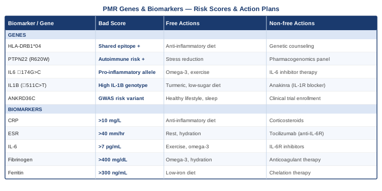
Having mapped both the measurable signals in your blood and the genetic architecture that shapes your inflammatory baseline, it is worth turning to one of the most frequently discussed topics in modern inflammation science — the dual role of interleukin-6 itself, and what cutting-edge research reveals about how to manage it at the behavioral level.
What Andrew Huberman's Research on IL-6 and Inflammation Reveals About PMR
Andrew Huberman, a neuroscientist and professor at Stanford University School of Medicine, has dedicated substantial content across his podcast and research collaborations to the biology of inflammation, IL-6 specifically, and the behavioral tools that modify inflammatory signaling. His work synthesizes mechanistic research in a way that is directly applicable to people managing chronic inflammatory conditions like PMR. The following ten insights, drawn from his podcast discussions and the underlying peer-reviewed literature he cites, are particularly relevant.
Insight 1: IL-6 Has Two Completely Different Identities
One of the most important clarifications Huberman makes about IL-6 is the distinction between its two biological roles that are frequently conflated in popular health discussions. When IL-6 is measured in the bloodstream of someone sitting at rest, it reflects a different biological process than when it is measured immediately after exercise. The resting, elevated IL-6 in PMR patients is secreted primarily by activated macrophages and immune cells in inflamed tissues — this is inflammatory IL-6, signaling distress. The IL-6 that appears during and after muscle contraction is secreted by the muscle fibers themselves, acting as what physiologists call a myokine — a hormone-like signal from muscle to other organs. These two contexts produce IL-6 from different cellular sources, acting through different receptor patterns, with largely opposite downstream effects. Understanding this distinction prevents the error of concluding that any exercise that raises IL-6 is harmful to PMR patients — it is specifically the resting, immune-driven IL-6 that is the problem.
Insight 2: Exercise-Induced IL-6 Stimulates Anti-Inflammatory IL-10
Huberman has highlighted research showing that the transient IL-6 released by contracting muscle during moderate aerobic exercise acts as a trigger for interleukin-10 (IL-10) production shortly after exercise ends. IL-10 is a potent anti-inflammatory cytokine that suppresses TNF-alpha, IL-1beta, and subsequent rounds of IL-6 production from macrophages. The brief IL-6 spike from a 30-minute brisk walk does not worsen PMR inflammation — it stimulates the body's own anti-inflammatory regulatory response. The key qualifier is intensity: high-intensity training that drives systemic stress hormones to damaging levels does the opposite. For PMR patients, this means regular moderate aerobic activity is not just safe — it is an active anti-inflammatory intervention through this specific myokine mechanism.
Insight 3: One Night of Poor Sleep Raises Next-Day IL-6
Huberman has discussed research demonstrating that even a single night of partial sleep deprivation (four to six hours) is sufficient to elevate next-morning circulating IL-6, along with NF-κB activity in immune cells and reductions in natural killer cell activity. For PMR patients who already have elevated baseline IL-6 from disease activity, disrupted sleep functions as an inflammatory amplifier. Critically, this effect is measurable on a single-night basis — meaning it is not about chronic sleep debt alone, but about each individual night. A practical implication is that PMR patients should view every sleep effort as a direct medical intervention, not a comfort measure.
Insight 4: Cold Exposure and Norepinephrine Suppress IL-6
Cold exposure protocols — whether cold water immersion, cold showers, or cold-water swimming — produce rapid and substantial norepinephrine release from the brainstem and from peripheral sympathetic nerve terminals. Research by Søberg and colleagues published in Cell Reports Medicine demonstrated that deliberate cold exposure protocols produce 200 to 300 percent norepinephrine increases that persist for hours. Norepinephrine binds beta-adrenergic receptors on macrophages and lymphocytes, where it signals via cAMP pathways that suppress NF-κB activation and IL-6 transcription. For PMR patients, practical application starts conservatively: a 30-second cold shower ending is a low-barrier entry point, progressing over weeks to 2 to 4 minutes of sustained cold water exposure three to five times per week. This should be approached cautiously in those with significant cardiovascular disease.
Insight 5: Chronic Stress and HPA-Driven IL-6 Elevation
The relationship between psychological stress and inflammatory cytokines runs through the hypothalamic-pituitary-adrenal (HPA) axis — a mechanism Huberman has covered extensively. Under chronic stress, the HPA axis becomes dysregulated — cortisol receptor sensitivity in immune cells decreases (glucocorticoid resistance), so the normal anti-inflammatory effect of cortisol is lost while the sensitization of NF-κB through upstream stress pathways remains. The net result is chronically elevated IL-6, TNF-alpha, and CRP in people with chronic psychological stress — an effect size comparable to poor diet or sedentary behavior. For PMR patients managing significant life stress alongside their diagnosis, this is a clinically meaningful inflammatory driver. Structured autonomic downregulation practices — physiological sighs, NSDR (non-sleep deep rest, essentially yoga nidra), and forward locomotion like walking — measurably shift HPA tone when practiced consistently.
Insight 6: Time-Restricted Eating Reduces Inflammatory Cytokines
Huberman has discussed research on time-restricted eating (TRE) — specifically 10 to 12-hour eating windows — and its effects on inflammatory markers including IL-6. The mechanism involves reduced nighttime insulin signaling allowing adipose tissue to undergo more complete overnight lipolysis, which reduces adipokine secretion from fat cells. It also involves circadian alignment of feeding with metabolic gene expression in the liver, reducing hepatic inflammatory activity. A study by Sutton and colleagues at the University of Alabama found that a 6-week early TRE protocol reduced blood pressure, oxidative stress, and inflammatory markers without caloric restriction. For PMR patients on corticosteroids who have developed truncal adiposity — a recognized glucocorticoid side effect and independent inflammatory amplifier — TRE is a practical intervention that requires no caloric counting or dietary restriction beyond timing.
Insight 7: Sunlight and Vitamin D Affect IL-6 Transcription
Huberman has dedicated considerable discussion to the biology of sunlight — not merely vitamin D synthesis but the broader effects of light exposure on neuroendocrine signaling. The active form of vitamin D (1,25-dihydroxyvitamin D3) binds vitamin D receptors on immune cells and directly downregulates IL-6 gene transcription through a vitamin D response element in the IL6 promoter region. In plain terms, adequate vitamin D levels reduce the amount of IL-6 your immune cells produce in response to a given inflammatory stimulus. Given that vitamin D deficiency is nearly universal in PMR patients on long-term corticosteroids, and that most PMR patients are in the age group with lowest sun exposure, this represents a pervasive and correctable inflammatory amplifier. Ten to twenty minutes of midday skin sun exposure daily combined with D3 supplementation to achieve 50 to 80 ng/mL serum 25-OH-D is the pragmatic approach.
Insight 8: Loneliness Raises Inflammatory Markers Including IL-6
Huberman has discussed the neuroscience of social connection and its biological consequences, drawing on research showing that subjective loneliness — independent of objective social contact — raises circulating IL-6, fibrinogen, and CRP to levels comparable to smoking or sedentary behavior. The mechanism involves perceived social threat activating inflammatory gene expression networks (particularly NF-κB) through limbic system-to-HPA axis pathways. For PMR patients who may have withdrawn from social activities due to pain, fatigue, or loss of independence, social isolation is not just a psychological concern — it is a biologically active driver of the same inflammatory markers they are trying to suppress. Restoring social engagement, even gradually and within physical limitations, is therefore a genuinely anti-inflammatory prescription.
Insight 9: The Paradox of Why Acute Exercise IL-6 Is Beneficial
This insight resolves what appears to be a paradox that confuses many patients: if IL-6 is bad in PMR, why is exercise — which transiently raises IL-6 — good? The answer, as Huberman explains through the work of Pedersen and colleagues in Denmark, is that the post-exercise anti-inflammatory cascade initiated by myokine IL-6 more than compensates for the transient pro-inflammatory spike. The net effect of repeated exercise sessions is a lowering of the resting, immune-cell-derived IL-6 between sessions — achieved through upregulation of IL-10, downregulation of TNF-alpha, and epigenetic remodeling of inflammatory gene expression in macrophages toward a more anti-inflammatory phenotype. In practical terms: the PMR patient who walks briskly for 30 minutes five days per week will, after 6 to 8 weeks, have measurably lower resting IL-6 than before they started — despite each individual walk transiently raising IL-6 during the session.
Insight 10: Stacking Interventions for Chronically Elevated IL-6 in PMR
Huberman consistently emphasizes the synergistic effect of stacking multiple behavioral interventions rather than relying on any single approach. For a PMR patient with chronically elevated IL-6, the optimal behavioral stack addresses all the mechanisms above simultaneously: morning outdoor sunlight exposure on waking (circadian alignment and vitamin D); a warm-to-cold contrast shower protocol; exercise within the first 6 hours of the day — brisk walking, pool walking, or gentle cycling for 20 to 40 minutes at conversational intensity; meals confined to a 10 to 12-hour window starting with breakfast; an afternoon 10 to 20-minute NSDR or body scan practice to reduce HPA-driven inflammatory tone; deliberate social interaction — even brief; and consistent sleep time with complete darkness and screen removal 45 minutes before bed. This is not a rigid prescription but a framework that addresses IL-6 through sleep, circadian biology, exercise, stress physiology, and social neuroscience simultaneously — the kind of multi-system approach that produces results no single intervention achieves alone.
The behavioral interventions described through these insights align closely with a body of structured evidence from complementary medicine approaches that have been formally studied in inflammatory and autoimmune conditions. The next section covers four of the most rigorously examined modalities specifically relevant to PMR.
Evidence-Based Complementary Approaches for Polymyalgia Rheumatica
Complementary approaches are most valuable when they are chosen based on their mechanistic relevance to the specific condition and when they are integrated alongside — not instead of — standard medical care. The four modalities below have the strongest available evidence for the biological mechanisms underlying PMR: immune dysregulation, systemic inflammation, HPA axis dysregulation, and the pain-fatigue cycle.
The Autoimmune Protocol — Sarah Ballantyne
The Autoimmune Protocol (AIP) is a dietary and lifestyle framework developed by immunologist and research scientist Sarah Ballantyne, PhD, designed specifically to address the gut-immune axis dysfunction that underlies autoimmune conditions. Its premise is that intestinal permeability — colloquially called "leaky gut" — allows dietary compounds and partially digested proteins to enter systemic circulation and trigger immune activation in genetically susceptible individuals. In PMR specifically, the gut-immune axis is relevant because the large-vessel macrophage activation that drives the disease is part of a broader innate immune dysregulation that is modulated by gut microbiome composition and intestinal barrier integrity. The AIP removes all foods documented to increase intestinal permeability or directly stimulate immune activation — grains, legumes, nightshades, eggs, dairy, nuts, seeds, and processed foods — during an elimination phase, then systematically reintroduces them to identify individual triggers.
The clinical evidence for AIP in autoimmune conditions includes a pilot study by Konijeti and colleagues (published in Clinical Gastroenterology and Hepatology, 2017) in Crohn's disease showing significant clinical and endoscopic remission rates after 6 weeks of AIP, and a similar trial in inflammatory bowel disease demonstrating reductions in fecal calprotectin and CRP. While RCTs specific to PMR have not yet been published, the mechanistic pathways — IL-6 reduction, NF-κB suppression through removal of dietary lectins and saponins, and gut microbiome modulation — are directly relevant to PMR's pathophysiology. Ballantyne's work in The Paleo Approach provides the clinical framework and the reintroduction protocol.
For PMR patients, practical AIP application requires attention to several real-world constraints. The elimination phase (typically 30 to 90 days) should not coincide with periods of high social demand or travel. During active PMR with significant pain and fatigue, meal preparation can be genuinely difficult — batch cooking simple AIP-compliant meals (braised meats, roasted root vegetables, bone broth-based soups) at lower-effort moments is a practical adaptation. Reintroduction should be done methodically, one food at a time with a 72-hour monitoring window, as PMR patients on corticosteroids may have blunted immediate inflammatory responses that make reactions slower to appear than in unmedicated individuals.
Mindfulness Meditation and MBSR
Mindfulness-Based Stress Reduction (MBSR) is an 8-week structured program developed by Jon Kabat-Zinn at the University of Massachusetts that combines body scan meditation, sitting meditation, mindful movement, and psychoeducation about stress physiology. For PMR, its relevance operates through multiple intersecting mechanisms: reduction of HPA-driven inflammatory cytokine production (particularly IL-6), improvement in pain catastrophizing, improvement in sleep quality, and enhancement of parasympathetic tone — all of which directly modulate disease burden. Unlike generalized relaxation practices, MBSR has been specifically studied in inflammatory disease populations with measurable biological outcomes.
A randomized controlled trial by Creswell and colleagues (published in Brain, Behavior, and Immunity, 2012) demonstrated that MBSR reduced serum IL-6 in lonely older adults — a population whose inflammatory profile closely resembles that seen in PMR — with greater reductions in those who showed the highest mindfulness skill acquisition. A systematic review in JAMA Internal Medicine found that mindfulness programs produced statistically significant reductions in self-reported pain, anxiety, and fatigue across multiple chronic pain and inflammatory conditions, with effects persisting at 6-month follow-up in studies that tracked it.
PMR patients applying MBSR should begin with the body scan practice rather than extended sitting meditation — 20 to 45 minutes of lying down with guided attention to body sensations. This is better tolerated during active disease phases when sitting is uncomfortable. Free evidence-based resources include the MBSR curriculum available through the Center for Mindfulness at UMass Medical School. The mindful movement component should be adapted for shoulder and hip mobility limitations — a qualified MBSR instructor experienced with chronic illness participants is worth seeking if available.
Tai Chi
Tai chi is a traditional Chinese movement practice characterized by slow, flowing, weight-shifting sequences performed with conscious breath coordination and meditative attention. Its relevance to PMR operates through several mechanisms simultaneously: gentle range-of-motion maintenance in the shoulders, hips, and spine — the primary sites of PMR stiffness — without impact or high-intensity loading; balance and proprioceptive training that reduces fall risk in a population already at heightened risk from corticosteroid effects on bone density and muscle mass; and neurological and HPA regulatory effects comparable to other mind-body practices. Tai chi is unique among exercise modalities in that it is simultaneously the most joint-gentle form of resistance and range-of-motion work available while also delivering meaningful autonomic nervous system regulation.
A systematic review and meta-analysis by Wang and colleagues (published in BMJ Open, 2016) analyzing 35 randomized controlled trials across rheumatic conditions including rheumatoid arthritis found that tai chi significantly reduced pain, improved physical function, and reduced inflammatory markers compared to usual care. A trial by Irwin and colleagues (published in the Journal of Gerontology) found that a 16-week tai chi program reduced NF-κB-driven inflammatory gene expression in peripheral blood mononuclear cells — the same cells that drive PMR's acute-phase response. The PMR-specific benefits thus extend well beyond pain management to include direct anti-inflammatory gene expression effects.
PMR patients beginning tai chi should seek Yang style beginner-level classes, which use larger, more upright postures that minimize deep knee flexion while maintaining all the upper-body and weight-shifting movements most relevant to PMR. Classes specifically for arthritis or older adults — widely available through community centers, YMCAs, and online platforms — provide appropriate modifications. Even a 20-minute practice three times per week produces measurable benefits; daily 15-minute practice is a practical target once the sequences are learned. During active flares, seated chair tai chi adaptations allow continuation of practice without exacerbating pain.
Breathing-Based Therapies
Breathing-based practices span a spectrum from ancient pranayama traditions to the contemporary Wim Hof Method (WHM), all of which share a common mechanism: deliberate alteration of arterial CO2 and O2 levels, autonomic nervous system tone, and interoceptive awareness through controlled breath patterns. For PMR, the most evidence-relevant application is the anti-inflammatory potential of specific breathing patterns demonstrated in clinical trials. A landmark 2014 study by Kox and colleagues (published in PNAS) demonstrated that healthy volunteers trained in WHM could voluntarily attenuate the innate immune response to intravenously administered endotoxin, producing significantly lower circulating TNF-alpha, IL-6, and IL-8 compared to untrained controls. This was the first demonstration that voluntary autonomic regulation could modulate systemic cytokine responses — directly relevant to PMR's cytokine-driven pathology.
Pranayama, the yogic breathwork tradition, has its own evidence base. Alternate nostril breathing and coherent breathing at 5 to 6 breath cycles per minute have both been shown in randomized trials to improve heart rate variability, reduce cortisol, and reduce salivary inflammatory markers. A meta-analysis in Psychosomatic Medicine found that yogic breathing interventions significantly reduced pro-inflammatory cytokines including IL-6 in populations with chronic stress and inflammatory conditions. The mechanism is primarily through vagus nerve activation increasing parasympathetic tone, which suppresses splenic macrophage TNF-alpha and IL-6 production through a well-described cholinergic anti-inflammatory reflex.
PMR patients should approach the Wim Hof breathing protocol with specific caution: the hyperventilation phase (30 to 40 deep breaths followed by a breath retention) can cause lightheadedness and in rare cases brief loss of consciousness — it must never be practiced in water, while driving, or without a seated or lying position. For those cleared by their physician, beginning with the basic 3-round WHM breathing sequence on waking and progressing over 4 to 6 weeks is a safe progression. Pranayama-based approaches are gentler and appropriate for all PMR patients from the outset — 5 minutes of coherent breathing (5-second inhale, 5-second exhale) practiced twice daily produces measurable HRV improvements within 2 weeks and is completely safe for all fitness levels.
Conclusion
Polymyalgia rheumatica is a condition that rewards attention to detail. It is not a simple, straightforward inflammatory process that responds only to corticosteroids and patience — it is a complex, immune-mediated disease shaped by genetic predispositions, amplified by lifestyle factors, and tracked reliably through a set of accessible blood biomarkers. The seven biomarkers covered in this article — CRP, ESR, IL-6, fibrinogen, ferritin, platelets, and alkaline phosphatase — each illuminate a different facet of what is happening in your blood vessels and immune system, and each responds to specific behavioral and nutritional interventions. The six genes — HLA-DRB1, PTPN22, IL6, TNFA, CTLA4, and NFKB1 — tell you something about the underlying architecture of your inflammatory susceptibility and point toward the interventions most likely to be relevant for your particular biology.
The core practical message is this: consistent monitoring creates opportunity. A patient who tracks their hs-CRP, ESR, and IL-6 every 4 to 6 weeks during a prednisolone taper is not more anxious about their disease — they are better equipped to catch a relapse before it becomes severe, to show their rheumatologist a clear trend rather than a single data point, and to identify which of their lifestyle interventions are actually moving the numbers. The supplements, dietary approaches, and mind-body practices described here are not replacements for medication — they are adjunctive tools that, used consistently and with appropriate medical supervision, reduce the inflammatory load that medication has to manage and may ultimately support a more successful and durable remission.
Your next practical step is straightforward: bring the biomarker list to your next rheumatology appointment, identify which ones you are not currently tracking, and ask whether adding them makes sense for your individual case. Review whatever genetic testing you may already have through consumer genomics platforms for the variants described here. Begin with the lowest-barrier lifestyle changes — a Mediterranean dietary pattern, consistent sleep, gentle daily movement, and a breathing practice — and build from there. PMR is a long-term project, not a short-term crisis, and the people who manage it best are those who approach it with that measured, informed, and persistent mindset.
Musculoskeletal: Joint Conditions Muscle Conditions
Cardiovascular: Vascular Conditions
Autoimmune: Inflammatory Conditions