This article contains AI generated content.
· UpdatedPolymyositis - 5 Genes And 7 Biomarkers To Track
Introduction
Living with polymyositis means living with a condition that most people have never heard of — and that even many clinicians see rarely. The progressive proximal muscle weakness, the fatigue that goes far beyond tiredness, the uncertainty about flares: these experiences are real and specific, and they deserve more than a generic framework built around suppressing symptoms and waiting to see what happens next.
The challenge with polymyositis is not just that it is rare. It is that the standard clinical picture — immunosuppression, monitoring by symptoms, watching CK levels — leaves out a large portion of what is actually measurable and actionable. The condition sits at the intersection of immunology, muscle biology, and genetics, and the people who navigate it best tend to be the ones who understand what is actually driving their disease, not just what category it falls into.
Generic lifestyle advice — reduce stress, eat well, rest when tired — is not wrong, but it is not enough. Polymyositis has specific biomarker signatures, specific antibody profiles, and documented genetic risk factors that create a much more targeted roadmap for intervention. The difference between knowing your CK is elevated and knowing why it is elevated, which antibody subtype you carry, or whether your IL-6 is driving the cycle matters enormously for what you do next.
This article approaches the problem from two angles. The first is a detailed breakdown of seven key biomarkers — what each one measures, how to track it, and what both lifestyle and supplement-based strategies can do when results are abnormal. The second is a look at five genes with documented roles in polymyositis susceptibility, along with specific plans for working around their effects whether or not supplementation is part of your approach. Neither section replaces medical care, but together they offer the kind of precise, targeted information that can make clinical conversations far more productive.
7 Biomarkers to Track in Polymyositis
Biomarkers are biology made measurable. In polymyositis, the right panel of tests tells a story that symptoms alone cannot: how active muscle damage is right now, which part of the immune system is most involved, whether cardiac or pulmonary tissue is at risk, and whether treatment is actually working. The seven markers below cover the full relevant spectrum — from the most basic to the most sophisticated — and each one earns its place in a serious monitoring strategy.
1. Creatine Kinase (CK)
Why it matters: Creatine kinase is the cornerstone biomarker in inflammatory myopathy. It is an intracellular enzyme found primarily in skeletal muscle, and when muscle fibers are damaged or actively inflamed, CK leaks into the bloodstream in large quantities. In active polymyositis, CK can be elevated 10 to 50 times the upper reference limit — occasionally even higher. It is used for initial diagnosis, for gauging disease severity, and for tracking treatment response over time. A falling CK typically signals that inflammation is being controlled; a rising CK can precede a clinical flare by weeks.
How to measure it: A serum CK test is included in most muscle enzyme or metabolic panels and costs between $10 and $40 USD. Results are available within 24 hours at virtually any clinical lab. Reference ranges vary slightly by laboratory and by sex — typical values are 22–198 U/L for women and 38–308 U/L for men. Importantly, CK should not be interpreted immediately after vigorous exercise, recent intramuscular injection, or a fall, as these can transiently elevate results independent of disease. A 2016 study in rheumatology referral populations confirmed that CK above 1000 U/L in non-exercise contexts warrants systematic evaluation for inflammatory myopathy.
If the score is high — plan without supplements: The most impactful non-pharmacological move when CK is elevated is exercise modification. This does not mean complete rest — in fact, prolonged inactivity worsens muscle atrophy in polymyositis — but it means replacing vigorous resistance training and high-impact activity with gentle range-of-motion work, hydrotherapy, and walking at low to moderate intensity. A Mediterranean-style dietary pattern (olive oil, oily fish, legumes, leafy greens, minimal processed food) consistently reduces the systemic inflammatory burden that drives ongoing muscle damage. Consistent sleep of seven to nine hours per night reduces pro-inflammatory cytokine output that directly contributes to CK elevation.
If the score is high — plan with supplements or equipment: Vitamin D3 and K2 address one of the most common and impactful deficiencies in polymyositis patients. A study published in the Journal of Rheumatology confirmed low serum vitamin D in idiopathic inflammatory myopathies, with muscle weakness correlating directly with deficiency. A typical protocol is 3000–5000 IU D3 daily with 100–200 mcg K2 (MK-7 form), taken with a fatty meal. Blood levels should be checked before and after, targeting 50–70 ng/mL. CoQ10 at 200–400 mg daily supports mitochondrial function in skeletal muscle and is well-tolerated long-term. Omega-3 fatty acids (2–4 g EPA+DHA daily, enteric-coated to minimize GI side effects) have anti-inflammatory effects in skeletal muscle tissue. These can all be taken continuously; recheck CK and vitamin D levels at 3-month intervals.
2. Myositis-Specific Antibodies (MSAs)
Why it matters: Myositis-specific antibodies are immune proteins directed against intracellular components — primarily RNA-processing enzymes — and they are among the most diagnostically and prognostically important markers in all of inflammatory myopathy. The most common is anti-Jo-1, present in roughly 20–30% of polymyositis cases, which targets histidyl-tRNA synthetase. Anti-Jo-1 positivity signals antisynthetase syndrome — a clinical cluster that includes interstitial lung disease, mechanic's hands, Raynaud's phenomenon, and inflammatory arthritis. Other antibodies carry distinct risk profiles: anti-SRP is associated with severe, rapidly progressive myopathy; anti-MDA5 with rapidly progressive interstitial lung disease and high mortality risk; anti-Mi-2 with a generally better treatment response. Knowing your MSA status shapes monitoring strategy and prognostic expectation. A genetic study linking HLA-DRB1*03 specifically to anti-Jo-1-positive inflammatory myopathy underscores how antibody subtype connects to the deeper biological picture.
How to measure it: A full MSA panel is a specialized immunology test typically ordered by a rheumatologist. Costs range from $100 to $500 USD depending on how many antibodies are included in the panel. Results provide a one-time baseline for risk stratification — these antibodies tend to remain relatively stable over time, so the test does not need frequent repetition, but antibody titers can sometimes shift with treatment or disease course. Comprehensive panels from reference labs such as Quest or LabCorp cover 10 or more MSAs in a single draw.
If antibodies are positive — plan without supplements: A positive MSA result — especially anti-Jo-1, anti-SRP, or anti-MDA5 — is a signal to pursue systematic monitoring beyond muscle markers alone. Baseline pulmonary function testing (spirometry, DLCO) and high-resolution CT of the chest should be established even before respiratory symptoms appear, as interstitial lung disease often begins subclinically. Avoiding cigarette smoke is critical — it significantly accelerates lung fibrosis in at-risk individuals. Diaphragmatic breathing exercises practiced 10–15 minutes daily support respiratory muscle endurance. Home peak flow monitoring (device cost: $20–$30) provides a simple early-warning system for declining lung function between clinic visits.
If antibodies are positive — plan with supplements or equipment: NAC (N-acetylcysteine) at 600 mg twice daily has antioxidant and mucolytic properties with documented relevance to interstitial lung disease. A 3-month trial with pulmonary function reassessment is reasonable. Vitamin D3 (as above) is especially important in anti-Jo-1-positive disease, where deficiency correlates with worse pulmonary outcomes. Astaxanthin (8–12 mg daily with a fatty meal) has emerging evidence for reducing oxidative stress in lung tissue. A portable spirometer ($50–$100) allows weekly FVC and peak flow checks at home, capturing trends that monthly clinic visits might miss.
3. C-Reactive Protein (CRP) and Erythrocyte Sedimentation Rate (ESR)
Why it matters: CRP and ESR are general markers of systemic inflammation. Neither is muscle-specific, but both reflect the overall inflammatory environment that polymyositis creates — and both have established relationships with cardiovascular risk, which is elevated in chronic inflammatory conditions. An important clinical nuance: CRP is often only mildly elevated or even normal in some polymyositis patients despite significant muscle inflammation, so these markers should always be interpreted alongside CK and aldolase, never in isolation. When CRP is disproportionately elevated, it may suggest a concurrent infection, a flare driven by systemic rather than primarily muscular inflammation, or an intercurrent autoimmune process.
How to measure it: Standard CRP and ESR tests are available at any clinical lab and cost $10–$30 USD each. High-sensitivity CRP (hsCRP) provides better resolution at lower inflammatory levels and is preferred for cardiovascular risk assessment; it typically costs $20–$50. Optimal hsCRP for cardiovascular risk is below 1.0 mg/L; values above 3.0 mg/L indicate elevated inflammatory and vascular risk. ESR normal ranges vary by age and sex but a significantly elevated ESR alongside elevated muscle enzymes supports active disease.
If the score is high — plan without supplements: Sleep quality and duration are among the most powerful non-pharmacological modulators of systemic CRP. Seven to nine hours of consistent, high-quality sleep reduces inflammatory cytokine production significantly over time. Dietary approaches with documented CRP-lowering effects include the Mediterranean diet, reducing refined carbohydrate and seed oil intake, and increasing colorful vegetables and polyphenol-rich foods. Time-restricted eating (eating within a 10–12 hour window, with a 12–14 hour overnight fast) has shown CRP-reducing effects in several clinical trials. Regular moderate aerobic exercise — walking 30 minutes at 60–70% maximum heart rate, five days per week — consistently reduces hsCRP in chronically elevated populations over 8–12 weeks.
If the score is high — plan with supplements or equipment: Omega-3 fatty acids at 2–4 g EPA+DHA daily are among the most consistently documented CRP-lowering supplements in clinical trials across multiple chronic inflammatory conditions. Curcumin with piperine (500–1000 mg curcumin with 5 mg piperine at each dose, twice daily with a fatty meal) has demonstrated statistically significant CRP reductions in multiple randomized controlled trials. A 12-week course is typical, with a 4-week break before reassessment to prevent tolerance. Magnesium glycinate at 300–400 mg nightly improves sleep quality and has indirect anti-inflammatory effects through NLRP3 inflammasome modulation. Side effects are minimal; avoid magnesium oxide due to poor bioavailability.
4. Aldolase
Why it matters: Aldolase is a glycolytic enzyme found in skeletal muscle that rises when muscle tissue is damaged or inflamed. It is most valuable as a complement to CK — particularly in cases where CK is paradoxically normal or only mildly elevated despite ongoing active disease, which can happen in longstanding polymyositis once significant muscle bulk has been lost. When CK and aldolase track together, the picture is consistent; when they diverge, it prompts important clinical questions about disease type, duration, or confounding medication effects.
How to measure it: Aldolase is a standalone serum test available at most clinical labs, typically costing $15–$40 USD. Normal range is approximately 1.0–7.5 U/L in adults. It must be specifically requested — it is rarely included in standard panels. Testing frequency should mirror CK monitoring: every 4–8 weeks during active disease or treatment adjustment, every 3–6 months during stable remission.
If the score is high — plan without supplements: Elevated aldolase in the context of polymyositis warrants the same activity-modification and anti-inflammatory dietary approach described for CK. But it also signals an important check on medications: corticosteroids at higher doses can independently cause steroid myopathy — a form of muscle damage that elevates aldolase and CK while not representing disease activity. This distinction is critical for treatment decisions. Supervised, gradual corticosteroid tapering — when medically appropriate — may itself reduce aldolase by removing the drug's direct myotoxic effect.
If the score is high — plan with supplements or equipment: Creatine monohydrate at 3–5 g daily has specific evidence for improving muscle function and reducing muscle enzyme leak in inflammatory myopathy. A double-blind, randomized, placebo-controlled trial published in Arthritis & Rheumatism showed functional improvements with creatine supplementation in patients with idiopathic inflammatory myopathies. It can be taken continuously without meaningful cycling. L-carnitine at 1–2 g daily supports skeletal muscle energy metabolism and reduces exercise-induced muscle damage. Both are well-tolerated and can be started simultaneously.
5. Lactate Dehydrogenase (LDH)
Why it matters: LDH is a broadly distributed enzyme involved in cellular energy metabolism. In polymyositis, it rises alongside CK and aldolase as a marker of muscle cell damage, and normalizes in parallel with clinical improvement. Its added value lies partly in its sensitivity as a monitoring tool, and partly in its role as a flag: when LDH is disproportionately elevated relative to the other muscle enzymes, it prompts consideration of concurrent issues — liver disease, hemolysis, or malignancy. Occult malignancy is associated with polymyositis at an elevated rate, and monitoring LDH as part of a broader panel supports early detection if a cancer workup becomes necessary.
How to measure it: LDH is included in most comprehensive metabolic panels and costs $10–$30 USD. Normal range is approximately 100–250 U/L in adults, though this varies by laboratory method. Baseline testing should be done at diagnosis; follow-up frequency mirrors CK and aldolase monitoring.
If the score is high — plan without supplements: Before attributing elevated LDH to polymyositis activity, rule out confounders: alcohol consumption, recent strenuous exercise, statin use, and active infection all elevate LDH independently. If confounders are excluded, the focus is on the same core interventions supporting CK reduction: dietary anti-inflammatory pattern, sleep optimization, moderate physical activity. If LDH remains disproportionately elevated compared to other muscle enzymes, discuss a broader cancer screen with your rheumatologist — this is not alarming, but it is a reasonable clinical step at baseline and periodically thereafter.
If the score is high — plan with supplements or equipment: Alpha-lipoic acid (300–600 mg daily) is a mitochondrial antioxidant with documented capacity to reduce oxidative-stress-driven enzyme release in metabolically stressed cells. An 8-week-on, 4-week-off cycling pattern is recommended to prevent potential biotin depletion with long-term use. B-complex vitamins support cellular energy metabolism across multiple pathways and are commonly depleted in chronic inflammatory conditions, particularly in patients on methotrexate (which depletes folate). A full-spectrum B-complex daily is a low-risk, broadly supportive addition.
6. Interleukin-6 (IL-6)
Why it matters: IL-6 is a cytokine — a molecular messenger — that sits upstream of much of the inflammatory cascade in polymyositis. It drives T cell activation, promotes B cell differentiation into antibody-producing plasma cells, and stimulates the liver to produce acute-phase proteins including CRP. Elevated IL-6 in polymyositis reflects a state of immune overdrive that cannot be fully captured by CRP or CK alone. It has become a validated therapeutic target: tocilizumab, an IL-6 receptor antagonist, is used in refractory cases of inflammatory myopathy. Research on IL-6 blockade in polymyositis models confirms its central pathogenic role. Measuring it gives an independent window into upstream immune activity.
How to measure it: Serum IL-6 is available through specialized reference labs and some direct-to-consumer platforms (e.g., Vibrant Wellness, Ulta Lab Tests). Costs range from $50 to $150 USD. It is not a standard rheumatology order, so it typically requires specific request or self-pay ordering. Optimal serum IL-6 is below 7 pg/mL; values above 20–30 pg/mL correlate with active systemic inflammatory disease. Re-testing every 3–6 months during active monitoring phases is appropriate.
If the score is high — plan without supplements: Time-restricted eating (eating within a 10–12 hour window daily) reduces fasting IL-6 in clinical studies, likely through effects on visceral adiposity and circadian rhythm regulation of immune cells. Moderate aerobic exercise — consistent walking or swimming at 60–70% maximum heart rate, 30 minutes per session, five days per week — transiently increases but chronically reduces circulating IL-6 through long-term adaptations in skeletal muscle and adipose tissue. Cold water exposure (ending a shower with 2–3 minutes of cold water, or a cold plunge at 10–15°C for 3 minutes, three times per week) has been shown in small trials to increase the anti-inflammatory cytokine IL-10 while dampening IL-6. Start conservatively and increase duration gradually; avoid in active cardiac disease.
If the score is high — plan with supplements or equipment: Curcumin with piperine (500–1500 mg curcumin twice daily with a fatty meal and piperine) inhibits NF-κB — the primary transcription factor driving IL-6 gene expression — and has shown statistically significant IL-6 reductions in multiple RCTs. Quercetin (500 mg twice daily) has additive effects through a complementary NF-κB inhibition pathway and is well-tolerated. Vitamin D3 at adequate serum levels directly suppresses IL-6 gene transcription through vitamin D receptor signaling. A continuous glucose monitor (CGM) — available for $30–$80 per month — can reveal glycemic variability patterns, as post-meal glucose spikes are a potent driver of cytokine release and can be reduced through simple dietary timing adjustments.
7. Cardiac Troponin I or T
Why it matters: Polymyositis is not limited to skeletal muscle. Cardiac involvement — including myocarditis, arrhythmia, and cardiomyopathy — occurs in an estimated 30–40% of patients and is a major contributor to disease-related mortality. A clinical review of cardiac involvement in polymyositis and dermatomyositis confirmed that subclinical cardiac inflammation often precedes clinical symptoms by months to years. High-sensitivity cardiac troponin (hs-cTnT or hs-cTnI) is the most sensitive available marker for myocardial cell injury. Any elevation warrants investigation, not reassurance — the assumption that all troponin elevation in polymyositis reflects cross-reacting skeletal troponin isoforms is a dangerous oversimplification.
How to measure it: High-sensitivity troponin testing costs $20–$80 USD and is available at most hospital labs and some direct-to-consumer services. When troponin is elevated, further workup — ECG, echocardiogram, and if indicated, cardiac MRI — provides the full picture. Monitoring frequency depends on individual risk: annually at minimum for all polymyositis patients; more frequently in anti-MDA5-positive disease or if there are any cardiac symptoms (palpitations, exertional dyspnea, chest tightness).
If the score is high — plan without supplements: Minimize cardiovascular stressors: avoid abrupt high-intensity exercise while troponin is elevated, treat any concurrent hypertension or dyslipidemia actively, and address sleep-disordered breathing (common in inflammatory myopathy patients, and a major independent cardiac stressor). Corticosteroid-induced metabolic changes — hyperglycemia, weight gain, dyslipidemia — all stress the myocardium independently and should be monitored and managed. A consumer-grade ECG monitor (Kardia Mobile, ~$100 one-time cost, or Apple Watch with ECG capability) enables at-home rhythm monitoring and can detect atrial fibrillation or other arrhythmias between clinic appointments.
If the score is high — plan with supplements or equipment: CoQ10 (200–400 mg daily) has specific evidence for myocardial protection in inflammatory states and supports mitochondrial energy production in cardiac muscle. Magnesium glycinate (300–400 mg nightly) supports cardiac rhythm stability and is frequently depleted in patients on corticosteroids. Omega-3 fatty acids at 3–4 g EPA+DHA daily have anti-arrhythmic and cardioprotective effects with robust clinical evidence. A home pulse oximeter (~$20) tracks nocturnal oxygenation for early detection of respiratory contributions to cardiac stress. All of these can be used long-term; recheck troponin at 3-month intervals while actively monitoring.
With those seven markers forming a comprehensive biological picture, the following table brings together both the biomarkers and the genetic variants covered in this article — a single-glance reference for tracking and action.
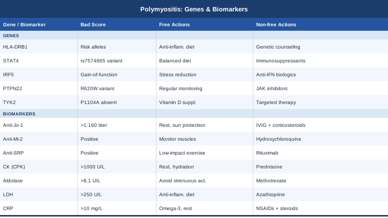
The Genetic Side: 5 Variants That Shape Your Risk
Genetics in polymyositis does not mean destiny. What the research does show is that certain gene variants shift the immune system toward autoimmune activation under the right environmental conditions — and that understanding those variants points toward specific, modifiable exposures and biological pathways. Most of the genetic risk in inflammatory myopathy is probabilistic rather than deterministic, and each of the variants below has lifestyle, dietary, and supplement-based counterweights worth knowing about.
1. HLA-DRB1 — The Strongest Genetic Risk Factor
What it does: HLA-DRB1 encodes a component of the major histocompatibility complex (MHC) class II, a protein that presents antigens to helper T cells. The HLA-DRB1*03:01 allele is the most consistently replicated genetic risk factor for polymyositis and antisynthetase syndrome. It is thought to influence which self-antigens get displayed to the immune system, potentially triggering the aberrant autoreactive T cell responses that drive muscle inflammation. Carrying this allele does not mean you will develop polymyositis — but in combination with environmental triggers, it significantly raises the probability. Research specifically linking HLA-DRB1*03 to anti-Jo-1-positive inflammatory myopathy supports the clinical relevance of this connection.
If the variant is present — plan without supplements: The goal is to minimize the environmental triggers that convert genetic susceptibility into active autoimmune disease. Avoid prolonged UV exposure without protection — UV-driven DNA damage generates neoantigens that HLA-DRB1*03:01 may present with particular efficiency. Address persistent viral infections aggressively, as Epstein-Barr virus and influenza are documented precipitants of polymyositis onset in genetically susceptible individuals. An autoimmune elimination diet (removing gluten, dairy, processed food, and high-omega-6 seed oils for 8–12 weeks) serves both as a diagnostic and therapeutic trial — many patients with HLA-DRB1 risk alleles report significant symptom changes with dietary intervention. Monitor anti-Jo-1 antibody status annually even during remission.
If the variant is present — plan with supplements or equipment: Vitamin D3/K2 (3000–5000 IU D3 with 150–200 mcg K2, targeting serum 25-OH vitamin D above 50 ng/mL) is arguably the single most relevant supplement for HLA-DRB1 risk — vitamin D receptor signaling directly modulates antigen-presenting cell behavior and downregulates autoimmune T cell activation. Selenium (100–200 mcg daily as selenomethionine) supports regulatory T cell function and helps dampen the autoimmune T cell activity that HLA risk variants promote. Do not exceed 400 mcg per day long-term due to toxicity at higher doses. Multi-strain probiotics (including Lactobacillus rhamnosus GG and Bifidobacterium longum) support gut barrier integrity, reducing the antigen load that drives immune activation through HLA-dependent mechanisms.
2. HLA-DQA1 — Closely Linked and Antisynthetase-Associated
What it does: HLA-DQA1 sits on chromosome 6 in close proximity to HLA-DRB1 and the two are typically inherited together as a haplotype. HLA-DQA1 contributes to the same antigen-presentation system, and specific HLA-DQA1 alleles have been independently associated with anti-Jo-1 antibody production and antisynthetase syndrome in genetic studies. Its clinical relevance is reinforced by the fact that antisynthetase syndrome — with its lung disease risk — represents a more complex and monitored subset of polymyositis, and knowing that this genetic background is present should heighten respiratory vigilance.
If the variant is present — plan without supplements: The strategy overlaps significantly with HLA-DRB1: an autoimmune-focused anti-inflammatory dietary approach, stress reduction (chronic psychological stress activates the same HPA-immune axis pathways that HLA variants amplify), and regular lung function monitoring. Establish baseline pulmonary function tests annually even in the absence of respiratory symptoms. Avoid occupational or residential mold exposure, which activates toll-like receptor pathways that HLA-DQA1 risk variants interface with. Breathing exercises — specifically diaphragmatic breathing practiced 10–15 minutes daily — support respiratory muscle endurance proactively.
If the variant is present — plan with supplements or equipment: Butyrate (sodium butyrate, 300–600 mg daily) supports the production of colonic regulatory T cells (Tregs), which help suppress the autoreactive immune activity that HLA variants amplify. Berberine (500 mg twice daily with meals) modulates gut microbiome composition and NF-κB signaling simultaneously — cycle 8 weeks on, 4 weeks off to prevent tolerance and preserve gut microbiome diversity. A home spirometer ($50–$100) for weekly FVC and peak flow monitoring provides granular data on lung function trends between rheumatology appointments.
3. PTPN22 — The Autoimmune Susceptibility Switch
What it does: PTPN22 encodes a lymphoid-specific tyrosine phosphatase that normally acts as a brake on T cell and B cell receptor signaling. The R620W variant (rs2476601) is one of the most widely replicated autoimmune susceptibility variants in the human genome — associated with rheumatoid arthritis, type 1 diabetes, systemic lupus erythematosus, and inflammatory myopathies including polymyositis. A landmark study documenting differential PTPN22 associations across autoimmune diseases confirmed its broad relevance. Carriers have a lower activation threshold for autoreactive lymphocytes — meaning immune responses to self-antigens are easier to trigger and harder to terminate.
If the variant is present — plan without supplements: Gut health is especially relevant here because gut-derived antigens are a primary stimulus for T and B cell activation, and PTPN22 variants affect how efficiently those cells are braked after stimulation. A diet rich in fermented foods (kefir, sauerkraut, kimchi, miso), diverse fiber, and polyphenol-rich plants helps maintain a microbiome composition that promotes regulatory rather than pro-inflammatory immune responses. Smoking cessation is critical — smoking activates the same T cell receptor pathways that PTPN22 variants fail to adequately suppress, and smoking is independently associated with worsening autoimmune disease in PTPN22 variant carriers. Avoid pesticide-heavy food when possible, as organophosphate residues can directly activate toll-like receptors linked to PTPN22-pathway immune escalation.
If the variant is present — plan with supplements or equipment: Vitamin D3 (maintaining serum levels above 50 ng/mL) is particularly important for PTPN22 variant carriers because vitamin D receptor signaling provides an alternative pathway for immune regulation that partially compensates for the impaired PTPN22 brake function. Zinc bisglycinate (15–25 mg daily, separate from calcium-rich foods which inhibit zinc absorption) supports regulatory T cell development and thymic function — zinc deficiency is common in inflammatory conditions and directly impairs the Treg populations that counterbalance PTPN22-driven hyperactivation. Spore-forming probiotics (e.g., Bacillus coagulans or Bacillus subtilis-based products) survive the gastric environment more reliably than standard strains and modulate immune signaling from the colonic epithelium.
4. STAT4 — Driving Th1 and Th17 Inflammation
What it does: STAT4 encodes signal transducer and activator of transcription 4, a downstream mediator of IL-12 and IL-23 signaling. These cytokines drive the differentiation of T helper cells toward the Th1 and Th17 phenotypes — exactly the inflammatory T cell subtypes that are found infiltrating muscle tissue in polymyositis. Risk variants in STAT4 are associated with multiple autoimmune conditions, and an overactive STAT4 pathway promotes the kind of aggressive, tissue-infiltrating immune response that characterizes active polymyositis. Elevated IL-6 (discussed above as a key biomarker) and STAT4 variants form a converging biological picture.
If the variant is present — plan without supplements: Reduce dietary and lifestyle inputs that promote Th1/Th17 immune skewing. This means limiting high-omega-6 vegetable oils (sunflower, soybean, corn oil) that shift the prostaglandin balance toward inflammation, prioritizing omega-3-rich foods (oily fish, flaxseed, walnuts), and ensuring consistent, high-quality sleep — sleep deprivation is one of the most potent promoters of Th1-dominant immune responses through IL-12 upregulation. Consistent moderate aerobic exercise supports Th2 and Treg balance over time, counteracting the Th1 skewing that STAT4 variants promote. Reducing chronic psychological stress is especially relevant because the cortisol rebound after stress exposure chronically amplifies Th1 activity.
If the variant is present — plan with supplements or equipment: EGCG from green tea extract (400–800 mg daily standardized extract) has documented inhibitory effects on STAT4 signaling and Th17 differentiation in mechanistic studies; human RCT evidence is limited but the mechanistic case is strong. Take with food to avoid nausea. Melatonin (0.5–2 mg taken 30–60 minutes before sleep) serves not primarily as a sedative but as an immune modulator — it has documented effects on Th1/Th2 cytokine balance and may dampen STAT4-driven inflammatory cycles during the overnight repair window. Tart cherry concentrate (30 ml daily) contains anthocyanins with anti-inflammatory and cytokine-modulating effects shown in clinical trials for inflammatory conditions.
5. IRF5 — The Interferon Amplifier
What it does: IRF5 encodes interferon regulatory factor 5, a transcription factor central to type I interferon production and pro-inflammatory cytokine expression (including IL-6, IL-12, and TNF-α). Gain-of-function variants in IRF5 are documented across lupus, rheumatoid arthritis, inflammatory bowel disease — and polymyositis. A robust interferon gene expression signature is detectable in both peripheral blood and muscle biopsies of many polymyositis patients, and IRF5 variants likely contribute to this pattern. This is directly relevant to fatigue — type I interferons are potent fatigue-inducing molecules, and the profound tiredness many polymyositis patients experience beyond simple muscle weakness may have an interferon-driven component.
If the variant is present — plan without supplements: Minimizing interferon pathway triggers is the core non-supplemental strategy. Avoid prolonged UV overexposure — UV activates toll-like receptor 7 and 9 pathways that feed directly into IRF5-driven interferon production. Early morning outdoor light exposure (10–30 minutes in the first hour after waking, before the UV index rises) entrains circadian rhythms without the same immune-activating UV dose. Sleep quality is arguably the most impactful lever: poor sleep amplifies both IRF5 expression and type I interferon signaling, creating a fatigue-inflammation feedback loop. Tracking morning fatigue patterns with a simple daily scale (1–10, logged on a phone app) can serve as a practical proxy for interferon pathway activity over time.
If the variant is present — plan with supplements or equipment: Vitamin D3 (maintaining serum 25-OH vitamin D above 50 ng/mL) directly downregulates IRF5 transcriptional activity through vitamin D response elements in the IRF5 gene promoter. NAC (N-acetylcysteine) (600 mg twice daily) reduces oxidative stress-driven toll-like receptor activation that is an upstream input to IRF5 signaling. Astaxanthin (8–12 mg daily with a fatty meal) is a fat-soluble carotenoid antioxidant with documented inhibitory effects on IRF-pathway-driven cytokine release in early clinical studies. A wearable HRV monitor (Oura Ring, WHOOP, or Garmin) tracks heart rate variability as a continuous proxy for autonomic and inflammatory balance — useful for identifying patterns between symptom flares and recovery.
A Book That Challenges the Standard Approach to Autoimmune Disease
The Autoimmune Solution by Amy Myers, MD, has become one of the most referenced lay-medical books on autoimmunity since its publication. Myers is a trained functional medicine physician who draws extensively on human clinical research to argue that the current standard of care — suppress the immune system, manage symptoms — addresses the downstream consequences of autoimmune disease while leaving the upstream drivers in place. Her framework is grounded in several well-documented mechanisms, and while the book is not specific to polymyositis, its core thesis maps closely onto what the emerging science of inflammatory myopathy supports.
The book's central claim is that autoimmune disease in most people is driven by a recognizable cluster of upstream factors: intestinal permeability, gut dysbiosis, nutritional deficiencies (especially vitamin D), environmental toxins, chronic infections, and unmanaged psychophysiological stress. Myers argues these factors are not incidental — they are mechanistically upstream of the immune dysregulation that produces tissue-specific autoimmune attacks.
10 Things Worth Knowing from This Book
1. Intestinal Permeability Is Not a Fringe Concept
Myers describes how disruption of tight junctions in the intestinal epithelium allows microbial fragments and food proteins to enter the bloodstream and trigger persistent immune activation. Gut dysbiosis has been specifically documented in inflammatory myopathy patients in recent small studies, lending credibility to this upstream mechanism in the polymyositis context.
2. The Three-Legged Stool Model
Myers proposes that autoimmune disease requires three simultaneous conditions: genetic susceptibility, intestinal permeability, and an environmental trigger. Removing any one of the three legs — particularly permeability — may interrupt the cycle even when the genetic predisposition is fixed. This reframes the problem from "I have a genetic condition" to "I have a genetic predisposition that requires specific environmental conditions to activate."
3. Gluten Is Not Only a Problem for Celiac Disease
Myers presents research showing that gluten-derived proteins trigger zonulin release — a tight junction regulator — in non-celiac individuals, transiently increasing intestinal permeability. She recommends a complete 90-day elimination for all autoimmune diagnoses as both a diagnostic and therapeutic trial. The evidence for this is strongest in gluten-sensitive enteropathy but the mechanistic argument extends broadly.
4. Vitamin D as Immune Regulator, Not Just Bone Nutrient
Myers dedicates significant space to the genomic effects of vitamin D on immune cell gene expression. Her recommended target of 60–80 ng/mL 25-OH vitamin D is above the conventional sufficient range but is within the bounds of what immunology research supports for autoimmune suppression. This aligns directly with the vitamin D data cited throughout this article.
5. Toxin Load Is a Recognized Autoimmune Trigger
Environmental toxins — heavy metals (mercury, lead), pesticides, and mycotoxins from mold — activate toll-like receptors and interferon pathways. Myers recommends a comprehensive heavy metals panel and mycotoxin urine test in any autoimmune patient where standard triggers have not explained disease onset. For polymyositis patients with refractory or unexplained flares, this is a logical investigative step.
6. Chronic Infections Can Perpetuate Disease Through Molecular Mimicry
Molecular mimicry occurs when a pathogen's proteins resemble host proteins closely enough that antibodies generated against the pathogen cross-react with self-tissue. In polymyositis, Epstein-Barr virus, Coxsackie B virus, and SARS-CoV-2 have all been identified as potential precipitants through this mechanism. Myers argues that chronic low-level viral reactivation — measurable through specific IgG titers — may be silently perpetuating immune activation long after the acute infection resolves.
7. HPA Axis Dysregulation Is Measurable, Not Just Theoretical
Myers goes beyond the usual "reduce stress" advice to describe how chronic stress dysregulates the hypothalamic-pituitary-adrenal axis, producing patterns of cortisol that initially suppress inflammation but chronically promote Th1 and Th17 immune dominance. She recommends morning salivary cortisol testing as a practical, inexpensive tool for assessing whether the stress-HPA axis is contributing to inflammatory cycles.
8. Thyroid Status Must Be Assessed in Every Autoimmune Patient
Autoimmune thyroid disease (Hashimoto's thyroiditis or Graves' disease) co-occurs with inflammatory myopathy at rates significantly above population baseline. Unrecognized hypothyroidism produces its own form of myopathy that can both mimic and worsen polymyositis. Myers insists on a full thyroid panel (TSH, free T3, free T4, reverse T3, anti-TPO, anti-thyroglobulin) — far beyond the TSH-only test that is typically ordered.
9. The 4R Gut Restoration Protocol
Myers presents a structured four-step approach: Remove inflammatory foods and pathogens, Replace digestive enzymes and stomach acid if deficient, Reinoculate with targeted probiotics and prebiotics, Repair the gut lining with glutamine (5 g daily), zinc carnosine (75 mg daily), and collagen. This provides a practical, sequenced gut-restoration framework directly applicable to the upstream immune drivers in polymyositis.
10. Food Functions as Gene-Regulatory Information
Myers' most paradigm-challenging point — and one supported by epigenetic research — is that dietary compounds act as signaling molecules that activate or silence gene expression in immune cells. For someone carrying HLA, PTPN22, STAT4, or IRF5 variants, this means food choices are directly influencing which inflammatory genes are expressed in muscle-infiltrating immune cells. The "just take your medication" framework implicitly treats diet as irrelevant to immune gene regulation — the science says otherwise.
Complementary Approaches With Clinical Relevance
The biomarker and genetic strategies above focus on what you can measure and directly modify. The approaches below are distinct modalities — not replacements for medical care, but additions that address aspects of polymyositis management that standard pharmaceutical treatment leaves largely untouched. Each one has meaningful human evidence in autoimmune or related inflammatory conditions.
The Autoimmune Protocol by Sarah Ballantyne
The Autoimmune Protocol (AIP), developed by Sarah Ballantyne, PhD (author of The Paleo Approach), is a structured dietary and lifestyle elimination framework designed specifically for autoimmune conditions. It removes potential immune-activating dietary compounds — gluten, dairy, legumes, grains, eggs, nightshade vegetables, alcohol, seed oils, and food additives — while emphasizing nutrient-dense foods that support gut healing, hormonal balance, and immune regulation. The rationale is grounded in identified mechanisms: gut permeability, molecular mimicry via dietary lectins and prolamins, and nutritional deficiencies that impair immune regulation.
Human evidence includes several published pilot studies. A 2017 pilot trial published in Inflammatory Bowel Diseases documented significant clinical remission in Crohn's disease and ulcerative colitis patients following the AIP. While direct RCT evidence specific to polymyositis is not yet available, the mechanistic overlap — gut barrier restoration, reduced antigen-driven immune activation, correction of nutrient deficiencies — is directly applicable. For a condition where gut dysbiosis and dietary triggers are documented contributors, this is among the most evidence-informed dietary frameworks available.
To apply it: begin with the 30–60 day elimination phase, removing all excluded foods strictly. After the elimination phase, conduct structured reintroductions one food at a time over 10 days each, tracking symptom changes and relevant biomarkers (CRP, CK, fatigue scale) during each reintroduction. The lifestyle component — sleep optimization, gentle movement, stress reduction, social connection — is equally emphasized and not optional to the protocol's design. Work with a registered dietitian familiar with AIP to ensure nutritional adequacy during the elimination phase.
Mindfulness-Based Stress Reduction (MBSR)
MBSR is an 8-week structured program developed by Jon Kabat-Zinn that combines mindfulness meditation, body scanning, and gentle yoga to cultivate a sustained non-reactive awareness of physiological and emotional experience. Its relevance to polymyositis is twofold: first, it directly addresses the HPA axis dysregulation that drives Th1/Th17-dominant inflammatory immune responses; second, it provides tools for managing the chronic pain, fatigue, and psychological burden that characterize life with inflammatory myopathy.
The clinical evidence is substantial. A meta-analysis published in JAMA Internal Medicine (Goyal et al., 2014) covering 47 trials found moderate evidence that mindfulness meditation reduces anxiety, depression, and pain in chronic disease populations. Specific to autoimmune conditions, a randomized trial in rheumatoid arthritis patients documented reductions in disease activity scores and inflammatory cytokines after MBSR. While direct RCT evidence in polymyositis remains limited, the convergence of evidence across related inflammatory conditions is persuasive.
To apply it: a standard MBSR course involves 2.5 hours of group sessions weekly for 8 weeks plus a full-day retreat, with daily home practice of 30–45 minutes. Both in-person and digital formats (Sounds True MBSR, Palouse MBSR online) are available. Start with 10–15 minutes of daily breath-focused meditation if a full program is not immediately accessible; the key is consistent daily practice rather than session length. Track morning fatigue and a simple pain or wellness score over the 8 weeks to gauge individual response.
Tai Chi for Inflammatory Muscle Conditions
Tai chi is a low-impact, mind-body movement practice involving slow, deliberate sequences of postures coordinated with controlled breathing. It is uniquely well-suited to polymyositis management because it provides meaningful physical activity — improving muscle strength, balance, and proprioception — without the muscle damage risk associated with higher-intensity exercise. It also addresses the fatigue and psychological dimension of chronic disease through its meditative structure.
Human evidence for tai chi in inflammatory rheumatic conditions is growing. A 2013 systematic review published in Arthritis Care & Research (Wang et al.) found consistent improvements in pain, fatigue, and physical function across rheumatic conditions including autoimmune arthritis. A subsequent RCT in fibromyalgia — a condition with overlapping fatigue and musculoskeletal features — documented improvements in functional capacity and CRP at 24 weeks. Evidence specific to inflammatory myopathy is limited, but the safety profile and documented effects on fatigue, strength, and immune modulation support its use.
To apply it: begin with a gentle beginner tai chi class (Yang-style short form is the most widely taught), 2–3 sessions per week of 30–45 minutes each. Chair-based tai chi is available for patients with significant lower limb weakness. Many hospital rheumatology programs offer group tai chi classes — ask specifically for programs designed for autoimmune or arthritis populations. Progression should be gradual and guided by symptom response; monitor CK during the first 4–6 weeks to confirm the activity level is not driving enzyme elevation.
Breathing-Based Therapies
Breathing-based interventions have direct relevance to polymyositis for two distinct reasons. First, for patients with anti-Jo-1 or anti-MDA5 antibodies — who are at risk for interstitial lung disease — targeted respiratory muscle training helps maintain diaphragmatic and intercostal muscle function as a protective strategy. Second, controlled breathing techniques demonstrably activate the vagus nerve and shift autonomic balance toward parasympathetic dominance, reducing inflammatory cytokine output through cholinergic anti-inflammatory pathways.
Clinical evidence supports both mechanisms. A systematic review in Respiratory Medicine confirmed that inspiratory muscle training (IMT) improves respiratory function and quality of life in patients with neuromuscular and inflammatory respiratory conditions. Separately, a randomized trial of slow-paced breathing (resonance frequency breathing at 6 breaths per minute) in patients with inflammatory bowel disease documented reductions in inflammatory markers and improved quality of life after 8 weeks.
To apply it: for respiratory muscle training, a threshold inspiratory muscle trainer (device cost: $25–$60, brands include Threshold IMT by Philips or PowerBreathe) provides calibrated resistance for diaphragm and intercostal muscles. Begin at 30% of maximum inspiratory pressure, 30 breaths twice daily, with progress assessed monthly by spirometry. For autonomic and anti-inflammatory effects, practice slow-paced breathing (5-second inhale, 5-second exhale, equaling 6 cycles per minute) for 20 minutes daily — an application like Paced Breathing or Kardia can pace the rhythm. This practice is suitable for all polymyositis patients regardless of lung involvement.
Massage Therapy
Therapeutic massage in polymyositis must be approached differently than in healthy muscle tissue — deep tissue techniques that work well for sports injury or general muscular tension can be damaging when muscle fibers are already inflamed. Gentle therapeutic massage, however, serves several relevant functions: it improves lymphatic drainage, reduces peripheral sensitization and pain signaling, and supports the parasympathetic activation that dampens inflammatory cytokine output.
A randomized trial published in Evidence-Based Complementary and Alternative Medicine found that moderate-pressure massage significantly reduced cortisol and increased serotonin and dopamine levels in patients with inflammatory conditions, with downstream effects on immune cell counts and inflammatory markers. A review of massage in autoimmune conditions in Rheumatology International supported its role as a complement — not a substitute — for pharmacological management.
To apply it: work with a licensed massage therapist who has experience with inflammatory muscle conditions. Specify that you have polymyositis and require gentle, effleurage-style techniques rather than deep tissue or trigger point work. Sessions of 30–60 minutes every 1–2 weeks during active disease; monthly during remission. Avoid massage during acute flares when CK is significantly elevated, as it can transiently increase enzyme leakage. Focus on the hands, feet, and neck — areas where gentle work provides benefit without direct contact with the most affected proximal muscle groups.
Conclusion
Polymyositis is a complex condition, but it is not an incomprehensible one. The biomarkers covered here — CK, myositis-specific antibodies, CRP, aldolase, LDH, IL-6, and cardiac troponin — give a working biological picture that goes well beyond what a single symptom assessment can provide. The five genetic variants — HLA-DRB1, HLA-DQA1, PTPN22, STAT4, and IRF5 — point toward specific immune pathways that can be supported through targeted lifestyle and supplementation strategies, regardless of whether genetic testing is available to you.
None of this replaces a skilled rheumatologist. What it does is give you the language and the framework to ask better questions, track what matters, and bring more specific information into each clinical conversation. The next smart step is to review which biomarkers you have not yet measured, discuss adding them to your monitoring panel, and identify one or two dietary or lifestyle changes — better sleep, an anti-inflammatory diet shift, a consistent exercise pattern — that can begin moving your biology in the right direction today.
Cardiovascular Respiratory Autoimmune
Musculoskeletal: Muscle Conditions
Autoimmune: Inflammatory Conditions Connective Tissue Conditions