This article contains AI generated content.
· UpdatedPopliteal Artery Entrapment Syndrome — 5 Genes And 7 Biomarkers To Track
Introduction
Popliteal artery entrapment syndrome occupies a frustrating corner of vascular medicine. It overwhelmingly affects young, physically active people — the demographic that doctors tend to reassure first and investigate last. The exertional calf pain, the pulses that disappear under positional stress, the progressive tightening mid-run that no amount of stretching seems to resolve: these are not vague complaints. They reflect a precise mechanical problem, and the people living with them deserve a precise response that goes beyond "rest and see how it goes."
Most people who eventually receive a PAES diagnosis have spent months or years in the gap between symptoms and answers. Once the structural finding is confirmed — a popliteal artery compressed by an aberrant gastrocnemius attachment, fibrous band, or hypertrophied muscle belly — the clinical conversation typically moves straight to surgical planning. That may well be the right call. What rarely follows, however, is a deeper look at the biological environment in which that artery lives: the inflammatory markers that accelerate local wall damage, the clotting factors that increase thrombotic risk at the compression site, and the genetic variants that may explain why the arterial wall is more vulnerable to injury in the first place.
None of that is meant to suggest you can supplement your way out of a structural problem. You cannot. But the biochemical context matters deeply for how PAES presents, how it progresses, and what recovery looks like after surgical correction. A person with high LP(a), elevated homocysteine, and a Factor V Leiden variant has a meaningfully different risk profile than someone with identical anatomy but clean labs. That difference is actionable, and knowing it leads to better conversations with the specialists managing your care.
This article covers two complementary lenses. The first — and more immediately useful — focuses on seven biomarkers that reflect vascular inflammation, clotting risk, endothelial integrity, and atherogenic load: all factors that interact directly with the compressed, stressed popliteal artery. The second examines five genetic variants that may predispose someone to connective tissue fragility, vascular smooth muscle dysfunction, or hypercoagulability. Together, they give a fuller picture of what is happening in the artery, not just around it — and that fuller picture is where better decisions get made.
7 Key Biomarkers to Monitor With Popliteal Artery Entrapment Syndrome
PAES is fundamentally a structural diagnosis, but its trajectory — how fast the arterial wall degrades, whether thrombus forms, how completely the artery recovers after surgical decompression — is shaped by biochemistry. The seven biomarkers below were selected for their direct relevance to arterial integrity, coagulation risk, and vascular inflammation. They are not generic cardiovascular markers thrown together; each one connects specifically to a mechanism that matters in the context of a repeatedly compressed popliteal artery.
Ankle-Brachial Index (ABI)
Why it matters and what it may reveal: The ankle-brachial index compares systolic blood pressure at the ankle to systolic pressure at the arm. It is the most direct, non-invasive measure of peripheral arterial flow available in clinical practice. A resting ABI below 0.9 indicates significant arterial obstruction. In PAES, the resting ABI is often normal — which is why it has historically been underused in this condition. The real diagnostic value lies in stress ABI: measuring the index immediately after exercise or during provocative plantarflexion or dorsiflexion. A drop of 0.15 or more from resting to post-exertional ABI is a strong indicator of dynamic arterial compression. Serial ABI measurements over time also document disease progression or surgical success.
How to Measure It
ABI is measured with a handheld Doppler probe and a standard blood pressure cuff at the ankle and arm. Cost: $0–$80 in outpatient vascular settings; it may be included in a routine vascular assessment. Stress or exercise ABI protocols cost $100–$300 and are performed by vascular technicians. Request a bilateral test with plantarflexion and dorsiflexion maneuvers — this is the clinically meaningful version for PAES.
If the Score Is Bad: Plan Without Supplements
A stress-abnormal ABI is primarily a structural and movement signal. The most important free interventions are: modifying or temporarily reducing the specific activity that triggers compression (typically running at race pace or on hard surfaces); substituting Zone 1–2 swimming or cycling to maintain cardiovascular fitness without triggering positional artery compression; daily gentle gastrocnemius and soleus stretching (3 sets × 30 seconds per side, twice daily) to reduce muscular bulk-driven compression in functional PAES; and improving foot-strike mechanics with a qualified running analyst to reduce repetitive plantarflexion loading.
If the Score Is Bad: Plan With Supplements or Equipment
Graduated compression socks (15–20 mmHg) during recovery walking support venous return and reduce stasis. Note: these are not for use during exercise if arterial flow is compromised — confirm appropriateness with your vascular physician. Beetroot-derived nitrate supplementation (400–500 mg nitrate equivalent, 2–3 hours before activity) has human evidence for improving peripheral blood flow via nitric oxide pathway activation. Use daily for 4–8 weeks; a 2-week break after 8 weeks is reasonable. Side effects: harmless red discoloration of urine; avoid if taking phosphodiesterase inhibitors.
Homocysteine
Why it matters and what it may reveal: Homocysteine is an amino acid produced during methionine metabolism. At elevated concentrations (above 10–15 µmol/L), it damages endothelial cells, promotes arterial stiffness, activates coagulation pathways, and impairs nitric oxide signaling. For someone with PAES, the popliteal artery is already under repeated mechanical stress. Elevated homocysteine accelerates the arterial wall damage that compression alone is already causing. It also substantially increases thrombotic risk — a critical concern given that untreated or late-diagnosed PAES can progress to downstream thrombus formation and acute limb ischemia. Elevated homocysteine is closely linked to MTHFR gene variants (covered in the genetics section) and to deficiencies in vitamins B6, B12, and folate.
How to Measure It
Homocysteine is measured in fasting plasma. Cost: $30–$80 via standard labs. Optimal is below 8–10 µmol/L; above 15 µmol/L is clinical hyperhomocysteinemia. Always test B12 and folate simultaneously — they explain most cases of elevation and directly guide the correction strategy.
If the Score Is Bad: Plan Without Supplements
Increase dietary folate (legumes, leafy greens, asparagus), B12 (animal protein, eggs, shellfish), and B6 (poultry, fish, potatoes, bananas). Address gut absorption: low stomach acid — common with age, PPI use, or H. pylori infection — impairs B12 absorption significantly. Reduce very high methionine intake (primarily very large servings of red meat) in individuals with persistent elevation despite adequate B vitamin intake.
If the Score Is Bad: Plan With Supplements or Equipment
Methylfolate (5-MTHF): 400–800 mcg daily, taken in the morning. Always prefer the methylated form over folic acid, particularly with MTHFR variants. No cycling required; this is safe for long-term use. Methylcobalamin (B12): 500–1000 mcg daily, sublingual preferred for absorption. Pyridoxal-5-phosphate (P5P): 25–50 mg daily. Do not exceed 100 mg/day to avoid peripheral neuropathy risk with prolonged use. Betaine (TMG): 500–1500 mg daily provides an alternative methylation pathway (betaine-homocysteine methyltransferase), particularly valuable when MTHFR variants limit the folate pathway. Recheck homocysteine at 8–12 weeks after starting supplementation to assess response.
High-Sensitivity C-Reactive Protein (hsCRP)
Why it matters and what it may reveal: hsCRP is a validated marker of systemic and arterial wall inflammation. In PAES, the popliteal artery sustains repeated mechanical trauma. A systemic inflammatory background — elevated hsCRP — amplifies that local damage, slows endothelial repair, and accelerates arterial wall remodeling. Peter Attia consistently highlights hsCRP as one of the most actionable inflammatory biomarkers precisely because it responds to lifestyle intervention faster than most cardiovascular markers. Optimal is below 0.5 mg/L; below 1.0 mg/L is acceptable; above 3.0 mg/L indicates meaningful arterial risk. Test only during periods free of acute illness, which can spike it 10–100-fold above baseline.
How to Measure It
hsCRP is available in most standard panels at no extra cost in many healthcare systems. Cost: $15–$50 if ordered separately. Test at baseline and retest after 8–12 weeks of intervention to assess response. Avoid testing within 2–3 weeks of infection, surgery, or significant injury.
If the Score Is Bad: Plan Without Supplements
Consistent Zone 2 aerobic training (150–180 minutes per week at a pace where you can hold a full sentence) reduces hsCRP reliably over 8–12 weeks. In PAES, substitute swimming or cycling for running if symptoms are triggered by impact. Sleep quality and duration are among the most powerful free levers: below 6 hours of sleep per night is one of the most consistent drivers of elevated hsCRP in population studies. Ultra-processed food and refined sugar reduction is the fastest dietary lever. Visceral fat reduction is a longer-term but structurally important intervention: adipose tissue produces IL-6, the primary driver of hepatic CRP synthesis.
If the Score Is Bad: Plan With Supplements or Equipment
Omega-3 fatty acids (EPA + DHA): 2–4 g/day of combined EPA+DHA. Anti-inflammatory at this dose range; multiple randomized controlled trials confirm hsCRP reduction. Take with a fat-containing meal to reduce GI upset. No cycling required. Side effects: fishy burps (use enteric-coated capsules); at above 3 g/day, monitor lipids as LDL may slightly increase in some individuals. Bioavailable curcumin (BCM-95 or phosphatidylcholine complex): 500–1000 mg/day. Standard curcumin has poor absorption. Evidence for hsCRP reduction in RCTs is moderate but consistent. Cycle 8 weeks on, 2 weeks off. Sauna (traditional or infrared): 20 minutes, 3–4 sessions per week has evidence for reducing systemic inflammatory markers. Avoid immediately post-surgery or during active PAES flare.
Lipoprotein(a) — LP(a)
Why it matters and what it may reveal: LP(a) is one of the most underappreciated independent vascular risk markers. It is nearly entirely genetically determined, changes little with diet or lifestyle, and is elevated in approximately 20% of the population. What makes it directly relevant to PAES is that LP(a) simultaneously promotes arterial wall inflammation and thrombogenesis — the two mechanisms behind the worst PAES outcomes: progressive arterial wall degradation and downstream clot formation. Thomas Dayspring and Allan Sniderman have both identified LP(a) as the single most commonly missed major vascular risk factor in routine clinical care. The threshold above which risk increases significantly is approximately 50 mg/dL or 100–125 nmol/L.
How to Measure It
LP(a) is not included in standard lipid panels and must be specifically requested. Cost: $30–$100 depending on the lab. Request measurement in nmol/L rather than mg/dL when possible, as the molar unit accounts for particle size variation and is more accurate. LP(a) needs to be measured only once (or twice, years apart) because it is highly stable throughout adult life.
If the Score Is Bad: Plan Without Supplements
LP(a) is largely unresponsive to diet and lifestyle change — an important point so patients do not feel blamed for an elevation that is genetically determined. The most impactful free action is knowing the number and communicating it clearly to your vascular surgeon and cardiologist so they can factor it into post-PAES monitoring frequency, choice of antiplatelet therapy, and imaging follow-up intervals. Aggressively managing all other modifiable risk factors — particularly ApoB, hsCRP, homocysteine, and blood pressure — reduces the net arterial risk burden when LP(a) itself cannot be lowered.
If the Score Is Bad: Plan With Supplements or Equipment
Niacin (extended-release vitamin B3): 500–1500 mg/day is one of the few agents with consistent evidence for reducing LP(a) by 20–30%. However, niacin carries significant side effects at therapeutic doses — flushing, elevated fasting glucose, potential hepatotoxicity — and requires medical supervision. Assess every 3 months. Do not self-prescribe at doses above 500 mg/day. PCSK9 inhibitors (prescription-only evolocumab or alirocumab) reduce LP(a) by 20–30% in addition to their primary LDL-lowering effect, and are indicated for very high-risk patients. Emerging note: RNA-based therapies targeting LP(a) synthesis (including pelacarsen) are in late-stage clinical trials and may become the first LP(a)-specific pharmacotherapy. If your LP(a) is significantly elevated, this is a therapeutic development worth tracking with your cardiologist.
D-Dimer
Why it matters and what it may reveal: D-dimer is a fibrin degradation product that rises when the body is actively dissolving blood clots. Its most important role in PAES is as a red flag marker: a significantly elevated D-dimer in a person with PAES symptoms may indicate that arterial thrombus formation is already underway — which represents a surgical emergency. Beyond acute risk, persistently mildly elevated D-dimer suggests chronic low-grade thrombotic activity and ongoing endothelial stress at the compression site. In the context of PAES, this marker is best viewed as an alarm signal rather than a management target: you do not optimize D-dimer, you investigate and eliminate its cause.
How to Measure It
D-dimer is a standard blood test available in emergency and outpatient settings. Cost: $20–$60. Normal is generally below 0.5 µg/mL FEU (reference ranges vary by lab). Important context: D-dimer rises physiologically with age, pregnancy, infection, surgery, and cancer. Interpretation always requires clinical context and should not be done in isolation.
If the Score Is Bad: Plan Without Supplements
If D-dimer is significantly elevated in a PAES patient, urgent vascular imaging takes absolute priority over all other interventions. Duplex ultrasound or CTA of the popliteal segment should be arranged same-day or within 24 hours to rule out acute arterial thrombosis. For mild, persistently elevated D-dimer without acute findings: ensure adequate daily hydration (2–3 L/day); avoid prolonged immobility; maintain gentle low-impact movement (Zone 1 walking, swimming); implement early mobilization post-surgery per the vascular team's protocol.
If the Score Is Bad: Plan With Supplements or Equipment
Nattokinase: 100–200 mg/day (2000 FU), taken on an empty stomach between meals, provides fibrinolytic activity via its serine protease mechanism. Cycle 8–12 weeks on, 4 weeks off. Critical caution: nattokinase has real anticoagulant effects. Do not combine with anticoagulant or antiplatelet medications without explicit physician approval. Omega-3 fatty acids at 2–4 g/day provide mild antithrombotic effects as above. Compression garments during travel and recovery reduce stasis risk. Medical oversight is non-optional for any management of elevated D-dimer in a PAES patient.
Fibrinogen
Why it matters and what it may reveal: Fibrinogen is simultaneously a clotting factor and an acute-phase inflammatory protein. Elevated fibrinogen (above 350–400 mg/dL) increases blood viscosity, promotes platelet aggregation, and accelerates thrombus formation. In the context of PAES, elevated fibrinogen compounds the arterial risk directly at the compression site: thicker, more coagulable blood flowing through an already-compromised artery is a meaningful and underappreciated combination. Allan Sniderman has highlighted fibrinogen as an overlooked component in comprehensive cardiovascular risk profiling, particularly in patients with other prothrombotic factors.
How to Measure It
Fibrinogen is included in extended coagulation panels or advanced cardiovascular risk assessments. Cost: $30–$80. Normal range is 200–400 mg/dL; levels above 400 mg/dL warrant attention in vascular patients. Test in a clinically stable state, as acute illness elevates fibrinogen significantly.
If the Score Is Bad: Plan Without Supplements
Consistent aerobic exercise (150+ minutes per week at Zone 2 intensity) is the most reliable lifestyle modifier for fibrinogen levels. Smoking cessation is one of the most powerful fibrinogen-lowering interventions available — fibrinogen falls measurably within weeks of quitting. A Mediterranean-pattern diet (olive oil, fatty fish, legumes, vegetables, low refined carbohydrate) is associated with lower fibrinogen in cohort studies. Alcohol moderation: heavy alcohol elevates fibrinogen despite the temporary opposite effect seen at very low doses.
If the Score Is Bad: Plan With Supplements or Equipment
Omega-3 fatty acids (EPA + DHA): 2–4 g/day consistently reduces fibrinogen across multiple clinical trials (dosing and safety as above under hsCRP). Mixed tocopherol vitamin E: 200–400 IU/day of a mixed tocopherol product (not alpha-tocopherol alone, which at high doses may increase risk in some populations). Take with a fat-containing meal. Cycle: 12 weeks on, 4 weeks off. Bioavailable curcumin as above also demonstrates anti-fibrinogen activity in anti-inflammatory studies.
ApoB (Apolipoprotein B)
Why it matters and what it may reveal: Apolipoprotein B is the structural protein on every atherogenic lipoprotein particle — LDL, VLDL, IDL, and LP(a). Each of these particles carries exactly one ApoB molecule, making ApoB the most accurate available measure of the number of circulating atherogenic particles. Peter Attia, Thomas Dayspring, and Allan Sniderman all advocate ApoB as the primary cardiovascular risk marker — consistently more predictive than LDL-C in isolation. For PAES specifically, the arterial wall injury created by repeated compression creates a local site that is more vulnerable to accelerated lipoprotein deposition. High ApoB in the context of PAES means both a higher systemic risk background and faster progression of any arterial lesion that forms at the compression site.
How to Measure It
ApoB is not included in standard lipid panels and must be specifically requested. Cost: $20–$60. Optimal for lower-risk individuals: below 80 mg/dL. For higher-risk individuals — PAES with demonstrated arterial wall injury, elevated LP(a), or family history of premature cardiovascular disease — a target below 60–70 mg/dL is increasingly advocated by preventive cardiology specialists.
If the Score Is Bad: Plan Without Supplements
Reduce saturated fat intake — the strongest dietary lever for LDL and ApoB reduction. Replace with unsaturated fat sources: olive oil, nuts, avocado, fatty fish. Increase soluble fiber: 10–15 g/day from oats, legumes, and psyllium reduces intestinal cholesterol reabsorption. Reduce refined carbohydrate and added sugar: these drive VLDL overproduction, which raises total ApoB particle count. Combined aerobic and resistance training improves lipoprotein profiles across all risk categories.
If the Score Is Bad: Plan With Supplements or Equipment
Psyllium husk: 5–10 g/day before meals with ample water. Well-tolerated, consistent evidence for LDL and ApoB reduction, safe for long-term use. Expect a 2-week GI adaptation period (temporary bloating); drink sufficient water. Berberine: 500 mg two to three times daily with meals inhibits PCSK9 by a mechanism partially analogous to statins. Evidence for LDL lowering is moderate; evidence for ApoB specifically is more limited. Cycle 3 months on, 1 month off. GI side effects (nausea, diarrhea) are common in the first 2–3 weeks; start with one dose/day and titrate up. Statin therapy (prescription): for PAES patients with elevated ApoB and demonstrated arterial wall injury, statin therapy is strongly supported by evidence and should be explicitly discussed with the managing physician. Red yeast rice: 1200–2400 mg/day contains naturally occurring monacolin K (chemically identical to lovastatin) and carries equivalent LDL-lowering potential at full dose — along with equivalent risks (myopathy, liver monitoring). Do not use at high doses without medical oversight.
With these seven biomarkers forming a clear biochemical picture, the table below consolidates the genes and markers covered in this article into a single reference for tracking and action planning.
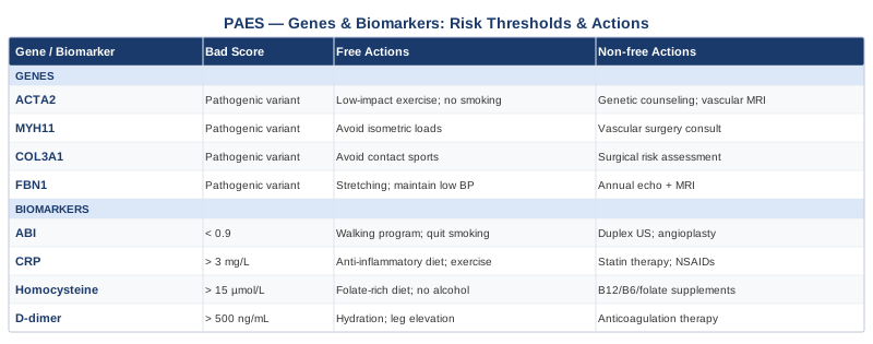
Understanding what your biomarkers reveal is only part of the picture. The genetic variants below may help explain why a given individual is more vulnerable to the structural, inflammatory, or thrombotic mechanisms that define PAES and its complications.
5 Genetic Variants That May Shape Vulnerability to PAES
Genetics in the context of PAES is not about a single gene that causes the condition. PAES is primarily a developmental and structural problem — the artery sits in the wrong anatomical position, or the muscle grows around it in an abnormal pattern. What genes do contribute is a set of underlying vulnerabilities: to connective tissue fragility, to vascular smooth muscle dysfunction, and to hypercoagulability. These vulnerabilities do not create PAES on their own, but they shape how badly it manifests and how much damage accumulates before and after treatment.
ACTA2 — Smooth Muscle Alpha-Actin
What it does and how it may affect PAES: ACTA2 encodes alpha-smooth muscle actin, the dominant contractile protein in vascular smooth muscle cells. Missense mutations in ACTA2 — including R149C and R179H variants — disrupt the actin cytoskeleton in arterial smooth muscle, impairing the cell's ability to maintain normal arterial wall tone, respond to mechanical stress, and regulate vessel diameter. In the popliteal artery, which is already subjected to mechanical compression, impaired smooth muscle function accelerates local wall remodeling and increases the risk of post-stenotic dilation. Research linking ACTA2 variants to thoracic aortic aneurysm also suggests broader arterial fragility across the body.
If the Gene Is Bad: Plan Without Supplements
Blood pressure control is the most impactful non-pharmaceutical intervention: targeting systolic BP below 120 mmHg reduces mechanical wall stress that smooth muscle cells with compromised function cannot adequately buffer. Avoid high-intensity isometric exercise (heavy Valsalva-inducing lifting) — this transiently raises arterial pressure dramatically and is particularly stressful on arterial walls with smooth muscle dysfunction. Consistent Zone 2 aerobic training supports endothelial nitric oxide production, which compensates partially for impaired smooth muscle contractile function. 30–45 minutes, 4–5 days per week.
If the Gene Is Bad: Plan With Supplements or Equipment
Magnesium glycinate: 300–400 mg elemental magnesium daily (taken in the evening). Magnesium acts as a physiological calcium channel antagonist in vascular smooth muscle, supporting relaxation and reducing arterial stiffness. Long-term use is safe; GI upset at higher doses (switch to glycinate or threonate form if this occurs). L-citrulline: 3–6 g/day supports endogenous nitric oxide production via the arginine pathway, partially compensating for impaired vascular smooth muscle regulation. Take in the morning. Cycle: 8–12 weeks on, 2–4 weeks off. Side effects minimal; mild GI discomfort possible at higher doses.
COL3A1 — Type III Collagen
What it does and how it may affect PAES: COL3A1 encodes type III collagen, the primary structural protein in blood vessel walls, skin, and hollow organs. Mutations in COL3A1 cause Vascular Ehlers-Danlos Syndrome (vEDS), characterized by arterial fragility, spontaneous arterial rupture, and impaired wound healing. Even subclinical COL3A1 variants — less severe than full vEDS — may confer reduced arterial wall tensile strength. In PAES, the popliteal artery is subjected to repetitive mechanical loading at the compression point; a wall with reduced type III collagen structural integrity is significantly more vulnerable to post-stenotic dilation, dissection, and wall thinning under that repeated stress.
If the Gene Is Bad: Plan Without Supplements
Avoid high-impact activities that load the popliteal region repetitively until PAES is surgically addressed — arterial wall fragility from COL3A1 variants raises the stakes of delayed treatment. Paced, progressive loading protocols in rehabilitation (rather than aggressive return-to-sport timelines) allow connective tissue to remodel at a safer rate. Rigorous blood pressure management as above — wall fragility is directly amplified by high mechanical load.
If the Gene Is Bad: Plan With Supplements or Equipment
Vitamin C (ascorbic acid): 500–1000 mg/day is essential as a cofactor for prolyl and lysyl hydroxylase enzymes that catalyze collagen cross-linking. Collagen synthesis is essentially impaired without adequate vitamin C. Long-term use is safe; above 2 g/day may increase oxalate kidney stones in susceptible individuals. Glycine: 5–10 g/day provides the most abundant amino acid in collagen and has been used in connective tissue support protocols. Take before bed. Safe for long-term use; evidence in humans is still emerging. Collagen peptide hydrolysate: 10–15 g/day (Type I/III blend with vitamin C co-supplementation) taken 30–60 minutes before loading activity has emerging evidence for connective tissue support in tendons and may extend to arterial wall maintenance. Cycle is not required; long-term use appears safe.
MYH11 — Smooth Muscle Myosin Heavy Chain
What it does and how it may affect PAES: MYH11 encodes the myosin heavy chain isoform expressed in vascular smooth muscle. Loss-of-function variants in MYH11 are associated with thoracic aortic aneurysm and a broader arteriopathy characterized by impaired smooth muscle contractility and defective vascular wall response to mechanical stress. In the popliteal artery context, MYH11 variants may reduce the artery's intrinsic capacity to maintain appropriate wall tension under the mechanical stress of compression — accelerating the progression from simple anatomical entrapment to wall damage, aneurysm, or thrombus.
If the Gene Is Bad: Plan Without Supplements
The approach parallels ACTA2 recommendations: strict blood pressure control (systolic target below 120 mmHg), avoidance of high-load isometric exercise, and regular aerobic training for vascular smooth muscle health. Additionally, pursue surgical PAES correction without delay if indicated — in the presence of MYH11 variants, conservative management for longer than necessary is higher risk than in a structurally normal arterial wall.
If the Gene Is Bad: Plan With Supplements or Equipment
Magnesium glycinate as above (300–400 mg/day). Taurine: 1–2 g/day has emerging evidence as a vascular smooth muscle stabilizer and cardiovascular protective amino acid; it supports calcium handling in vascular smooth muscle cells, partially compensating for impaired myosin contractile function. Long-term use at these doses appears safe. Home blood pressure monitoring device: given arterial wall fragility with MYH11 variants, owning and regularly using a validated upper-arm cuff (daily morning readings) enables prompt detection of blood pressure excursions. This is an equipment recommendation with direct clinical relevance.
MTHFR — Methylenetetrahydrofolate Reductase
What it does and how it may affect PAES: MTHFR converts 5,10-methylenetetrahydrofolate to 5-methyltetrahydrofolate, the active folate form required for homocysteine remethylation. The C677T variant (rs1801133) is the most studied; homozygous TT genotype reduces enzyme activity by approximately 70%, causing elevated homocysteine in the presence of suboptimal B vitamin status. This directly connects to the homocysteine biomarker discussed above: MTHFR variants are among the most common genetic explanations for elevated homocysteine. Research on MTHFR in vascular disease has been extensively reviewed — Frosst et al. (1995) established the C677T variant as a risk factor for cardiovascular disease via homocysteine elevation. In PAES, this genetic driver of endothelial damage and thrombotic risk is particularly significant because it is also one of the most correctable.
If the Gene Is Bad: Plan Without Supplements
Maximize dietary methylfolate intake from whole food sources: dark leafy greens, legumes, asparagus, avocado. Avoid high-dose folic acid supplementation in isolation — individuals with TT genotype convert synthetic folic acid inefficiently and may accumulate unmetabolized folic acid, which has been linked to immune modulation concerns in some research. Limit alcohol, which depletes folate rapidly. Address digestive absorption if B12 or folate levels are low despite reasonable dietary intake.
If the Gene Is Bad: Plan With Supplements or Equipment
Methylfolate (5-MTHF): 400–800 mcg/day. The methylated form bypasses the MTHFR enzyme entirely. This is a direct correction for the genetic deficit. Long-term use is safe; no cycling required. Methylcobalamin: 500–1000 mcg/day sublingual. P5P (active B6): 25–50 mg/day. TMG (betaine): 500–1000 mg/day provides the betaine pathway as a backup. This combination is specifically designed to normalize homocysteine in MTHFR TT genotype carriers. Recheck homocysteine at 8–12 weeks after starting; target below 10 µmol/L.
F5 — Factor V Leiden (R506Q)
What it does and how it may affect PAES: The Factor V Leiden mutation (R506Q) makes activated Factor V resistant to cleavage and inactivation by activated Protein C, one of the body's primary anticoagulant mechanisms. This resistance causes hypercoagulability: heterozygous carriers have a 3–8-fold increased risk of venous thromboembolism; homozygous carriers carry 50–80-fold increased risk. As described by Bertina et al. (1994) in the landmark identification of this mutation, it is among the most common inherited thrombophilias in populations of European descent. In PAES, the combination of arterial compression, stasis, turbulent flow at the stenosis, and endothelial injury creates precisely the conditions where Factor V Leiden amplifies risk dramatically — particularly the risk of acute arterial thrombosis downstream of the compression point.
If the Gene Is Bad: Plan Without Supplements
Prevent stasis and dehydration rigorously: adequate hydration, regular movement breaks, avoidance of prolonged immobility. Communicate the mutation to your vascular surgeon explicitly — it changes the risk-benefit calculation for timing of surgical intervention, post-operative anticoagulation protocol, and duration of antiplatelet therapy. Avoid combined oral contraceptives if applicable (estrogen-containing contraceptives multiply the thrombotic risk of F5 Leiden substantially). Track D-dimer and fibrinogen more frequently as surveillance tools.
If the Gene Is Bad: Plan With Supplements or Equipment
Omega-3 fatty acids: 3–4 g/day of EPA+DHA for antithrombotic effect. Nattokinase as above (100–200 mg, between meals, with explicit physician review before use given the F5 Leiden mutation and any concurrent medications). Compression stockings during travel (class 1 graduated, 15–20 mmHg) reduce stasis-related clot formation significantly in thrombophilia carriers. Anticoagulation therapy during high-risk periods (surgery, prolonged immobilization, post-partum) should be explicitly discussed with a hematologist or vascular physician in F5 Leiden carriers with PAES.
What Outlive by Peter Attia Reveals About Vascular Health
Outlive: The Science and Art of Longevity (2023) is one of the few recent books that directly challenges the conventional cardiovascular risk framework — and it does so with a level of scientific rigor and clinical specificity that makes it genuinely useful rather than simply provocative. Attia argues that the tools most doctors use to assess cardiovascular risk (Framingham score, total cholesterol, standard lipid panels) are fundamentally inadequate for catching arterial disease early enough to matter. Several of his core arguments map directly onto the biomarker strategy described above.
1. ApoB Is the Number That Matters Most
Attia dedicates substantial attention to what he calls the primacy of ApoB over LDL-C. He argues that LDL particle count — not the cholesterol cargo within each particle — determines arterial wall penetration risk. ApoB is the only direct measure of particle count available in standard labs. He advocates for an ApoB target below 60 mg/dL in high-risk individuals, which is significantly more aggressive than most current guidelines recommend.
2. LP(a) Should Be Tested in Everyone, Once
Attia describes LP(a) as perhaps the most egregious oversight in standard cardiovascular care. He notes that approximately one in five people has a significantly elevated LP(a) and will never know it because it is not on a standard lipid panel. His recommendation: test it once, in everyone, since it is almost entirely genetic and tells you something no other biomarker tells you about lifelong thrombogenic and inflammatory arterial risk.
3. Zone 2 Training Is a Non-Negotiable Vascular Intervention
Attia treats Zone 2 aerobic exercise — roughly the intensity at which you can hold a conversation but are breathing somewhat harder — as the highest-leverage single intervention for vascular and metabolic health. At this intensity (150–180 minutes per week minimum), mitochondrial biogenesis increases, endothelial nitric oxide synthase activity improves, and systemic inflammatory markers fall. For PAES patients who cannot run, he would endorse swimming or cycling as equivalent substitutes.
4. The Framingham Risk Score Is Designed to Catch Late Disease
One of Attia's most useful points is that the tools conventionally used to assess cardiovascular risk were validated in populations where the disease was already advanced. By the time a Framingham score flags significant risk, arterial disease has often been developing for decades. Early ApoB and LP(a) testing in younger individuals — including athletes, the primary PAES population — catches a risk window that Framingham simply misses.
5. Insulin Resistance and Vascular Disease Are the Same Story
Attia argues that insulin resistance is the upstream driver of most cardiovascular disease — not simply a diabetes precursor. Hyperinsulinemia promotes endothelial dysfunction, increases VLDL production (raising ApoB), and contributes to inflammatory signaling. For PAES patients, fasting insulin and HOMA-IR are therefore worth adding to the standard panel as upstream markers of vascular risk.
6. Sleep Is a Vascular Medicine
Outlive draws on extensive research linking poor sleep to elevated cortisol, inflammatory cytokines, and LDL receptor downregulation — all of which raise ApoB and hsCRP. Attia's recommendation of consistent 7–9 hour sleep with a fixed rise time and cool sleeping environment is framed as a cardiovascular intervention, not just a wellness practice.
7. Blood Pressure Targets Should Be Lower Than Current Guidelines
Attia's reading of the SPRINT trial and related data leads him to advocate for systolic BP targets below 120 mmHg — more aggressive than the 130 mmHg threshold in many current hypertension guidelines. He argues that arterial wall stress is cumulative and that starting blood pressure control earlier and targeting lower produces meaningfully better outcomes over decades. For PAES patients with smooth muscle gene variants, this argument is even more compelling.
8. The Ankle-Brachial Index Deserves Routine Use
Attia discusses ABI as a neglected tool in primary care — simple, inexpensive, and directly predictive of cardiovascular mortality, yet rarely performed until symptoms appear. He frames it as part of a broader early-detection strategy and notes that a low ABI is among the most informative single findings you can get from a $30 test.
9. Metabolic Health Is Tracked Through Five Markers, Not One
Attia identifies the five metabolic health markers as: waist circumference, fasting triglycerides, HDL-C, blood pressure, and fasting glucose. He notes that in population studies, a majority of adults are impaired on at least one — meaning most people are already on the metabolic disease spectrum. For vascular patients, tracking all five alongside ApoB and LP(a) gives a comprehensive risk picture.
10. Preventive Action Needs to Happen 20 Years Earlier Than It Usually Does
Perhaps the most paradigm-shifting argument in Outlive is that cardiovascular disease is not something you prevent in your 60s by starting statins. It is something you prevent in your 30s and 40s by understanding your ApoB, LP(a), homocysteine, and metabolic markers — and acting on them while the arterial wall is still recoverable. For PAES patients, this means that a diagnosis of arterial entrapment at age 25 or 35 is an opportunity to investigate and correct the underlying vascular biology at exactly the right time.
Complementary Approaches With Clinical Backing for PAES
The following approaches do not replace surgical correction in structural PAES, nor do they substitute for biomarker management. They address the surrounding biology: managing pain, supporting tissue repair, improving vascular tone, and maintaining function during the often extended periods of investigation and recovery that characterize this condition.
Low-Level Laser Therapy (Photobiomodulation)
Photobiomodulation (PBM) uses red and near-infrared light (typically 660–850 nm) to stimulate mitochondrial cytochrome c oxidase in target tissues, increasing ATP production, reducing oxidative stress, and supporting endothelial cell repair. In the context of PAES, its relevance is twofold: it may support arterial wall healing at the compression site, and it has demonstrated anti-inflammatory effects in vascular tissue. A systematic review by Chow et al. confirmed anti-inflammatory and tissue repair effects of PBM in vascular-adjacent tissues. Evidence specific to popliteal artery injury is limited — most vascular PBM research concerns wound healing and peripheral neuropathy — but the mechanistic basis is solid.
For PAES, the practical protocol involves applying a PBM device (660 nm and 850 nm LEDs or laser probe) to the posterior knee region for 10–15 minutes per session, 3–4 times per week. A device with 60–100 mW/cm² of power density is appropriate for tissue-depth penetration at the popliteal level. Sessions should not be applied directly over an acute hematoma or within 2 weeks of vascular surgery. Begin conservatively (5 minutes per session) and increase to 15 minutes over 2–3 weeks. Devices cost $150–$600; prescription-grade clinical devices are more powerful and available through physiotherapy clinics.
Massage Therapy
Deep tissue massage and myofascial release of the posterior lower limb — specifically targeting the gastrocnemius and soleus — has direct mechanical relevance for functional PAES, in which arterial compression results from muscular hypertrophy or increased resting tone rather than from a fixed anatomical anomaly. Reducing the volume and tension of these muscles through regular manual therapy may decrease the degree of popliteal artery compression during activity. A small but consistent body of research supports the role of therapeutic massage in reducing muscle stiffness and improving peripheral circulation. Evidence for PAES specifically is anecdotal, but the physiological rationale is strong.
A specific protocol for PAES-related muscle work involves weekly 45–60 minute sessions focusing on the gastrocnemius, soleus, and popliteus with deep longitudinal stripping and cross-fiber friction techniques. Trigger point work targeting the medial head of the gastrocnemius is particularly relevant given its role in anatomical PAES variants. Sessions should be performed by a therapist with sports vascular or neuromuscular experience. Do not perform deep massage during active arterial thrombosis, acute inflammation, or within 4 weeks of popliteal surgery. Monthly maintenance sessions are appropriate once symptoms have stabilized.
Yoga
Yoga addresses two mechanisms relevant to PAES: posterior compartment flexibility and vascular tone. Specific poses targeting the gastrocnemius, soleus, hamstrings, and popliteal region can reduce the structural compression component in functional PAES over time. The controlled breathing and parasympathetic activation components of yoga also improve vagal tone and support endothelial nitric oxide production. A study by Hagins et al. demonstrated measurable improvements in cardiovascular variables with regular yoga practice in adults with cardiovascular risk factors. Evidence specific to PAES is absent; evidence for peripheral vascular function improvement is emerging.
A practical yoga protocol for PAES focuses on: downward dog (sustained calf and popliteal stretch), standing calf stretch against wall (2 minutes per side daily), supine hamstring stretch, and gentle Yin yoga postures targeting the posterior lower limb held for 2–5 minutes. Practice for 20–30 minutes, 4–5 times per week. Avoid forceful inversions if blood pressure is elevated or if acute vascular compromise is present. Hot yoga environments may cause vasodilation that temporarily exacerbates symptoms in some PAES patients — prefer moderate-temperature practice.
Biofeedback
Biofeedback — specifically thermal biofeedback targeting peripheral circulation — teaches patients to consciously influence blood flow to the extremities by modulating autonomic nervous system activity. This is relevant to PAES because autonomic regulation of vascular tone affects the baseline tension of the gastrocnemius and the degree of vasospasm that can co-occur with compression-related arterial injury. A clinical study by Freedman et al. demonstrated that thermal biofeedback training produced significant improvements in peripheral blood flow in patients with Raynaud's phenomenon — a related peripheral vascular condition. Evidence in PAES is limited to mechanistic extrapolation.
Thermal biofeedback sessions involve monitoring finger or toe temperature with a temperature sensor and training to raise peripheral temperature by 1–2°C through relaxation and attention focusing. Sessions run 30–40 minutes, with 8–12 sessions over 4–6 weeks being a standard protocol in clinical biofeedback programs. Home biofeedback devices are available for $100–$400 and allow daily practice between clinical sessions. The effect is modest but cumulative and carries no side effects. Most relevant for functional PAES and for managing sympathetically-mediated vasospasm post-surgery.
Breathing-Based Therapies
Slow, diaphragmatic breathing at 5–6 breaths per minute (resonance frequency breathing) activates the baroreflex arc and significantly increases heart rate variability — a direct marker of vagal tone and vascular regulatory capacity. Improved vagal tone is associated with better endothelial function, reduced systemic vascular resistance, and lower inflammatory signaling. For PAES patients managing chronic pain and vascular stress, breathing-based training offers a low-cost, daily-practice intervention that works both structurally (improving oxygen delivery at rest) and autonomically. A randomized trial by Giardino et al. confirmed HRV improvements with resonance frequency breathing in clinical populations. Evidence specifically for peripheral artery conditions is limited; autonomic vascular benefits are well-supported.
A daily protocol: 5–10 minutes of slow diaphragmatic breathing at 5.5–6 breaths per minute (4–5 second inhale through nose, 4–6 second exhale through nose or pursed lips). Use a metronome or a guided breathing app to maintain rate precision. Perform daily, ideally in the morning before activity and again in the evening. This can be combined with progressive muscle relaxation for amplified parasympathetic response. No equipment is strictly necessary; a paced breathing app costs nothing. Consistent daily practice over 4–8 weeks produces measurable HRV improvements. No side effects; occasional light-headedness with overbreathing resolves immediately by returning to normal breathing.
Conclusion
Popliteal artery entrapment syndrome is a structural problem — but what happens to the artery before, during, and after that compression is biochemical. The seven biomarkers covered here (ABI, homocysteine, hsCRP, LP(a), D-dimer, fibrinogen, and ApoB) give you a direct, measurable window into the vascular environment that determines whether PAES stays mechanical or becomes progressive. The five genetic variants (ACTA2, COL3A1, MYH11, MTHFR, F5) offer a layer of explanation for why your arterial wall may be more vulnerable to that compression than another person's.
None of this replaces the structural evaluation and, where indicated, the surgical management that PAES requires. But the person who arrives at that conversation knowing their ApoB is elevated, their LP(a) is high, their MTHFR genotype is TT, and their homocysteine is 18 µmol/L is in a fundamentally different position than the person who knows only that their popliteal artery is in the wrong place. A useful next step: request the ABI with provocative testing and the non-standard lab markers (LP(a), ApoB, homocysteine, hsCRP) at your next vascular appointment, bring the results in, and ask your physician how each one should factor into your monitoring plan and post-surgical recovery strategy.
Musculoskeletal: Sports Injuries
Cardiovascular: Blood Vessel Conditions Vascular Conditions
Autoimmune: Inflammatory Conditions Connective Tissue Conditions