This article contains AI generated content.
· UpdatedPopliteal Cyst Dissection – 5 Genes and 6 Biomarkers to Track
Introduction
If you have been told you have a Baker's cyst — or a dissecting popliteal cyst, where fluid has spread into the back of the calf — you already know the frustration. It may have been drained, it may have come back, and the explanation you received was probably brief: excess joint fluid, caused by something irritating the knee. Rest, maybe compression, possibly aspiration. The mechanics of why your knee keeps producing that fluid in the first place usually go unexamined.
That matters because a dissecting popliteal cyst is almost never a standalone problem. It's a consequence of intra-articular pathology: inflammation from rheumatoid or psoriatic arthritis, cartilage breakdown from osteoarthritis, a meniscal tear, or crystal deposition from gout. The cyst is the downstream signal; the biology driving it upstream varies considerably from person to person. Two people with identical MRI findings may have completely different root causes — and almost certainly different optimal approaches.
Generic advice — rest, ice, anti-inflammatories, physical therapy — is not wrong, but it's incomplete without knowing what's actually driving the joint environment. That's where biomarkers and genetic data become useful. Measuring the specific molecules involved in your joint's inflammatory and degradative activity gives you something to work with: a target, a baseline, and a way to verify whether your interventions are actually moving the needle.
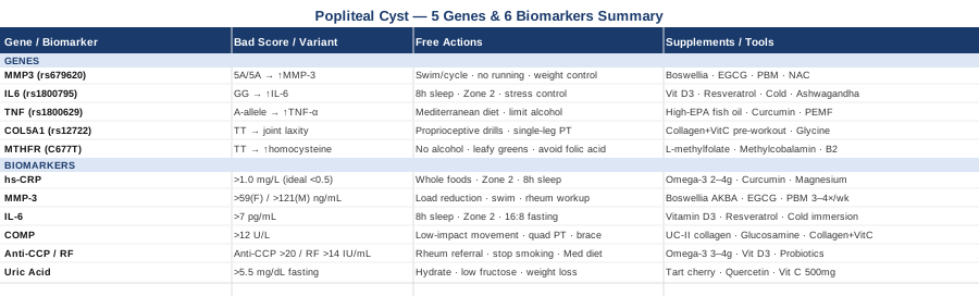
This article takes that more targeted approach. The primary focus is the six most informative biomarkers for tracking the biology behind a popliteal cyst — including how to measure them, what each one reveals, and what to do when the numbers are off. You'll also find a more compact look at the five genetic variants most relevant to joint vulnerability, a summary of an episode that reframes connective tissue recovery, and complementary modalities with real clinical evidence. None of this replaces a rheumatologist or orthopedic surgeon — but all of it gives you better questions to ask and clearer data to act on.
6 Biomarkers to Track if You Have a Popliteal Cyst
The joint environment that produces a Baker's cyst is not invisible — it leaves measurable traces in your blood and, when assessed, in the fluid itself. The following biomarkers offer the clearest diagnostic and monitoring utility, whether you're trying to understand the root cause, gauge the intensity of the inflammatory process, or evaluate whether lifestyle and treatment changes are actually working.
Biomarker 1: High-Sensitivity C-Reactive Protein (hs-CRP)
Why it matters: C-reactive protein is the liver's acute-phase response to inflammatory cytokine signaling. It rises when the immune system is active, and in the joint context, it reflects the degree of synovial membrane irritation driving excess fluid production. Elevated hs-CRP is one of the most consistently reported biomarkers in inflammatory arthritis — the most common systemic driver of popliteal cysts. It also correlates with flare severity and, over time, with structural damage progression.
Standard CRP is too coarse for this purpose. High-sensitivity CRP (hs-CRP) detects chronic low-grade inflammation in a range the standard test misses, which is where most joint disease lives between overt flares. Peter Attia uses hs-CRP as a primary inflammatory surveillance marker in his longevity practice precisely because of its sensitivity to background inflammatory states.
How to measure it: Any standard blood panel — request hs-CRP specifically. Available at all labs. Cost: $10–$30. Optimal target: below 0.5 mg/L; below 1.0 mg/L is acceptable; above 3.0 mg/L indicates elevated risk and warrants investigation.
If the score is bad, the plan without supplements: Prioritize removing pro-inflammatory dietary inputs: ultra-processed foods, industrial seed oils (sunflower, safflower, soybean used in cooking), and high-glycemic carbohydrate loads. Moderate aerobic exercise at Zone 2 intensity (conversational pace, 30–40 minutes, 4–5 days per week) consistently reduces hs-CRP over 8–12 weeks in clinical studies. Sleep quality is non-negotiable: even one night of fragmented sleep measurably elevates CRP within 24 hours. Address chronic psychological stress using structured breathing or MBSR — the HPA-immune axis connection is real and well-documented.
If the score is bad, the plan with supplements or equipment: - Omega-3 fatty acids (EPA + DHA): 2–4g combined EPA+DHA daily with meals. Reduces CRP through lipid-mediated resolution pathways (resolvins, protectins). Onset: 8–12 weeks for measurable CRP effect. Cycle: continuous. Side effects: fishy aftertaste, loose stools at high doses; check interactions with blood thinners. - Curcumin (BCM-95 or theracurmin form for bioavailability): 500–1,000mg/day. Inhibits NF-κB, the master transcription factor upstream of CRP production. Cycle: 12 weeks on, 4 weeks off. Side effects: GI discomfort at high doses; avoid in gallstone disease. - Magnesium glycinate: 300–400mg at night. Magnesium deficiency independently associates with elevated hs-CRP; correction is low-cost and broadly beneficial. Cycle: continuous. Side effects: loose stools if overdosed.
Biomarker 2: Matrix Metalloproteinase-3 (MMP-3)
Why it matters: MMP-3, known as stromelysin-1, is an enzyme produced by synovial fibroblasts and macrophages in response to inflammatory signals. It degrades cartilage extracellular matrix components — type II collagen, proteoglycans, fibronectin — and is a direct mediator of structural joint damage. Serum MMP-3 correlates closely with synovitis activity, and in rheumatoid arthritis it's used as a measure of disease progression distinct from CRP.
The specific relevance to popliteal cysts: an inflamed synovial membrane doesn't just passively allow fluid to accumulate — it actively produces and maintains that inflammatory environment. Elevated MMP-3 tells you the lining of the joint is actively destructive, not merely irritated. If MMP-3 is high and CRP is also elevated, you're looking at a more aggressive joint process that warrants prompt attention.
How to measure it: Serum MMP-3 ELISA, available through specialty and functional medicine labs. Less routinely ordered than CRP but increasingly accessible. Cost: $40–$100. Reference ranges vary by lab; generally above 59 ng/mL in women and 121 ng/mL in men is considered elevated, though lower values warrant attention in the context of joint symptoms.
If the score is bad, the plan without supplements: The first priority is addressing the root cause driving synovial activation — which often means pursuing a rheumatologic workup if not already done. Load management is the mechanical lever: reducing compressive and shearing forces on the affected compartment through activity modification (replace running with swimming or cycling), weight loss if applicable, and physical therapy targeting quadriceps and hip abductor strength to offload the medial compartment.
If the score is bad, the plan with supplements or equipment: - Boswellia serrata (standardized to AKBA, the active component): 100–250mg AKBA/day. Inhibits leukotriene synthesis and directly downregulates MMP-3 expression in synovial tissue in human studies. Cycle: 12 weeks on, 4 weeks off. Side effects: mild GI discomfort; rare skin reactions. - EGCG (green tea extract): 400–800mg/day with meals. Inhibits MMP-1, MMP-3, and MMP-9 expression in synovial fibroblasts through AP-1 and NF-κB pathway suppression. Cycle: 8 weeks on, 2 weeks off. Side effects: caffeine sensitivity in some; hepatotoxic risk at very high doses — stay within studied ranges. - Near-infrared photobiomodulation device (660nm + 850nm): 10–15 minutes over the popliteal area, 3–4x/week. Reduces MMP activity in inflamed tissue via mitochondrial cytochrome c oxidase stimulation and downstream anti-inflammatory signaling. Home panels available for $200–$500. Side effects: minimal; do not use directly over eyes or areas of suspected infection.
Biomarker 3: Interleukin-6 (IL-6)
Why it matters: IL-6 is one of the central cytokines in the inflammatory cascade. It drives the liver to produce CRP, promotes synovial fibroblast proliferation, and amplifies cartilage matrix degradation. In rheumatoid arthritis — one of the most common causes of recurrent Baker's cysts — IL-6 blockade is a primary treatment strategy: the drug tocilizumab is specifically designed to neutralize IL-6 receptors. That therapeutic target tells you how pivotal this cytokine is.
Elevated serum IL-6 in patients with knee joint pathology correlates with larger effusion volumes, higher pain scores, and accelerated structural progression. If a cyst keeps reforming despite drainage, persistently high IL-6 is often part of the explanation — the inflammatory stimulus that drives fluid production has not been addressed.
How to measure it: Serum IL-6 via high-sensitivity ELISA or multiplex cytokine panel. Available through specialty and functional labs. Cost: $50–$120. Normal range: below 7 pg/mL in healthy adults; values above 10–15 pg/mL in a non-acute setting merit investigation.
If the score is bad, the plan without supplements: Among the most potent non-pharmacological drivers of chronic IL-6 elevation are sleep disruption and psychological stress — often more impactful than diet. Eight hours of quality sleep per night, with consistent sleep and wake times, is foundational. Brief moderate exercise reduces IL-6; prolonged strenuous exercise transiently spikes it. Time-restricted eating (16:8 protocol) has shown modest but consistent IL-6 reduction in clinical studies without caloric restriction, suggesting a circadian and metabolic mechanism beyond simple weight change.
If the score is bad, the plan with supplements or equipment: - Vitamin D3 (with K2): If serum 25-OH vitamin D is below 40 ng/mL, supplementing to reach 50–70 ng/mL reduces IL-6 production in immune cells. Dose: typically 3,000–5,000 IU/day depending on baseline. K2 (MK-7 form, 100–200mcg/day) for calcium routing. Cycle: continuous with annual blood monitoring. Side effects: rare at these doses; monitor if you have hypercalcemia risk. - Resveratrol: 250–500mg/day with a fat-containing meal (bioavailability is fat-dependent). Inhibits STAT3 and NF-κB pathways upstream of IL-6 transcription. Cycle: 12 weeks on, reassess. Side effects: GI discomfort; potential hormonal interactions at high doses. - Cold water immersion: 10–15 minutes at 10–15°C, 3x/week. The acute norepinephrine release (increases of up to 300% documented) and cold-adaptation pathways ultimately reduce chronic inflammatory cytokine tone, including IL-6, through sustained neural and metabolic signaling changes. No cost beyond a tub and cold water.
Biomarker 4: Cartilage Oligomeric Matrix Protein (COMP)
Why it matters: COMP is a structural glycoprotein embedded in the extracellular matrix of cartilage, tendon, and ligament. When cartilage is actively degrading — whether from osteoarthritis, inflammatory arthritis, or mechanical trauma — COMP fragments are released into synovial fluid and blood. Elevated serum COMP is a direct indicator of cartilage turnover rate and predicts structural joint damage progression ahead of clinical symptoms and radiographic change.
This matters because the most common structural causes of Baker's cysts — medial compartment OA and meniscal pathology — both drive elevated COMP before they become obvious on imaging. A high COMP level in someone with a recurrent popliteal cyst confirms that active cartilage destruction is occurring, not just transient inflammation. It also provides an objective endpoint for tracking whether interventions aimed at protecting cartilage are actually working. Peter Attia's longevity practice includes COMP in joint health assessments for this reason.
How to measure it: Serum COMP ELISA, available through specialty labs and functional medicine practitioners. Cost: $60–$150. Values above 12 U/L generally indicate elevated cartilage turnover, though ranges vary by lab and should be interpreted in clinical context.
If the score is bad, the plan without supplements: Mechanical load management is the primary lever. Cartilage depends on compression-decompression cycling for nutrition (it has no direct blood supply), so low-impact movement is both necessary and protective: swimming, cycling, and walking are preferred over running or jumping on the affected knee. Physical therapy specifically targeting quadriceps strength (the primary absorber of knee compressive force) and hip abductor strength (which controls valgus collapse) is the most evidence-based structural intervention. An unloader knee brace for the affected compartment can meaningfully reduce contact forces during daily activities.
If the score is bad, the plan with supplements or equipment: - Undenatured type II collagen (UC-II), 40mg/day: This is a tolerance-induction mechanism (oral tolerance), not a building-block supplement — the dose is very low intentionally. Modulates immune response to type II collagen fragments in joint tissue. Evidence from human RCTs shows benefit in OA symptoms. Cycle: continuous; reassess at 3 months. Side effects: minimal; occasional mild GI discomfort. - Glucosamine sulfate + chondroitin sulfate: 1,500mg/500mg daily. The GAIT trial (NEJM, 2006) showed significant benefit in the subgroup with moderate-to-severe OA pain. Mixed evidence overall, but the subset that responds appears to respond meaningfully. Cycle: 3-month trial; continue only if clinical response observed. Side effects: shellfish allergy risk with standard glucosamine; use non-shellfish sources if needed. - Collagen peptides (15g) + vitamin C (50mg), 30–60 minutes before low-load exercise: Amplifies circulating amino acid availability at the window of peak collagen synthesis stimulated by exercise. Supported by research from Keith Baar's lab at UC Davis. Cycle: daily or before each exercise session. Side effects: caloric addition; low individual risk.
Biomarker 5: Anti-Cyclic Citrullinated Peptide Antibodies (Anti-CCP) and Rheumatoid Factor (RF)
Why it matters: Rheumatoid arthritis is one of the most common systemic causes of recurrent, bilateral, or treatment-resistant Baker's cysts — and a significant proportion of people don't know they have early or seronegative RA until substantial joint damage has already accumulated. Anti-CCP antibodies are highly specific for RA (approximately 95% specificity) and can be detectable years before clinical diagnosis. In the context of a popliteal cyst with unexplained or recurring joint effusion, a positive anti-CCP changes the entire management framework.
RF (rheumatoid factor) has lower specificity (elevated in multiple conditions) but adds diagnostic weight when combined with anti-CCP. Identifying RA early is not academic — early DMARD treatment prevents irreversible structural damage that no later intervention can undo.
How to measure it: Anti-CCP IgG ELISA and RF latex agglutination, both available through any primary care or rheumatology lab. Cost: $30–$80 combined. Positive anti-CCP threshold: above 20 U/mL. Positive RF: above 14 IU/mL. Both positive together significantly increases RA likelihood.
If the score is bad, the plan without supplements: A positive anti-CCP result is a clear indication for rheumatology referral — this is not a biomarker to manage independently with lifestyle alone, especially in the early window where disease-modifying therapy prevents joint destruction. Within the lifestyle domain: complete smoking cessation is the most powerful modifiable RA risk factor, with smoking increasing both RA risk and disease severity via citrullination pathways. Anti-inflammatory dietary patterns (Mediterranean diet) reduce disease activity scores modestly but consistently in human trials. Eliminate sleep debt and manage chronic stress actively.
If the score is bad, the plan with supplements or equipment: - Omega-3 fatty acids (EPA + DHA, 3–4g/day): Multiple meta-analyses confirm that fish oil supplementation reduces RA disease activity scores (DAS28), morning stiffness, and NSAID use, and is safe alongside DMARDs. This is one of the highest-evidence supplement interventions for RA specifically. Effect onset: 12–16 weeks. - Vitamin D3 (optimize to 50–70 ng/mL): Low vitamin D correlates with higher RA severity and poorer treatment response. Supplementation alongside medical management improves outcomes in deficient patients. - Multi-strain probiotics (Lactobacillus rhamnosus + Lactobacillus acidophilus): Gut microbiome dysbiosis is mechanistically linked to RA autoimmune activation through molecular mimicry and intestinal permeability pathways. 20–50 billion CFU/day. Cycle: 3 months minimum. Side effects: transient bloating initially; safe with DMARDs.
Biomarker 6: Serum Uric Acid
Why it matters: Gout is significantly underdiagnosed as a cause of knee joint inflammation and Baker's cyst formation. When serum uric acid exceeds 6.8 mg/dL — the saturation threshold for monosodium urate crystal formation — crystals can deposit in the joint space and trigger intense synovitis with large effusions. The resulting intra-articular pressure is exactly the mechanism that fills a popliteal bursa.
Even below the diagnostic gout threshold, chronically elevated uric acid is associated with higher circulating IL-1β and IL-18 levels, impaired endothelial function, and metabolic dysregulation that worsens joint inflammatory environments. Thomas Dayspring includes uric acid in metabolic surveillance panels precisely because of its underappreciated systemic and inflammatory implications. If joint symptoms appear atypical for RA or OA — episodic, sudden-onset, highly painful — uric acid should be measured immediately.
How to measure it: Standard blood panel. Cost: under $15. Measure fasting for accuracy. Conservative target for joint protection: below 5.5 mg/dL (more stringent than the typical clinical threshold of 6.8 mg/dL).
If the score is bad, the plan without supplements: Reduce dietary purine burden: limit organ meats, anchovies, sardines, shellfish, and — critically — high-fructose corn syrup and fructose-rich beverages (fructose drives endogenous uric acid production via purine nucleotide degradation, independent of dietary purines). Increase hydration substantially — uric acid is renally excreted, and adequate fluid intake is the most impactful single non-pharmacological intervention. Weight loss if applicable: adipose tissue amplifies urate production through xanthine oxidase upregulation.
If the score is bad, the plan with supplements or equipment: - Tart cherry extract (Montmorency variety): 480–960mg standardized extract daily, or 240mL unsweetened concentrate. Inhibits xanthine oxidase and promotes renal uric acid clearance. Human studies show 20–25% reduction in gout flare frequency with regular use. Cycle: continuous. Side effects: prefer extract over juice to minimize sugar load. - Quercetin: 500–1,000mg/day. Inhibits xanthine oxidase activity (similar mechanism to allopurinol, lower potency) and has additive effect with tart cherry. Cycle: continuous. Side effects: rare; potential interaction with quinolone antibiotics and blood thinners. - Vitamin C, 500mg/day: Modestly reduces serum uric acid through competitive renal tubular reabsorption inhibition. Low-cost, safe addition as part of a broader uric acid management protocol.
What Your Genes Say About Joint and Connective Tissue Vulnerability
Biomarkers tell you what's happening now. Genetic variants offer a different kind of information: they describe your underlying tendencies — how efficiently your joints handle inflammatory challenges, how resilient your connective tissue architecture is, and which molecular pathways are most likely to become overactivated under stress. This is not deterministic risk; it's biological context. Understanding it helps you prioritize which interventions will matter most for your specific makeup.
The following five variants have the strongest evidence for influencing joint health, synovial tissue behavior, and connective tissue integrity — the triad directly implicated in popliteal cyst formation and dissection.
Gene 1: MMP3 (rs679620 — the 5A/6A Promoter Polymorphism)
MMP3 encodes stromelysin-1, the enzyme directly involved in synovial tissue remodeling and cartilage matrix degradation. A well-studied promoter polymorphism creates two alleles: the 5A allele produces significantly higher MMP3 expression than the 6A allele. Homozygous 5A/5A carriers show faster cartilage degradation in both RA and OA studies, higher serum MMP-3 levels at baseline, and greater joint destruction over time.
If the gene is bad, the plan without supplements: Load management and mechanical joint protection are the highest-leverage tools for high-MMP3 expressers. Avoid repetitive high-impact activities on the affected knee (running on hard surfaces, heavy loaded squats). Prioritize water-based movement, cycling, and elliptical for cardiovascular fitness. Maintain healthy body weight strictly — adipose-derived inflammatory signals amplify synovial MMP expression disproportionately. Structured physical therapy to optimize joint biomechanics and reduce shear forces at the medial compartment is more important for this genotype than average.
If the score is bad, the plan with supplements or equipment: Boswellia AKBA, EGCG, and photobiomodulation (as described in the MMP-3 biomarker section) all directly downregulate MMP expression and are the primary targeted interventions. Add N-acetylcysteine (NAC), 600–1,200mg/day: oxidative stress amplifies MMP transcription, and NAC as a glutathione precursor reduces this amplification at the cellular level. Cycle: 8 weeks on, 2 weeks off. Side effects: GI discomfort at higher doses; sulfur odor in breath and urine.
Gene 2: IL6 (rs1800795 — the -174 G/C Promoter Variant)
The IL6 promoter variant at position -174 strongly influences IL-6 transcription rate. The G allele is associated with higher IL-6 production under inflammatory challenge. GG homozygotes show measurably higher circulating IL-6 across multiple population studies and greater susceptibility to conditions characterized by elevated IL-6, including RA and OA. In joint disease specifically, GG carriers show larger synovial effusions and more aggressive inflammatory activity.
If the gene is bad, the plan without supplements: Sleep architecture optimization is the most underestimated intervention for IL6 GG carriers. Poor sleep is one of the most potent modulators of IL-6 elevation — and for high IL-6 expressers, the amplification effect is greater. Target 8 hours with consistent timing aligned to your circadian rhythm. Moderate, consistent aerobic exercise (Zone 2, 150–200 minutes/week) is anti-inflammatory; excessive high-intensity exercise spikes IL-6 acutely. Chronic stress management must be treated as a clinical priority, not a lifestyle optional extra.
If the score is bad, the plan with supplements or equipment: Vitamin D3, resveratrol, and cold water immersion as outlined in the IL-6 biomarker section. Add Ashwagandha (KSM-66 or Sensoril extract), 300–600mg/day: reduces cortisol-driven IL-6 amplification through HPA axis normalization. Particularly relevant for GG carriers who also carry high stress loads. Cycle: 8 weeks on, 2 weeks off. Side effects: rare mild sedation; potential thyroid interaction at very high doses — caution in thyroid disease.
Gene 3: TNF (-308 G/A, rs1800629)
TNF-alpha is a master regulator of joint inflammation — it's the direct target of some of the most effective biologic drugs used in RA (etanercept, adalimumab, infliximab). The -308 promoter variant significantly affects TNF-alpha transcription; the A allele (minor allele) upregulates TNF-alpha production substantially. A allele carriers show higher systemic inflammatory tone, faster synovial damage in RA, and worse prognosis in inflammatory joint conditions.
If the gene is bad, the plan without supplements: Dietary composition matters meaningfully for TNF A-allele carriers. A Mediterranean dietary pattern — extra-virgin olive oil, fatty fish, colorful vegetables, legumes, minimal processed food — has been shown in human trials to directly reduce circulating TNF-alpha. Industrial trans fats and excessive saturated fat from processed sources upregulate TNF signaling; moderate alcohol is acceptable, high alcohol intake is a potent TNF inducer. Prioritize dietary consistency rather than periodic cleanses.
If the score is bad, the plan with supplements or equipment: - High-EPA fish oil (3g+ EPA/day): EPA's metabolic products (E-series resolvins, protectins) actively dampen TNF signaling — a resolution mechanism rather than mere suppression. Critical distinction: use a high-EPA formula, not balanced EPA/DHA, for this specific application. - Curcumin (theracurmin or BCM-95 form): 500–1,000mg/day. Inhibits NF-κB upstream of TNF transcription — particularly effective alongside EPA for synergistic NF-κB suppression. - PEMF device (pulsed electromagnetic field therapy): 30-minute sessions over the affected knee, 4–5x/week. Multiple randomized controlled trials show PEMF reduces TNF-alpha expression in joint tissue and reduces synovitis activity. Home devices available for $200–$800. Safe for most individuals; avoid near electronic implants (pacemakers, cochlear implants). This is one of the few non-pharmacological tools with direct device-level evidence for TNF modulation in joints.
Gene 4: COL5A1 (rs12722)
COL5A1 encodes the alpha-1 chain of type V collagen, which plays a regulatory role in collagen fibril diameter and mechanical integrity across connective tissues including ligaments, joint capsule, and tendons. The TT genotype of rs12722 is consistently associated with greater connective tissue laxity and extensibility, higher rates of ligament and tendon injury across multiple sports medicine studies, and poorer mechanical joint stability.
For popliteal cysts specifically, loose joint mechanics increase the stress placed on the posterior capsule and popliteal bursa when intra-articular pressure rises from any cause. TT genotype individuals are more susceptible to having a cyst extend or dissect into surrounding tissue under pressure loads that a CT-genotype individual would tolerate without consequence.
If the gene is bad, the plan without supplements: Proprioceptive and neuromuscular stability training is the primary protective intervention. Balance board training, single-leg strength exercises (Bulgarian split squats, single-leg Romanian deadlifts at moderate loads), and hip stabilizer activation drills reduce dynamic joint laxity and decrease abnormal loading of the posterior capsule. Avoid abrupt transitions to high-load or high-impact activities without progressive preparation. Functional knee bracing during higher-demand activities may meaningfully reduce posterior capsule stress in TT individuals.
If the score is bad, the plan with supplements or equipment: - Collagen peptides (15g) + vitamin C (50mg), taken 30–60 minutes before exercise: Stimulates collagen synthesis in connective tissue at the window of peak anabolic demand during exercise. Supported by human research from Keith Baar's UC Davis laboratory. Cycle: daily before training. Side effects: minimal; small caloric addition. - Orthosilicic acid (stabilized, 10–20mg/day): Silica is a required cofactor for collagen hydroxylation and cross-linking — the process that gives collagen its mechanical stiffness. Often neglected in joint supplement protocols. Cycle: 3-month trial. Side effects: well tolerated. - Glycine (3–5g/day, separate from collagen): The most abundant amino acid in collagen; free glycine supplementation upregulates collagen gene expression independently of collagen peptide ingestion. Inexpensive, widely available. Side effects: generally well tolerated; high doses may cause drowsiness.
Gene 5: MTHFR (C677T, rs1801133)
The MTHFR C677T variant reduces the enzyme's ability to convert dietary folate into 5-methyltetrahydrofolate — the active form used in the methylation cycle. TT homozygotes have approximately 70% reduced enzyme function. Gary Brecka has extensively highlighted MTHFR as a bottleneck for systemic inflammatory regulation, and while some of his specific claims extend beyond the current evidence, the core mechanism is real and well-documented.
Poor MTHFR function leads to elevated homocysteine — and homocysteine directly damages connective tissue through oxidative mechanisms, promotes endothelial dysfunction that impairs tissue repair, and correlates with greater disease severity in inflammatory arthritis. It's not the most direct genetic factor in joint disease, but it's one of the most actionable because the correction is straightforward.
If the gene is bad, the plan without supplements: Eliminate dietary folate antagonists: alcohol is the most significant, depleting folate stores substantially even at moderate intake. Consume dietary sources of pre-methylated folate: dark leafy greens (especially spinach and romaine), liver, and asparagus. Avoid fortified foods using synthetic folic acid if you are a TT homozygote — unmetabolized folic acid accumulates and competitively inhibits natural folate metabolism.
If the score is bad, the plan with supplements or equipment: - L-methylfolate (5-MTHF form), 400–1,000mcg/day: The active form that bypasses the MTHFR bottleneck entirely. Do not use standard folic acid in TT carriers — it competes with 5-MTHF at folate receptors and may worsen the functional deficit. Cycle: continuous. Side effects: rare anxiety or overstimulation at high doses in some individuals — start low. - Methylcobalamin (active B12), 500–1,000mcg sublingual daily: Works synergistically with methylfolate in the methylation cycle; reduces homocysteine in combination. Cycle: continuous. Side effects: essentially none at these doses. - Riboflavin (B2), 100–400mg/day: A required cofactor for MTHFR enzyme function. Riboflavin supplementation independently reduces homocysteine in TT carriers even without folate supplementation — making it a uniquely targeted adjunct for this genotype. Cycle: continuous. Side effects: yellow urine (harmless riboflavin excretion).
The Andrew Huberman Work on Connective Tissue and Inflammation Recovery
Andrew Huberman's collaboration with Dr. Keith Baar — a molecular physiologist and tendon/connective tissue researcher at UC Davis — produced one of the most practically useful sets of protocols for joint and connective tissue recovery currently available to the public. The key discussion, covered in episodes on flexibility, connective tissue adaptation, and injury recovery, challenges several widespread assumptions in standard orthopedic rehabilitation. Here are the ten most impactful insights, directly relevant to reducing Baker's cyst recurrence and improving joint resilience.
1. The Collagen Synthesis Window Is Real and Specific
Collagen synthesis in connective tissue peaks approximately 4–6 hours after stimulation from low-load movement. Taking 15g of gelatin or collagen peptides plus 50mg of vitamin C, 30–60 minutes before low-impact exercise, provides substrate exactly when the anabolic machinery is primed. Keith Baar's group demonstrated this in human studies with measurable increases in tendon and ligament collagen synthesis. This simple timing protocol is likely the most underused connective tissue intervention available.
2. Low-Load Loading Beats Rest for Connective Tissue
Complete rest causes connective tissue stiffness, fiber disorganization, and mechanical weakening over time. Low-load isotonic or isometric loading at 20–40% of maximum — light enough to feel almost easy — is what drives collagen remodeling and tissue adaptation. For Baker's cyst, this means gentle range-of-motion and low-resistance knee work is actively therapeutic, not merely tolerated.
3. Heat Before Loading Improves Collagen Fibroblast Activity
Connective tissues have poor blood supply and are highly temperature-sensitive. Brief moist heat application (10–15 minutes) before low-load exercise increases collagen fibroblast activity and tissue compliance. This is a simple, free enhancement to any connective tissue rehabilitation protocol.
4. Cold Timing Relative to Exercise Is Critical
Cold applied immediately after resistance training suppresses the inflammatory signals necessary for connective tissue adaptation and muscle hypertrophy. Cold is better used several hours after exercise for pain management, or in dedicated non-exercise sessions for systemic inflammatory tone management. Timing changes the biological outcome.
5. Sleep Governs Structural Repair
Growth hormone secreted during slow-wave sleep is the primary hormonal driver of connective tissue repair. Missing sleep is not a neutral trade-off — it measurably reduces GH output and slows tissue healing. Consistent sleep timing (same bedtime and wake time) is as important as duration for maintaining slow-wave sleep architecture.
6. Omega-3s Have a Mechanobiological Effect — Not Just Anti-Inflammatory
EPA and DHA are incorporated into cell membranes and directly alter how cells respond to mechanical loading signals — a mechanobiological effect separate from their anti-inflammatory action. This means omega-3 sufficiency changes how synovial and connective tissue cells sense and adapt to joint stress, not just how inflamed they become.
7. The Sympathetic Nervous System Reduces Tissue Blood Flow
Chronic sympathetic activation (stress response) reduces blood flow to connective tissue and promotes inflammatory cytokine release through neurogenic pathways. Poorly vascularized tissues like joint capsules and ligaments are disproportionately affected. Reducing sympathetic tone through structured breathing practices is a direct joint health intervention, not merely stress management.
8. The Physiological Sigh Is the Fastest Autonomic Reset
A double nasal inhale followed by a long oral exhale — the physiological sigh — was shown in a Stanford-led randomized trial published in Cell Reports Medicine to reduce physiological stress markers faster and more durably than any other single respiratory maneuver tested. Three to five repetitions provide rapid parasympathetic activation without equipment. This has direct relevance to managing the chronic sympathetic tone that worsens joint inflammatory environments.
9. Zone 2 Cardio Is the Anti-Inflammatory Protocol
Sustained low-intensity aerobic exercise at the aerobic threshold — approximately 130–145 bpm for most adults, where you can speak in full sentences — reduces circulating inflammatory cytokines, improves mitochondrial density in joint-supporting muscle, and promotes synovial fluid circulation. Huberman recommends 150–200 minutes per week as a non-negotiable baseline for health. This is achievable on a bicycle or in a pool even with an active knee condition.
10. Neurological Pain Sensitization Outlasts the Structural Problem
After a chronic joint condition or recurring cyst, the nervous system can remain sensitized to pain inputs even after the structural issue has resolved. Central sensitization is biological, not psychological. Pain education, graded movement exposure, and reducing pain catastrophizing through mindfulness practices address this sensitization layer directly and are backed by neuroscience. Addressing only the structural component and ignoring the sensitization component is one of the most common reasons for incomplete recovery.
Complementary Approaches with Clinical Evidence
The following modalities were selected for having meaningful human clinical evidence applicable to the conditions commonly underlying a dissecting popliteal cyst: inflammatory joint disease, chronic knee pain, synovitis management, and connective tissue recovery.
Tai Chi
Tai chi is a slow, controlled movement practice combining joint-loading postures, weight shifting, and deep breathing in sequences that build neuromuscular stability and coordination without high compressive forces. For the inflammatory arthritis and osteoarthritis that most commonly drive Baker's cysts, it represents an unusual combination: a practice that simultaneously reduces systemic inflammatory tone, improves proprioception and joint stability, and maintains range of motion — all through low-impact movement.
A landmark randomized controlled trial by Chenchen Wang and colleagues, published in Annals of Internal Medicine (2016), compared tai chi directly to physical therapy for knee osteoarthritis over 12 weeks with a 52-week follow-up. Tai chi produced equivalent pain and functional improvement to PT, with additional benefits for depression and quality of life. A meta-analysis in Rheumatology International concluded that tai chi significantly reduces knee pain and improves function in knee OA — the most common structural cause of Baker's cysts. Evidence quality: high for knee OA; extrapolated but biologically plausible for inflammatory arthritis-related cysts.
Practical application: begin with a supervised beginner Yang-style tai chi class, 3 sessions per week, 30–45 minutes each. For the first 4–6 weeks, in-person instruction is strongly preferable to video-only to ensure correct weight distribution and to avoid deep squatting forms that increase posterior capsule pressure. Tai chi can generally be safely practiced even during mild cyst activity — stay within pain-free range and avoid any posture that compresses the popliteal space.
Low-Level Laser Therapy / Photobiomodulation
Photobiomodulation (PBM) uses specific wavelengths of red and near-infrared light (typically 630–1,000nm) to stimulate cytochrome c oxidase in mitochondria, enhance local ATP production, reduce oxidative stress, and modulate inflammatory cytokine expression in tissue. In joint applications, PBM has demonstrated the ability to reduce synovial inflammation, decrease MMP expression, improve local microcirculation, and promote tissue repair — a combination of effects directly relevant to the inflammatory environment maintaining a popliteal cyst.
A Cochrane review on low-level laser therapy for osteoarthritis found short-term benefit for pain outcomes compared to sham, with moderate evidence quality. A systematic review by Bjordal and colleagues across musculoskeletal applications found consistent short-term pain relief and functional improvement. The evidence base is moderate but improving as device parameters become more standardized across studies. The anti-inflammatory cellular mechanism is well-established in vitro and increasingly supported in human tissue studies.
Practical application: use a home panel device combining 660nm and 850nm wavelengths (devices from established brands are available for $200–$500). Apply 10–15 minutes over the popliteal area, 3–4 sessions per week. Maintain 6–12 inches from skin surface. Protect eyes from direct beam exposure. PBM is most useful as an adjunct during recovery and for ongoing maintenance of the joint inflammatory environment — it is not a standalone treatment for a large active cyst that may require aspiration. Combine with physical therapy and biomarker-guided interventions for best effect.
Massage Therapy
Therapeutic soft tissue work applied to the muscles and fascia surrounding the knee — particularly the gastrocnemius, popliteus, biceps femoris, and popliteal fascia — can reduce local muscular hypertonicity, improve lymphatic drainage from the popliteal space, and decrease pain-sensitized neural activity around the joint. In the context of a dissecting cyst where fluid has extended into the calf, gentle manual lymphatic drainage (MLD) techniques specifically guide fluid toward inguinal lymph nodes and may support resorption.
A systematic review in Journal of Physical Therapy Science found that massage therapy significantly reduced knee pain and improved range of motion in knee osteoarthritis patients. Manual lymphatic drainage has evidence for reducing lower extremity effusions in inflammatory and post-procedural contexts, with application to joint-related swelling supported in rheumatology literature. The evidence specific to Baker's cyst is limited but mechanistically coherent. Evidence quality: moderate for knee OA and surrounding tissue management; limited but plausible for cyst-specific fluid drainage support.
Practical protocol: weekly or biweekly sessions with a licensed massage therapist trained in MLD, focusing on effleurage strokes directed toward the inguinal nodes and release of posterior chain tension (hamstrings, calf musculature). Avoid direct deep pressure over the cyst itself — this risks pain and potential rupture. Home self-massage with a foam roller along the hamstrings and calf can be performed daily as a complement. Immediately stop and seek medical evaluation if massage is followed by sudden increase in calf pain, warmth, or swelling — these may indicate cyst rupture mimicking deep vein thrombosis.
Mindfulness Meditation / MBSR
Mindfulness-based stress reduction (MBSR) is the 8-week structured program developed by Jon Kabat-Zinn at the University of Massachusetts, training attention and non-reactive awareness through formal meditation, body scan practice, and gentle movement. Its relevance to popliteal cyst conditions is twofold: first, MBSR consistently reduces circulating inflammatory markers including CRP, IL-6, and NF-κB activity in well-designed clinical trials; second, it directly addresses central pain sensitization — the neurological amplification of pain signals that frequently outlasts the structural problem in chronic joint conditions.
A study by Black and colleagues in Brain, Behavior, and Immunity demonstrated that MBSR reduced gene expression of inflammatory signaling pathways in peripheral immune cells in older adults. A comprehensive meta-analysis in Annals of Behavioral Medicine confirmed MBSR's effectiveness for chronic pain — reducing pain intensity, pain unpleasantness, and psychological distress beyond active control conditions. The parasympathetic activation induced by mindfulness practice also reduces the sympathetic tone that amplifies joint inflammatory cytokine production via neural-immune pathways. Evidence quality: high for chronic pain and inflammatory marker reduction; increasingly supported for inflammatory joint conditions.
Practical application: a formal 8-week MBSR course is the best-studied format, available in-person through major medical centers or free online through Palouse Mindfulness (Jon Kabat-Zinn material). Daily practice of 20–45 minutes is the studied dose — specifically body scan meditation for the first weeks, transitioning to seated and movement-based practices. Begin with body scan: this trains non-reactive awareness of physical sensations, directly useful for chronic joint discomfort. Daily brief practice is more beneficial than infrequent long sessions.
Breathing-Based Therapies
Controlled breathing practices — including slow-paced breathing (approximately 6 cycles per minute), diaphragmatic breathing, and the physiological sigh protocol — directly modulate autonomic nervous system balance in ways that have measurable downstream effects on joint-relevant biology. Chronic sympathetic nervous system activation increases inflammatory cytokine production via neural-immune pathways, reduces microvascular perfusion to poorly vascularized connective tissues, and amplifies pain sensitization — all directly adverse to joint recovery. Correcting this through breathing is not metaphorical; it is mechanistic.
A Stanford-led randomized controlled trial published in Cell Reports Medicine (2023) compared cyclic sighing, box breathing, and mindfulness meditation across daily practice conditions and found that cyclic sighing (the physiological sigh protocol: two short nasal inhales, one extended oral exhale) produced the greatest sustained reductions in physiological stress markers, respiratory rate, and negative affect while improving positive affect — surpassing mindfulness meditation in several comparisons. Slow-paced breathing at 6 cycles per minute (5 seconds in, 5 seconds out) has separately demonstrated reductions in heart rate variability-indexed sympathetic tone and improvements in pain tolerance in chronic pain populations. Evidence quality: high for autonomic and pain modulation; biologically well-supported for indirect joint inflammatory benefit.
Practical application: 5–10 minutes of slow-paced breathing twice daily — morning and pre-sleep — using a free app (Breathwrk, Insight Timer) for pacing guidance. For acute pain spikes, joint flares, or procedural anxiety (aspiration, injections), 3–5 physiological sighs provide rapid autonomic downregulation in under 90 seconds with no equipment or cost. Make this the first tool reached for during flares before escalating to analgesics — it may resolve the autonomic component of pain amplification quickly.
Conclusion
A dissecting popliteal cyst is a downstream signal — not a standalone structural problem, but a consequence of a joint environment that is actively inflamed, degraded, or metabolically dysregulated. Standard treatment addresses the cyst; a more complete approach tracks and corrects what's driving it.
The six biomarkers covered here — hs-CRP, MMP-3, IL-6, COMP, anti-CCP, and uric acid — give you a biological map of that environment. The genetic variants offer a picture of your underlying vulnerabilities. Neither requires exotic access: most of these tests can be requested through a primary care physician or functional medicine practitioner, often at a combined cost well under $300.
Your next concrete step: identify two or three biomarkers you haven't tested recently, request them at your next blood panel, and bring specific target ranges to that conversation. Add one evidence-based lifestyle change based on the most relevant finding. Reassess in 12 weeks. Measured, incremental changes compound — but only when you actually track them. If any test returns a positive anti-CCP or significantly elevated inflammatory markers, move that rheumatology referral to the top of your priority list.
Endocrine & Metabolic Autoimmune
Musculoskeletal: Joint Conditions
Autoimmune: Inflammatory Conditions Connective Tissue Conditions