This article contains AI generated content.
· UpdatedPopliteofibular Ligament Tear: 6 Genes And 7 Biomarkers To Track
Introduction
The popliteofibular ligament sits deep in the posterolateral corner of the knee — a small but structurally critical structure that stabilizes the joint against rotational and varus forces. A tear here rarely happens in isolation; it often occurs alongside damage to the lateral collateral ligament, the popliteus tendon, or the posterior cruciate ligament. That combination makes recovery longer, more uncertain, and more prone to complications than a typical ligament injury. If you are navigating a PFL tear, you already know that the standard advice — rest, physical therapy, maybe surgery — does not feel specific enough to your situation.
What most rehabilitation protocols fail to account for is how differently individuals heal at the biological level. Two people with nearly identical injuries can have radically different outcomes based on their inflammatory response, their collagen remodeling capacity, their nutritional status, and even their genetic makeup. These variables are measurable. Tracking them changes how you can manage recovery — not as a passive patient, but as someone actively working with the biology of healing rather than against it.
Genetic factors explain part of this individual variation. Specific polymorphisms in collagen genes, matrix metalloproteinase genes, and inflammatory signaling genes affect how your body builds, breaks down, and repairs connective tissue. These are not destiny — they are tendencies that can often be compensated for when identified. Epigenetic factors, meaning how environment and daily habits switch genes on or off, add another layer of opportunity that most orthopedic consultations never address.
This article explores two parallel paths to a more informed recovery. The first examines seven key biomarkers you can track through standard and specialty labs — concrete, measurable signals of inflammation, collagen synthesis, nutritional sufficiency, and tissue remodeling balance. The second looks at six genetic factors that influence your baseline risk and healing capacity, along with specific strategies to compensate. Combined, these approaches do not replace your orthopedic surgeon or physiotherapist; they give you sharper questions to ask them, and a way to measure whether what you are doing is actually working.
7 Biomarkers To Track After A Popliteofibular Ligament Tear
Tracking the right biomarkers during recovery from a posterolateral knee injury replaces guesswork with measurable, objective data on inflammation levels, tissue remodeling activity, nutritional status, and healing capacity. The seven below represent a practical, evidence-grounded panel. Most can be ordered through a standard lab request; some require specialty panels available through sports medicine or integrative medicine physicians.
1. High-Sensitivity C-Reactive Protein (hs-CRP): Your Inflammation Gauge
Why it matters: CRP is produced by the liver in response to tissue injury and systemic inflammation. After a PFL tear — particularly one requiring surgical repair — inflammatory burden rises predictably. While some inflammation is necessary for initiating the repair cascade, persistent or excessive elevation impairs tissue remodeling, increases pain sensitization, and raises the risk of adjacent joint degeneration. Peter Attia, in his clinical work and in Outlive, consistently flags hs-CRP as one of the most actionable inflammation markers available in routine practice, noting that most patients have no idea where their baseline sits.
How to measure it: A standard blood draw, processed by any major laboratory. Cost ranges from $10 to $40. Request high-sensitivity CRP specifically — regular CRP lacks the precision needed at the lower end of the range, which is where healing-phase monitoring matters most. Optimal target for recovery: below 1.0 mg/L; below 0.5 mg/L is ideal. Above 3 mg/L during recovery signals a significant inflammatory burden that warrants active intervention. Test every 4–6 weeks while tracking recovery.
If the score is elevated — the plan without supplements: Sleep quality is the first lever — seven to nine hours in a cool, dark room with consistent timing. Eliminate ultra-processed foods, refined seed oils (soybean, corn, sunflower), and added sugars. Prioritize whole foods, colorful vegetables, and wild-caught fatty fish three to four times per week. Cold exposure — ten to fifteen minutes of cold water immersion or contrast showers — has shown modest anti-inflammatory effects in controlled settings, though evidence for CRP specifically is still developing. Reduce psychosocial stress, which potently elevates CRP through cortisol and sympathetic nervous system pathways.
If the score is elevated — the plan with supplements or equipment: Omega-3 fatty acids at two to four grams of combined EPA and DHA per day, taken with meals, have consistent evidence for lowering hs-CRP across multiple randomized trials. Curcumin with piperine (500–1000 mg curcumin, 20 mg piperine daily, taken with food containing fat) has also demonstrated efficacy in several clinical trials. Correcting vitamin D deficiency independently reduces hs-CRP. Cycle omega-3 supplementation with retesting every three to four months. Side effects at these doses are rare; enteric-coated fish oil reduces the risk of GI discomfort.
2. COMP (Cartilage Oligomeric Matrix Protein): The Tissue Damage Signal
Why it matters: COMP is a glycoprotein released into circulation when cartilage, ligament, or tendon matrix is being mechanically stressed or degraded. In acute ligament injuries, serum COMP rises significantly within hours of trauma, then gradually normalizes during repair. Persistently elevated COMP beyond the acute phase suggests ongoing matrix breakdown — signaling inadequate healing, excessive loading, or early joint degeneration in adjacent structures that bear compensatory load during recovery. Validated extensively in rheumatology and sports medicine research as a sensitive marker of joint tissue turnover, COMP is one of the few markers that directly reflects what is happening at the ligament matrix level.
How to measure it: Requires a specialty laboratory or a sports medicine rheumatology panel. Cost ranges from $80 to $200 depending on the laboratory. Measured through blood serum. Baseline values vary by individual, age, and sex, which makes trend monitoring — comparing serial values at four to six week intervals — more clinically meaningful than any single snapshot. Work with a sports medicine physician to interpret trends in the context of your rehabilitation phase and loading progression.
If the score is elevated — the plan without supplements: Reduce mechanical loading on the joint while maintaining circulation through non-weight-bearing or low-impact activity such as aquatic therapy or cycling with minimal resistance. Avoid repeated high-impact loading during early recovery phases. Structured physiotherapy that emphasizes neuromuscular control over raw strength reduces joint stress while still providing the mechanical stimulation that ligament tissue requires for organized healing. Aquatic therapy in particular allows controlled motion with dramatically reduced compressive load.
If the score is elevated — the plan with supplements or equipment: Type II collagen hydrolysate at ten grams daily has early evidence supporting its role in reducing COMP elevation and improving joint function; a six-month cycling protocol is reasonable with retesting midway. Hinged knee braces and lateral offloading orthoses reduce mechanical stress on the posterolateral compartment during weight-bearing activities. Prolotherapy, while not a supplement, is used clinically to stimulate ligament healing and has some supportive evidence for posterolateral knee injuries specifically; this requires a physician referral and evaluation.
3. 25-OH Vitamin D: The Healing Foundation
Why it matters: Vitamin D deficiency is extraordinarily common, and its consequences for musculoskeletal healing are well-documented. Vitamin D receptors are present on ligament fibroblasts, meaning the active hormone form directly influences the cells responsible for building and repairing ligament matrix. Low vitamin D has been associated with increased ligament and tendon injury risk in several athletic populations. Adequate levels support collagen gene expression, calcium-phosphorus balance, and immune modulation during the healing cascade. Research across multiple sports medicine cohorts has found high rates of insufficiency in injured athletes, raising the question of whether deficiency contributed to injury susceptibility in the first place.
How to measure it: Routine blood test available through any standard laboratory. Cost is $30–$60. The test is labeled as "25-hydroxyvitamin D" or "25(OH)D." Optimal range for active tissue recovery: 50–80 ng/mL (125–200 nmol/L). Most conventional laboratory reference ranges flag anything above 30 ng/mL as "sufficient," which is a conservative threshold calibrated for preventing deficiency disease, not for optimizing healing. Retest every eight to twelve weeks while supplementing.
If the score is low — the plan without supplements: Sun exposure of fifteen to twenty-five minutes on arms and legs between 10 a.m. and 2 p.m., daily when possible, ideally without sunscreen during that window. Increase intake of naturally vitamin D-rich foods: wild salmon, mackerel, sardines, egg yolks from pasture-raised hens, and beef liver. These dietary sources alone rarely bring levels into the optimal range without significant sun exposure, but they contribute meaningfully.
If the score is low — the plan with supplements or equipment: Vitamin D3 supplementation at 2,000–5,000 IU daily, taken with a fat-containing meal and always combined with vitamin K2 (MK-7 form, 100–200 mcg daily) to ensure appropriate calcium routing toward bone rather than soft tissue. At severely deficient starting levels (below 20 ng/mL), loading doses under medical supervision may be appropriate. Avoid exceeding 10,000 IU per day without monitoring serum levels. Side effects at therapeutic doses are rare when K2 is co-administered; hypercalcemia is a risk only at prolonged very high doses without monitoring.
4. MMP-3 (Matrix Metalloproteinase-3): The Remodeling Regulator
Why it matters: Matrix metalloproteinases are enzymes that break down components of the extracellular matrix — the scaffold that gives ligaments their structural integrity and tensile strength. MMP-3 in particular regulates collagen remodeling in connective tissue. During the acute phase of injury, elevated MMP-3 reflects the body's effort to clear damaged matrix and begin reorganization. Persistently elevated MMP-3 beyond the acute window, however, tips the balance toward net matrix degradation rather than net synthesis, directly impairing the structural quality of healing tissue. Research linking MMP-3 activity to soft tissue healing outcomes supports its clinical relevance as a recovery monitoring tool.
How to measure it: Serum MMP-3 requires a specialty laboratory or an inflammatory joint disease panel. Cost ranges from $80–$200. More commonly ordered in rheumatology for inflammatory arthritis monitoring, but sports medicine and integrative medicine physicians can request it as a standalone marker. Trends over three to six months are more informative than a single measurement. Pair it with COMP for the most complete picture of matrix turnover.
If the score is elevated — the plan without supplements: Load management is the primary tool. Excessive mechanical stress on healing connective tissue upregulates MMP expression. Structured progressive loading — with clearly defined rest intervals between rehabilitation sessions — allows MMP-3 to normalize between stimulus bouts. Avoid prolonged NSAID use without clinical guidance; the suppression of inflammation through this mechanism can paradoxically interfere with the anabolic signaling that regulates organized matrix repair and may blunt the adaptive response needed for tensile strength restoration.
If the score is elevated — the plan with supplements or equipment: Curcumin has demonstrated MMP-3 inhibitory effects in human joint tissue studies and is the most clinically studied option in this domain. Green tea extract standardized to EGCG (400–800 mg/day) has preclinical evidence for MMP modulation, with some human data in inflammatory joint conditions. Hydrolyzed collagen peptides may support matrix synthesis to counteract excessive MMP-driven breakdown. Protocol: curcumin 500–1000 mg/day with piperine, plus EGCG 400 mg/day, cycled eight weeks on with two weeks off. Avoid concurrent anticoagulant medications without physician guidance, as both curcumin and EGCG have mild blood-thinning properties.
5. Omega-3 Index: Your Anti-Inflammatory Reserve
Why it matters: The omega-3 index measures the percentage of EPA and DHA incorporated into red blood cell membranes — a reflection of long-term omega-3 status rather than a recent dietary snapshot. A high omega-3 index (above 8%) correlates with lower systemic inflammatory signaling, better cellular membrane function, and improved recovery from musculoskeletal injury. Thomas Dayspring, a leading lipidologist, emphasizes this marker not only for cardiovascular risk assessment but as a direct indicator of global inflammatory tone. In the context of ligament healing, an adequate omega-3 index supports the resolution of post-injury inflammation, limits excessive scar tissue formation, and improves the anti-inflammatory signal environment within which remodeling occurs.
How to measure it: Requires a fingerstick or venous blood draw processed by specialty laboratories such as OmegaQuant or Cleveland HeartLab. Cost: $50–$100 for a home test kit or through a physician order. Target: above 8%. Most Western adults test in the four to six percent range — a meaningful deficit. Retest every three to four months when actively supplementing to track response.
If the score is low — the plan without supplements: Consume three to four servings of fatty fish per week — wild salmon, sardines, mackerel, and herring are the highest EPA+DHA sources. Simultaneously reduce omega-6 intake by eliminating vegetable oils (soybean, corn, and sunflower oil) from cooking, replacing them with olive oil or avocado oil. This ratio shift — increasing omega-3 while reducing omega-6 competition — can modestly improve the index over several months without supplementation, though dietary changes alone rarely reach the eight percent threshold for most people.
If the score is low — the plan with supplements or equipment: High-quality triglyceride-form fish oil or algal omega-3s (for vegetarians) at two to four grams of combined EPA+DHA per day, taken with meals to maximize absorption. Improvement in the omega-3 index is slow — expect three to four months to meaningfully shift the percentage. Retest at that point. At higher doses, monitor for prolonged bleeding time if on anticoagulants. Once the index reaches eight percent, transition to a lower maintenance dose of one to two grams daily and retest every six months to confirm stability.
6. PINP (Procollagen Type I N-Terminal Propeptide): Are You Actually Building New Tissue?
Why it matters: PINP is released into circulation as a direct byproduct of new collagen synthesis — specifically type I collagen, which constitutes the structural backbone of ligaments and tendons. Tracking PINP over the course of recovery tells you whether the body is actively assembling new connective tissue matrix. When PINP is low relative to the expected healing phase, it signals that synthesis is lagging — which may reflect protein insufficiency, persistent inflammation suppressing anabolic signaling, vitamin C deficiency (a required cofactor), or suboptimal sleep. Most established in bone turnover research, PINP has growing relevance in soft tissue recovery monitoring when combined with breakdown markers like CTX-I to reveal the net collagen balance.
How to measure it: Available through most major laboratory networks. Cost: $50–$120. Often ordered in bone health panels but can be requested as a standalone marker. Interpreting PINP alongside a collagen degradation marker (such as CTX) gives a more complete picture: if PINP is low and CTX is high, net collagen balance is clearly negative. If both are elevated, active remodeling is underway. Best tracked at baseline post-injury, then every six to eight weeks.
If the score is low — the plan without supplements: Ensure adequate total protein intake — at a minimum of 1.6 grams per kilogram of bodyweight, ideally up to 2.2 g/kg during active recovery phases. Distribute protein evenly across three to four meals rather than concentrating it in one sitting, as muscle protein synthesis has a ceiling per meal. Time a protein-rich meal within one to two hours of any rehabilitation exercise session. Sleep architecture is critical here: growth hormone released during deep slow-wave sleep is a primary driver of collagen synthesis, and fragmented sleep directly suppresses PINP levels.
If the score is low — the plan with supplements or equipment: Hydrolyzed collagen peptides at fifteen to twenty grams taken thirty to sixty minutes before a rehabilitation session, combined with 200–500 mg of vitamin C at the same time, has clinical evidence for stimulating collagen synthesis in periarticular connective tissue. The timing relative to exercise loading is important — the combination appears to enhance incorporation of new collagen into mechanically stimulated tissue. Cycle this protocol over eight to twelve weeks with PINP retesting midway. Glycine (3–5 grams daily as a separate supplement) provides an additional substrate for collagen assembly. Side effects are minimal; mild digestive discomfort is occasionally reported with collagen peptides, typically resolving within one week.
7. Homocysteine: The Overlooked Connective Tissue Threat
Why it matters: Homocysteine is an intermediate amino acid that, when elevated, impairs the crosslinking of collagen fibers — the chemical process that gives ligaments their tensile strength and resistance to tearing. High homocysteine interferes with lysyl oxidase, the enzyme responsible for collagen and elastin crosslinking. The result is softer, structurally weaker connective tissue that heals poorly and carries higher risk of re-injury and poor mechanical outcomes. Thomas Dayspring consistently recommends tracking homocysteine as part of a comprehensive metabolic workup, noting its underappreciated role in tissue health well beyond cardiovascular risk — where most physicians currently use it. In the context of a ligament tear, persistently elevated homocysteine may be a silent saboteur of the crosslinking quality of newly synthesized collagen.
How to measure it: Standard blood draw available through any routine laboratory. Cost: $30–$60. Target: below 8 µmol/L for optimal connective tissue function; above 10 µmol/L warrants intervention; above 15 µmol/L is clinically significant and associated with meaningful risk across multiple tissues. Retest eight to twelve weeks after any dietary or supplement intervention.
If the score is elevated — the plan without supplements: Increase dietary intake of folate (dark leafy greens, lentils, asparagus, black-eyed peas), B12 (meat, eggs, dairy, shellfish), and B6 (poultry, fish, bananas, chickpeas). Reduce alcohol consumption, which interferes with B-vitamin absorption and accelerates homocysteine accumulation. Homocysteine is highly responsive to dietary B-vitamin status, and meaningful reductions are achievable through diet alone when deficiency is mild.
If the score is elevated — the plan with supplements or equipment: The core protocol combines methylfolate (400–1000 mcg daily), methylcobalamin (1000 mcg daily), and pyridoxal-5-phosphate, the active form of B6, at 25–50 mg daily. Avoid folic acid — the synthetic form — particularly if MTHFR polymorphism is suspected, as it can paradoxically elevate homocysteine in poor methylators. Riboflavin (B2, 400 mg/day) is sometimes added when MTHFR variants are confirmed. Retest after eight to twelve weeks. These supplements are generally well-tolerated; high-dose B6 above 200 mg/day can cause peripheral neuropathy — stay well within the therapeutic window described above.
With those seven markers as a measurable foundation, it is worth stepping back to look at the genetic layer underneath. Some people will find that their persistently elevated hs-CRP, poor PINP response, or elevated homocysteine reflects a heritable tendency — something that shifts which strategies will work best and what starting thresholds to target.
6 Genes That Shape Your Popliteofibular Ligament Recovery
While biomarkers reveal what is happening in real time, genetics explain part of why your baseline risk and healing trajectory differ from someone with a nearly identical injury. The following six gene variants have the most consistent body of evidence for ligament health, collagen metabolism, and inflammatory response. Most can be assessed through consumer DNA services (23andMe, AncestryDNA) or through clinical genetic panels, with interpretation supported by a sports medicine geneticist or integrative physician.
COL1A1 (rs1800012): The Collagen Architecture Gene
What it does: COL1A1 encodes the alpha-1 chain of type I collagen, the dominant structural protein in ligaments and tendons throughout the body. The Sp1 binding site polymorphism (rs1800012) alters gene expression and directly affects collagen fibril diameter and mechanical stiffness. The "T" allele variant has been consistently associated with higher susceptibility to soft tissue injuries including ligament tears in multiple sports medicine cohorts. This does not mean injury is inevitable — it means the structural reserve is lower, making load management and proactive nutrition more important.
If the gene is bad — the plan without supplements: Focus rehabilitation on eccentric loading protocols, which provide the strongest stimulus for collagen remodeling in tendons and ligaments. COL1A1 T-allele carriers should budget twenty to thirty percent longer rehabilitation timelines than standard protocols suggest. Rapid return-to-sport carries higher re-injury risk for this genotype. Consistent load periodization — alternating loading and recovery sessions — is more critical than training volume.
If the score is bad — the plan with supplements or equipment: The collagen peptides plus vitamin C pre-exercise protocol (15–20g collagen, 200–500 mg vitamin C, taken thirty to sixty minutes before rehabilitation) directly supports collagen synthesis at the gene expression level. Copper at two to three milligrams per day from food or supplement serves as a cofactor for lysyl oxidase, the crosslinking enzyme whose activity can partially compensate for weaker fibril architecture. A hinged knee brace during high-risk activities provides external structural reinforcement while endogenous collagen quality develops. Cycle collagen supplementation in eight-week blocks with PINP monitoring to confirm synthesis response.
COL5A1: The Ligament Laxity Regulator
What it does: COL5A1 encodes type V collagen, which regulates fibril diameter in type I collagen networks. Specific variants — particularly the BstUI RFLP and rs12722 — have been linked to increased joint laxity, altered ligament biomechanical properties, and elevated rates of knee ligament injuries in athletic populations. Collins and colleagues have published extensively on this gene in endurance and team sport athletes. Evidence is strong for cruciate ligament injuries and is biologically consistent for posterolateral corner structures, though PFL-specific data are limited.
If the gene is bad — the plan without supplements: Prioritize neuromuscular control training to compensate for reduced passive ligament stiffness — single-leg stability work, perturbation training, proprioception circuits, and controlled landing mechanics education. COL5A1 risk carriers often benefit most from balance and reaction-speed training because their passive restraints are less protective than average. This neuromuscular compensation must be maintained long-term, not just during acute rehabilitation.
If the score is bad — the plan with supplements or equipment: The same collagen plus vitamin C pre-exercise protocol applies here. Proprioceptive equipment — wobble boards, BOSU trainers, and air-filled discs — used three to four times weekly adds neuromuscular loading that partially offsets reduced passive joint stability. Some integrative practitioners recommend glycine supplementation at three to five grams daily as a collagen synthesis substrate in COL5A1 carriers; evidence is early but mechanistically plausible. Cycle eight weeks on, two weeks off, with no significant side effects expected at these doses.
MMP3 (rs679620): The Matrix Remodeling Variant
What it does: The rs679620 variant in the MMP3 gene affects baseline activity of matrix metalloproteinase-3, the enzyme that degrades collagen and extracellular matrix components during tissue remodeling. Higher-activity allele variants are associated with more aggressive matrix breakdown in response to mechanical stress or inflammation. This is directly relevant to PFL recovery, where the quality of healed tissue depends on controlled remodeling — not excessive enzymatic degradation that outpaces synthesis. Individuals with high-activity MMP3 variants may paradoxically heal more slowly despite an apparent abundance of biological activity.
If the gene is bad — the plan without supplements: Load management is the most powerful tool for MMP3 high-activity carriers. A strictly periodized rehabilitation program with clearly delineated rest phases — not ad-hoc rest based on symptoms alone — reduces the mechanical MMP stimulus between sessions. Objective strength testing milestones (single-leg hop symmetry, isokinetic testing) should guide return-to-sport progression, not subjective readiness.
If the score is bad — the plan with supplements or equipment: Curcumin (500–1000 mg/day with piperine) and EGCG from green tea extract (400 mg/day) form the evidence-based MMP-3 modulating protocol. Run for eight weeks, then recheck serum MMP-3. Avoid concurrent NSAID use without clinical guidance. Monitor for mild GI effects with curcumin; take with food and cycle with two-week breaks to prevent tolerance.
IL-6 (rs1800795): The Inflammatory Amplifier
What it does: Interleukin-6 is a pivotal cytokine in both acute injury-phase inflammation and the transition into tissue repair. The -174 G>C polymorphism (rs1800795) affects IL-6 production capacity; the GG genotype is associated with higher IL-6 output in response to tissue damage or mechanical stress. While IL-6 is necessary for initiating the repair cascade, chronically elevated IL-6 promotes central sensitization to pain, delays tissue maturation, and increases the risk of fibrotic rather than functional scar formation. Evidence for this gene's role in soft tissue healing trajectories is still building, but the biology is consistent with what is seen clinically in prolonged recovery cases.
If the gene is bad — the plan without supplements: Sleep is the most powerful modulator — IL-6 production spikes measurably with sleep deprivation. Cold water immersion after rehabilitation sessions (ten to fifteen minutes) has documented effects on attenuating excessive post-exercise IL-6 elevation. Psychological stress independently upregulates IL-6 through cortisol and sympathetic pathways; structured stress management is not optional for GG genotype carriers recovering from serious injury.
If the score is bad — the plan with supplements or equipment: Omega-3 fatty acids at two to four grams EPA+DHA daily attenuate IL-6 receptor signaling. Magnesium glycinate at 300–400 mg nightly supports sleep architecture and reduces sympathetic tone, which independently dampens IL-6 overexpression. Track both hs-CRP and a direct serum IL-6 measurement (available through specialty labs, $80–$180) for the most granular inflammatory picture in GG genotype carriers.
ACTN3 (R577X): The Muscle Stability Gene
What it does: ACTN3 encodes alpha-actinin-3, a structural protein found exclusively in fast-twitch muscle fibers. The common R577X polymorphism results in complete ACTN3 protein deficiency in XX homozygotes — estimated to affect roughly 18% of the population. While ACTN3 status primarily affects explosive power and force production, its downstream effect on dynamic knee stabilization is clinically relevant: insufficient fast-twitch fiber function in the biceps femoris and popliteus muscle reduces the reflexive, dynamic protection of the posterolateral corner, particularly during rapid direction changes, landing, and deceleration movements.
If the gene is bad — the plan without supplements: Emphasize reactive neuromuscular training — plyometric progressions phased appropriately to healing stage, reactive balance challenges, and speed-based strength work — to compensate for reduced fast-twitch reserve. XX genotype individuals typically require longer adaptation periods to develop equivalent dynamic knee stability and should not be rushed through return-to-sport testing even when static strength metrics appear adequate.
If the score is bad — the plan with supplements or equipment: Creatine monohydrate at three to five grams per day (no loading phase required) has robust evidence for improving high-intensity neuromuscular output and may partially offset ACTN3-related force deficits by increasing phosphocreatine availability in fast-twitch fibers. Use throughout rehabilitation; reassess at three-month intervals. Ensure adequate hydration. Neuromuscular electrical stimulation (NMES) devices applied to the lateral hamstring and popliteus during early rehabilitation phases can support fast-twitch fiber recruitment when active loading is impossible.
VEGF (rs2010963): The Healing Vascularization Gene
What it does: Vascular endothelial growth factor drives the growth of new blood vessels into injured tissue — a process that is essential for delivering oxygen, nutrients, growth factors, and repair cells to the healing ligament. Ligaments are notoriously poorly vascularized even under normal conditions; adequate VEGF signaling becomes especially critical after injury. The rs2010963 variant affects VEGF expression levels; lower-expression variants may impair the angiogenic response in injured connective tissue, prolonging the inflammatory phase and delaying transition into the proliferative repair stage.
If the gene is bad — the plan without supplements: Low-load aerobic activity during early recovery — walking, stationary cycling, aquatic exercise — promotes blood flow to the injury site without excessive mechanical stress. Heat application via warm compresses or infrared devices (twenty-minute sessions, avoided during the acute inflammatory phase) and contrast therapy support local circulation. Avoiding prolonged immobilization is especially important for VEGF-low genotypes, where the circulatory stimulus from movement has a proportionally greater impact on healing timeline.
If the score is bad — the plan with supplements or equipment: Photobiomodulation (LLLT) has demonstrated the ability to stimulate local VEGF expression and enhance angiogenesis in injured connective tissue — discussed in detail in the complementary approaches section below. Nitric oxide precursors including L-citrulline at six grams per day support vascular dilation and local blood flow; cycle eight weeks on with two weeks off and monitor for blood pressure changes. Beetroot juice or nitrate-rich foods may provide a simpler dietary equivalent for mild VEGF insufficiency.
Moving from genetics and biomarkers to the broader clinical picture, the work of Peter Attia offers one of the most practically applicable frameworks for thinking about tissue recovery in the context of whole-body health.
What Peter Attia's "Outlive" Teaches You About Healing That Most Doctors Don't
Outlive: The Science and Art of Longevity by Peter Attia (2023) synthesizes research from exercise physiology, endocrinology, nutrition science, and clinical medicine into a framework that directly challenges the conventional medical approach to injury recovery. Attia trained as a surgeon and is one of the most rigorous clinician-researchers working at the intersection of longevity science and functional medicine. While Outlive is not specifically a book about ligament tears, its protocols and principles apply precisely to the type of slow, complex recovery that a PFL injury demands. It references hundreds of peer-reviewed studies and challenges common assumptions — including the belief that rest alone is a healing strategy.
1. Stability Is a Distinct Skill From Strength — And More Relevant Here
Attia draws a clear distinction between muscular strength and true joint stability — the capacity to transfer force across joints in a controlled, precise manner under dynamic conditions. In the posterolateral knee, the PFL works alongside active stabilizers; when it is injured, the active stabilizers must be trained with even greater specificity. Standard strength exercises (leg press, knee extension) build mass without addressing the movement patterning that actually protects the joint under load. Attia's protocols emphasize single-leg loading, rotational resistance, and proprioceptive challenge — exactly the work that matters most for PFL rehabilitation.
2. Zone 2 Cardio Maintains the Anabolic Metabolic Environment
Attia argues that Zone 2 aerobic work — low-intensity, sustainable exercise where conversation remains possible — is foundational for mitochondrial function and whole-body metabolic efficiency. During PFL recovery, this translates to maintaining systemic metabolic health even when the injured limb is restricted, through upper body ergometers, seated cycling, or aquatic work. A metabolically healthy body produces better insulin signaling, lower inflammation, and a more favorable hormonal environment for tissue repair — all of which support healing at the ligament level.
3. Protein Intake in Recovery Is Systematically Underestimated
Standard clinical guidelines recommend 0.8 grams of protein per kilogram of bodyweight. Attia's clinical protocols target 1.6–2.2 g/kg, particularly during recovery from injury, surgery, or periods of immobilization. At these levels, muscle preservation improves, collagen synthesis substrate is adequate, and the catabolic effects of reduced activity are meaningfully blunted. He emphasizes distributing protein across three to four meals rather than concentrating it in one — a strategy supported by evidence on per-meal muscle protein synthesis limits.
4. Sleep Is the Primary Recovery Drug — Not Rest
Attia dedicates significant attention to sleep as the primary period of tissue repair, hormonal restoration, and immune calibration. Growth hormone release, which drives collagen synthesis, peaks during slow-wave sleep. Fragmented sleep — even from alcohol, late-night eating, or inconsistent sleep timing — directly suppresses this anabolic signal. He recommends prioritizing sleep architecture through environmental controls: consistent timing, bedroom temperature below 19°C (67°F), and eliminating alcohol within three hours of bedtime.
5. Inflammation Has a Goldilocks Zone During Healing
One of Outlive's counterintuitive points is that suppressing inflammation too aggressively — through chronic NSAID use, immediate ice application, or corticosteroid injections — can blunt the very signaling needed for organized tissue repair. Attia explains that the goal is not to eliminate post-injury inflammation but to ensure it resolves appropriately, transitioning from the inflammatory to the proliferative and remodeling phases. This perspective reframes how hs-CRP elevation should be interpreted: a temporary rise after injury may be appropriate, while persistence beyond six to eight weeks signals a problem worth investigating.
6. Objective Functional Testing Drives Return-to-Sport — Not Imaging
Attia consistently emphasizes that anatomical images (MRI findings, surgical reports) are poor predictors of functional outcome and readiness for return to activity. What matters is objective performance data: single-leg hop symmetry indices, isokinetic strength ratios (hamstring to quadriceps), and dynamic stability assessments. For PFL injuries, these functional criteria should set the timeline, not the calendar date or the surgeon's subjective impression.
7. Blood Flow Restriction Training Preserves Muscle During Restricted Loading
Attia covers blood flow restriction (BFR) training as a tool for maintaining muscle mass and neuromuscular function when direct loading is impossible. BFR applies a tourniquet-like cuff to the limb, restricting venous outflow and creating a metabolic stimulus equivalent to high-load training at a fraction of the mechanical stress. For PFL recovery — particularly in the weeks after surgery when direct joint loading is contraindicated — BFR allows meaningful strength maintenance with minimal risk to the healing ligament.
8. Psychological Resilience Is a Physiological Variable
Attia addresses the psychological dimension of serious injury as directly relevant to physiological outcomes, not as a soft add-on. Chronic psychological stress elevates cortisol, which suppresses collagen synthesis, impairs immune function, disrupts sleep architecture, and worsens every biomarker discussed in this article. He recommends structured psychological support — whether through therapy, structured mindfulness, or community — as a legitimate clinical intervention for anyone navigating a prolonged recovery.
9. Rethink the Default Surgical Timeline
Attia argues that the decision between surgical repair and conservative management is highly individual, and that outcomes depend heavily on what precedes and follows the surgical decision — rehabilitation quality, nutritional preparation, and functional testing standards. For PFL tears, this means ensuring that the prehabilitation phase (strengthening the surrounding musculature before any surgical intervention) is taken seriously, and that post-operative rehabilitation is supervised against objective functional criteria rather than arbitrary time benchmarks.
10. A Poorly Managed Injury Is a Longevity Problem
The central framework of Outlive is optimizing function over the long arc of life, not just the next six months. A poorly managed PFL tear that leads to chronic knee instability, accelerated lateral compartment arthritis, or compensatory injuries in the hip and ankle represents exactly the kind of avoidable functional decline that Attia's protocols are designed to prevent. Approaching recovery with genuine long-term thinking — accepting that five months of optimal rehabilitation is worth more than two months of inadequate care — is itself the most impactful clinical decision you will make.
Complementary Approaches With Meaningful Evidence
The five modalities below have the most relevant clinical evidence for soft tissue injury recovery in the knee and posterolateral musculoskeletal structures. They are adjuncts to standard orthopedic and physiotherapy care, not replacements. Evidence quality varies — this is noted where relevant.
Low-Level Laser Therapy / Photobiomodulation
Photobiomodulation (PBM) uses specific wavelengths of red and near-infrared light, typically 630–1000 nm, to stimulate cellular energy production via cytochrome c oxidase in the mitochondria. The downstream effects include reduced oxidative stress, accelerated ATP production, anti-inflammatory cytokine modulation, and enhanced tissue vascularization through VEGF upregulation. For posterolateral knee injuries, PBM is particularly relevant because ligament tissue is poorly vascularized by nature and depends on photobiological stimulation to compensate for this limitation during repair.
Systematic reviews and randomized controlled trials have found PBM to significantly reduce pain and improve functional outcomes in tendon and ligament injuries compared to sham treatment. Commonly studied parameters: wavelengths of 820–904 nm, energy densities of 1–4 J/cm², applied directly over the injury site. Most PFL-adjacent evidence comes from Achilles tendinopathy and knee soft tissue trials; the biological mechanism is consistent across ligamentous tissue. Evidence is good, though condition-specific PFL data remain limited.
In practice: a class III or class IV laser device applied through a physiotherapist or sports medicine clinic, two to three sessions per week over the posterolateral knee. Home red-light panels (660 nm and 850 nm) can supplement clinic-based treatment at lower cost. Avoid direct laser or bright-light exposure to the eyes. Treatment response typically begins at four to six sessions; twelve to sixteen sessions constitutes a reasonable initial course. There are no significant adverse effects at clinically established energy parameters.
Biofeedback
Biofeedback trains individuals to consciously modulate physiological signals that are normally automatic. In knee rehabilitation, electromyographic (EMG) biofeedback provides real-time visual or auditory feedback on muscle activation levels, allowing patients to consciously target muscles whose recruitment has been disrupted by injury. After a PFL tear, the proprioceptive signaling pathway from the posterolateral corner to the central nervous system is compromised; EMG biofeedback accelerates the neuromuscular re-education needed to restore protective muscle timing around the lateral knee.
Clinical trials in ACL and general knee rehabilitation have demonstrated that EMG biofeedback combined with standard physiotherapy improves quadriceps and hamstring activation symmetry more effectively than physiotherapy alone, with faster return to functional muscle balance. The mechanism — giving patients real-time information about internal muscle states — is directly applicable to the biceps femoris and popliteus re-education required after posterolateral corner injury. Evidence is good for knee rehabilitation generally.
EMG biofeedback is delivered through a physiotherapist using surface electrode systems in sessions of thirty to sixty minutes, two to three times weekly during the sub-acute and early functional rehabilitation phases. Portable consumer EMG biofeedback devices are available for home practice to reinforce clinic-based learning. Sessions should be integrated within the broader rehabilitation program; biofeedback alone without concurrent exercise loading is insufficient.
Massage Therapy
Manual therapy and massage support ligament rehabilitation by reducing muscle guarding in the surrounding structures, improving local and regional circulation, managing scar tissue formation, and restoring range of motion that is secondarily restricted by muscle tension. For the posterolateral knee, targeted soft tissue work on the biceps femoris, lateral gastrocnemius, popliteus, and iliotibial band addresses the compensatory tension patterns that develop when the body guards an injured posterolateral corner — tension that, left unaddressed, increases compressive load on healing tissue.
Systematic reviews of manual therapy in knee rehabilitation support its use as an adjunct to exercise-based protocols, with the strongest evidence for pain reduction and range-of-motion improvement in post-acute and post-surgical phases. Cross-friction massage applied directly to healing ligament scar tissue has a theoretical basis for improving collagen fiber alignment, though direct evidence in PFL-specific applications is limited.
Practical application: sessions with a sports massage therapist or manual physiotherapist at one to two times per week during the active rehabilitation phase, reducing to monthly maintenance once stability goals are met. Deep transverse friction to lateral knee structures should be performed only by a trained therapist who can assess the healing phase and apply appropriate pressure; excessive pressure during the proliferative phase can disrupt organized collagen deposition.
Mindfulness Meditation / MBSR
Mindfulness-based stress reduction (MBSR) is an eight-week structured program that trains deliberate, non-reactive attention to present-moment experience, including sensations of pain and discomfort. For anyone navigating a prolonged and uncertain recovery from a PFL tear, the psychological burden — frustration, fear of re-injury, identity disruption for athletes, and anxiety about surgical outcomes — activates cortisol and sympathetic nervous system pathways that measurably impair tissue repair biology. Addressing this psychological dimension is not a lifestyle add-on; it is a physiological intervention.
Randomized controlled trials of MBSR in chronic pain and musculoskeletal injury populations have demonstrated reductions in pain catastrophizing, pain intensity ratings, and inflammatory marker levels (including IL-6 and CRP) compared to control conditions. The neurobiological mechanisms include downregulation of the default mode network's pain-amplifying loops and modulation of hypothalamic-pituitary-adrenal axis activity. Evidence is good for pain management; direct data for ligament-specific healing timelines are limited but biologically plausible.
Practical application: online MBSR programs through Palouse Mindfulness or equivalent platforms offer the full eight-week curriculum. Daily practice of twenty to forty minutes is standard; even ten minutes of focused breath-attention practice before sleep has demonstrated measurable cortisol effects. Integrate MBSR during the sub-acute and chronic rehabilitation phases rather than the acute post-injury period, where pain may make sustained attention difficult. No adverse effects are associated with this practice.
Breathing-Based Therapies
Controlled breathing practices have direct physiological effects on autonomic nervous system tone, pain processing, and cortisol regulation that make them relevant tools during musculoskeletal recovery. Prolonged recovery from a posterolateral knee injury — particularly when it involves surgery, repeated physiotherapy, or persistent pain — chronically activates sympathetic dominance. This systemic state elevates inflammatory cytokines, suppresses anabolic hormone signaling, fragments sleep, and impairs the recovery environment at every biological level measured in this article.
Diaphragmatic breathing, box breathing, and resonance frequency breathing protocols have demonstrated measurable reductions in cortisol levels, perceived pain intensity, and inflammatory markers in both healthy populations and chronic pain patients. These effects reflect genuine autonomic modulation — increased heart rate variability (HRV), parasympathetic upregulation, and reduction in sympathetic drive — rather than placebo. Resonance frequency breathing (approximately 5.5 breaths per minute, which varies slightly by individual) appears to have the strongest autonomic modulation effects.
A practical daily protocol: ten minutes of slow diaphragmatic breathing (four-second inhale, four-second hold, four-second exhale, four-second hold — box breathing) before sleep and again before rehabilitation sessions. A free HRV monitoring app (such as Elite HRV) used alongside a compatible chest strap provides biofeedback on the autonomic response to practice, accelerating learning. The cost is minimal, the side effects are absent, and the downstream effects on sleep quality, pain threshold, and inflammatory biomarkers are often perceptible within one to two weeks of consistent practice.
Conclusion
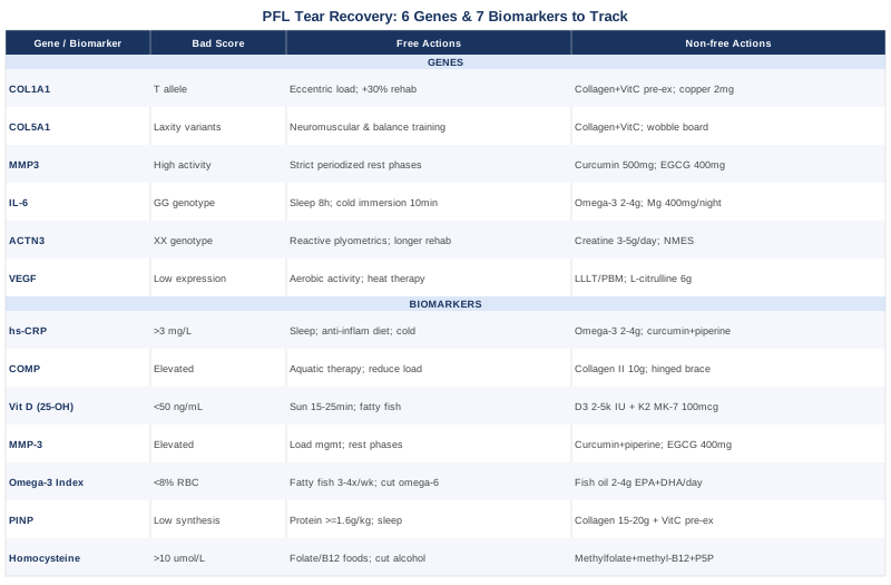
A popliteofibular ligament tear is not a simple injury, and its recovery is not well-served by generic advice alone. The seven biomarkers described here — hs-CRP, COMP, vitamin D, MMP-3, omega-3 index, PINP, and homocysteine — give you a measurable, trackable picture of your inflammatory burden, collagen synthesis activity, nutritional foundation, and tissue remodeling balance. The six genetic factors — COL1A1, COL5A1, MMP3, IL-6, ACTN3, and VEGF — explain part of why your recovery may diverge from expectation, and they point toward specific compensating strategies rather than leaving you guessing. Together, these tools shift recovery from a passive waiting process into an active, data-driven protocol.
The most practical next step is to request a targeted panel from your physician — starting with hs-CRP, 25-OH vitamin D, and homocysteine, which are affordable, widely available, and immediately actionable. If your recovery is progressing more slowly than expected, adding COMP and MMP-3 through a specialty lab provides a more granular picture of what is happening at the tissue level. Bring these results to your sports medicine physician or physiotherapist with specific questions about what the numbers mean for your rehabilitation phase and loading plan. Better information does not guarantee a faster recovery, but it dramatically improves the quality of every decision along the way.
Musculoskeletal: Joint Conditions Tendon & Ligament Conditions Sports Injuries
Autoimmune: Inflammatory Conditions Connective Tissue Conditions