This article contains AI generated content.
· UpdatedPorphyria Genes And Biomarkers — 7 Genes And 6 Biomarkers To Track
Introduction
Living with porphyria often means navigating a condition that most physicians encounter only a handful of times in their careers. The average time from first symptom to confirmed diagnosis is measured in years, not months. During that time, people are told they have anxiety, irritable bowel syndrome, or worse — that their symptoms are imagined. Even after a confirmed diagnosis, the guidance they receive is often generic: avoid triggers, eat regularly, stay out of the sun. It is accurate, but it rarely tells you where you actually stand.
The problem is that porphyria is not one disease. It is a family of at least eight distinct disorders, each caused by a different enzyme defect in the heme biosynthesis pathway, each with its own pattern of triggers, symptoms, and long-term risks. What is essential for managing Porphyria Cutanea Tarda is almost irrelevant for someone with Acute Intermittent Porphyria, and vice versa. Generic advice designed to cover all types often ends up being too vague to drive real decisions for any of them.
What changes the picture is specificity. Tracking a targeted set of biomarkers reveals what is actually happening in your body right now — whether a crisis is building, whether your liver is under strain, whether your iron stores are silently fueling your disease. Understanding which gene variants you carry explains the why behind your specific pattern of vulnerability and unlocks a more personalised prevention strategy. Together, these two angles transform porphyria management from crisis response into informed, forward-looking care.
This article covers both tracks. The first and most detailed covers six biomarkers — the ones most useful for real-world monitoring, with practical guidance on what to do when any of them lands outside a safe range. The second covers the seven genes most relevant to porphyria risk, with lifestyle and medical plans for each. An additional section connects the science of circadian biology to heme metabolism in ways most clinicians do not mention. Finally, a review of the best-supported complementary approaches rounds out the picture. Better information does not cure porphyria, but it reliably leads to better decisions.
Summary
This article breaks porphyria management into four actionable layers. The biomarker section covers six laboratory tests — urinary PBG, urinary ALA, plasma porphyrins, free erythrocyte protoporphyrin, iron and ferritin, and liver enzymes — explaining what each one reveals, how and where to measure it (with cost ranges), and exactly what to do when a result is abnormal, both with and without medical intervention. The genetics section unpacks seven key genes — HMBS, PPOX, CPOX, UROD, HFE, FECH, and ALAS2 — explaining the specific porphyria each drives and outlining practical plans for carriers whether or not they want to pursue supplements or medications. A separate section applies the most relevant findings from circadian biology research directly to heme synthesis, revealing ten non-obvious insights for daily management. Finally, five complementary approaches with meaningful clinical evidence are reviewed, each with a specific protocol and a realistic assessment of how to apply it.
If you only have time for two things: get your urinary PBG and ALA tested, and ask your geneticist about your specific gene variant. Everything else in this article builds on those foundations.
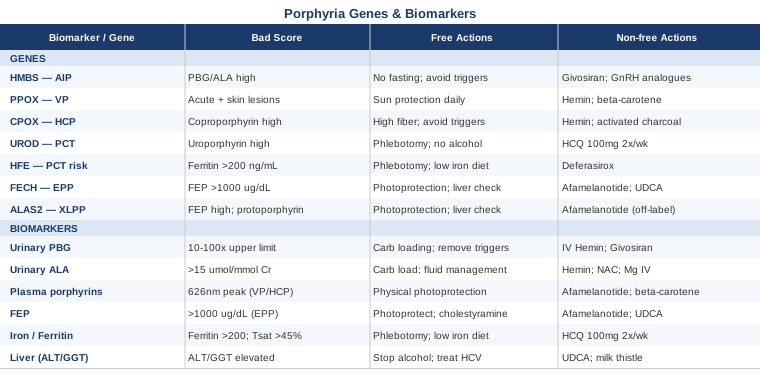
6 Biomarkers That Can Change How You Manage Porphyria
Biomarkers in porphyria serve two distinct purposes that are easy to conflate. Some — like urinary PBG — are activity markers, reflecting what the disease is doing right now. Others — like ferritin or liver enzymes — are consequence markers, reflecting cumulative damage that is building silently between episodes. Both matter, and monitoring only one category gives an incomplete picture. The six markers below were chosen because together they cover the acute, the cutaneous, and the hepatic dimensions of porphyria, at a cost and complexity level that most people can realistically sustain.
1. Urinary Porphobilinogen (PBG)
Urinary PBG is the single most important marker for any of the acute hepatic porphyrias — Acute Intermittent Porphyria (AIP), Variegate Porphyria (VP), and Hereditary Coproporphyria (HCP). When the enzyme that converts PBG to hydroxymethylbilane is deficient or overwhelmed, PBG spills into the urine at levels that are often 10 to 100 times the upper limit of normal during an acute attack. It is the reason emergency clinicians are taught to check urine color in unexplained abdominal crises.
Between attacks, PBG may return to near-normal in many patients, which is both reassuring and occasionally misleading. Some individuals with AIP never fully normalize, and persistently elevated inter-attack PBG correlates with higher risk of neuropathic complications over time. Tracking this marker quarterly when stable and immediately at the first sign of symptoms gives you real-time visibility into disease activity that no symptom diary alone can provide.
How to Measure It
A spot urine sample collected in a light-protected container (foil-wrapped) is the standard first-line test. Quantitative measurement by ion-exchange chromatography is preferred over the older Watson-Schwartz colorimetric test, which has false positives and is less sensitive. Cost range: $50–$200 at most US specialty labs and academic medical centers; some insurance plans cover it under ICD-10 E80 codes. During a suspected attack, a same-day spot urine result is informative enough to guide urgent management decisions. For longitudinal monitoring, a 24-hour urine collection provides more accurate quantification.
If the Score Is High: The Plan Without Supplements
A markedly elevated PBG during symptoms calls for immediate carbohydrate loading: 300–400g of glucose equivalent per day. This works because glucose suppresses the transcription of ALAS1 — the rate-limiting enzyme in heme synthesis — through an insulin/PGC-1α–dependent pathway. In mild-to-moderate attacks, oral glucose (glucose polymer drinks, regular meals with white rice or bread, or glucose tablets) can sometimes halt disease progression within 24–48 hours. Remove any recently started medications from the porphyria unsafe drug list (www.drugs.porphyria.com maintains the best available database), ensure adequate hydration, rest, and avoid fasting under any circumstances. Call your porphyria specialist — not a general emergency room unless you are already trained to explain your diagnosis clearly and have documentation ready.
If the Score Is High: The Plan With Supplements or Equipment
When oral carbohydrate loading fails to halt the attack within 24 hours, or when pain is severe or neurological symptoms appear, intravenous hemin (Panhematin) is the treatment of choice. The standard protocol is 4mg/kg IV once daily for four days, administered in a unit familiar with the protocol. Hemin provides exogenous heme, which suppresses ALAS1 through negative feedback and drops PBG within 24–48 hours. Side effects include thrombophlebitis (use a central line or large-bore vein with albumin pre-treatment), coagulopathy with high cumulative doses, and infusion site reactions.
For patients with recurrent attacks (four or more per year), the RNA interference drug givosiran (Givlaari) has changed the landscape. Given as a 2.5mg/kg subcutaneous injection monthly, it reduces hepatic ALAS1 mRNA and dramatically lowers both PBG levels and attack frequency. In the pivotal ENVISION trial, it reduced attack rates by 74%. Side effects include elevated liver enzymes, homocysteine rise (supplement with B6, B12, folate), and injection-site reactions. It is a specialist-initiated drug requiring porphyria centre oversight and costs $>$400,000/year without insurance — financial assistance programs exist. Cycling is continuous for eligible patients.
2. Urinary Delta-Aminolevulinic Acid (ALA)
ALA is the immediate precursor to PBG in the heme biosynthesis pathway, and in the acute porphyrias it accumulates alongside PBG. But ALA carries its own separate clinical significance: it is structurally similar to GABA, the brain's main inhibitory neurotransmitter, and at high concentrations it may act as a GABA-mimetic, potentially explaining the neuropsychiatric features of acute porphyria — the anxiety, the confusion, the autonomic instability — better than PBG does. This also makes ALA a particularly informative marker when neurotoxic symptoms dominate.
In the rare condition of ALA Dehydratase Porphyria (ADP), ALA rises in isolation without a corresponding rise in PBG, because the enzyme defect sits one step earlier in the pathway. Tracking ALA alongside PBG therefore ensures you do not miss this rarer variant or miss the severity of an acute episode where ALA-driven neurotoxicity is the primary concern.
How to Measure It
ALA is typically measured on the same 24-hour urine collection used for PBG, using high-performance liquid chromatography (HPLC). Most porphyria specialty labs offer a panel combining both markers. Cost range: $50–$200, often bundled with PBG at no extra cost. Reference values vary by laboratory, but ALA above 15 µmol/mmol creatinine in a symptomatic patient is clinically significant in most published guidelines.
If the Score Is High: The Plan Without Supplements
The first step is the same as for PBG: aggressive carbohydrate loading and trigger removal. However, because high ALA may contribute independently to neurological symptoms including peripheral neuropathy and SIADH (syndrome of inappropriate antidiuretic hormone), careful fluid management is important. Do not over-hydrate in the absence of guidance — hyponatremia is a real risk in acute attacks. Log neurological symptoms specifically (weakness, numbness, pain distribution) to monitor whether ALA-driven neurotoxicity is progressing.
If the Score Is High: The Plan With Supplements or Equipment
N-acetylcysteine (NAC) has a theoretical rationale here: ALA auto-oxidizes and generates reactive oxygen species, and NAC as a glutathione precursor may reduce this oxidative load. Animal model evidence supports this, but controlled human trials specifically in porphyria are limited. Doses of 600mg twice daily are commonly used in other oxidative stress contexts. If neurological progression continues despite hemin, magnesium infusion (for autonomic symptoms including tachycardia and hypertension, dosed at 2–4g IV) is often used supportively. For chronic low-grade ALA elevation contributing to peripheral neuropathy, long-term pain management may include gabapentin or pregabalin — notably, both are listed as likely safe in porphyria, though always verify against the most current drug database.
3. Plasma Porphyrin Scan (Fluorescence Emission Peak)
When PBG and ALA are elevated but you need to know whether you are dealing with AIP or one of the two forms with cutaneous involvement (VP or HCP), the plasma porphyrin fluorescence emission scan is the decisive test. At a plasma pH of approximately 8.5, VP shows a characteristic emission peak at around 626 nm that AIP does not — a difference that changes both prognosis and management, particularly regarding sun exposure.
For patients with erythropoietic porphyrias (EPP, XLPP), plasma porphyrins track disease activity in the skin and can correlate with photosensitivity severity. Unlike the acute-specific markers, plasma porphyrins are often persistently elevated between crises in these cutaneous types, making them suitable for routine monitoring rather than attack-specific testing.
How to Measure It
A protected blood sample drawn in a foil-wrapped EDTA tube is sent to a specialized porphyria laboratory for fluorescence scanning. This is not the same as a standard urine or blood porphyrin screen available at most routine labs — the fluorescence emission peak requires specialist equipment. The European Porphyria Network reference laboratories and major academic porphyria centers in the US (Mount Sinai, Mayo Clinic, UCSF) offer this test. Cost range: $100–$350.
If the Score Is Abnormal: The Plan Without Supplements
For elevated plasma porphyrins indicating cutaneous involvement, photoprotection becomes the central non-pharmacological intervention. This goes far beyond standard sunscreen: broad-spectrum physical blockers (zinc oxide and titanium dioxide, SPF 50+) rather than chemical filters (which are inadequate for the 400–500nm visible light wavelengths that trigger porphyrin photosensitivity), UV-protective clothing rated UPF 50+, anti-UV window film on car and home windows, and wearing gloves when driving. For EPP patients, who burn painfully in visible light including on cloudy days, even brief incidental sun exposure through glass can trigger a reaction. Gradual light desensitisation — very carefully increasing light exposure over weeks — is sometimes used but must be supervised, as it carries real risk.
If the Score Is Abnormal: The Plan With Supplements or Equipment
Afamelanotide (Scenesse), a synthetic analogue of alpha-melanocyte-stimulating hormone, is FDA-approved specifically for EPP and represents the most significant advance in cutaneous porphyria management in decades. It is given as a 16mg biodegradable subcutaneous implant every 60 days. In randomised trials it increased pain-free time in sunlight by roughly 70–90 additional minutes per day and dramatically improved quality of life. Side effects include nausea (usually brief), implant-site reactions, and skin darkening. It is a specialist-administered treatment.
Beta-carotene (high oral dose, 60–180mg/day) was the standard approach before afamelanotide and still offers modest benefit for some patients — evidence in randomised trials has been mixed, but many patients report some protection. It causes benign skin yellowing (carotenodermia) and is generally safe. Given for continuous use during months of peak light exposure; can be tapered in winter.
4. Free Erythrocyte Protoporphyrin (FEP)
Free erythrocyte protoporphyrin, also called free erythrocyte porphyrin or FEP, measures the protoporphyrin that accumulates inside red blood cells when ferrochelatase — the final enzyme in the heme pathway — is deficient. It is the defining biomarker for Erythropoietic Protoporphyria (EPP) and X-Linked Protoporphyria (XLPP), where values routinely exceed 1000 µg/dL (normal: <30 µg/dL in most labs).
One critical caveat: FEP also rises in iron deficiency, but for a different reason and in a different molecular form (zinc protoporphyrin predominates in iron deficiency; free protoporphyrin predominates in EPP). The distinction matters for diagnosis. Newer tests measure both free and zinc protoporphyrin fractions separately. Elevated FEP also correlates with liver disease risk in EPP — very high levels predict cholestatic liver injury, so this marker guides both photosensitivity management and hepatic surveillance simultaneously.
How to Measure It
An EDTA whole blood sample is needed, kept protected from light. Standard hematology labs can run total erythrocyte protoporphyrin; for the fractionated (free vs. zinc) measurement, a specialized lab is required. Cost range: $75–$200. Frequency of testing: every 6–12 months when stable, more frequently if liver enzymes are rising.
If the Score Is High: The Plan Without Supplements
Since FEP elevation in EPP reflects ongoing accumulation of photoreactive protoporphyrin in circulating red cells, the first non-pharmacological approach is the most comprehensive photoprotection strategy described above, combined with regular liver enzyme monitoring. Cholestyramine at 4–8g daily (a bile acid sequestrant) reduces the enterohepatic recirculation of protoporphyrins — because protoporphyrin is excreted in bile and can be reabsorbed from the gut. This is not a supplement in the usual sense; it is a prescription medication, but it is inexpensive and has decades of use in EPP management. Frequency: daily, continuous; main side effects are constipation and fat-soluble vitamin depletion (monitor and supplement vitamins A, D, E, K if used long-term).
If the Score Is High: The Plan With Supplements or Equipment
Beyond afamelanotide (described above), patients with markedly high FEP and rising liver enzymes may benefit from adding ursodeoxycholic acid (UDCA) at 300–600mg/day to provide hepatoprotective effects and reduce porphyrin-mediated cholestasis. This is commonly used in cholestatic liver diseases. Side effects are mild (loose stools at high doses); it is taken continuously when liver markers warrant it. Very severe EPP hepatopathy in a small percentage of patients ultimately requires liver transplantation — monitoring FEP trends helps identify this trajectory early enough to act.
5. Iron Panel and Serum Ferritin
Iron occupies a unique and central role in porphyria beyond simple nutrition. In Porphyria Cutanea Tarda (PCT) — the most common form of porphyria overall — iron excess in the liver directly promotes the oxidative inhibition of uroporphyrinogen decarboxylase (UROD), the deficient enzyme. Without excess iron, PCT often does not activate even in people with the UROD genetic variant. This is why the NIDDK identifies phlebotomy as the primary treatment for PCT — depleting iron removes the driver of the disease at its source.
The full iron panel (serum iron, total iron-binding capacity, transferrin saturation, and ferritin) is not interchangeable. Ferritin alone can be elevated by inflammation and miss the pattern of mild iron loading that still promotes PCT. Transferrin saturation above 45% paired with elevated ferritin above 200 ng/mL is the pattern most clinically relevant for PCT and hemochromatosis overlap. Since the HFE gene variants C282Y and H63D dramatically increase PCT risk, this panel should be measured in any confirmed PCT patient regardless of symptoms.
How to Measure It
Standard fasting morning blood draw. Include serum ferritin, serum iron, TIBC, and calculate transferrin saturation. Cost range: $30–$100 — frequently included in comprehensive metabolic panels. For PCT monitoring, check every 3–6 months during phlebotomy treatment, then annually once remission is achieved.
If the Score Is Abnormal: The Plan Without Supplements
Therapeutic phlebotomy is the most effective single intervention in PCT and the first-line treatment. The standard protocol is 450mL of whole blood removed every 2–4 weeks (same as blood donation), with the target being a ferritin level below 20–30 ng/mL. Most patients require 5–15 sessions to achieve remission. Skin lesions typically begin to resolve within 3–6 months of reaching target ferritin. Concurrent changes: complete alcohol cessation (alcohol promotes hepatic iron absorption and independently inhibits UROD), avoid iron supplements, reduce heme-iron foods (red meat), and discontinue estrogen-containing contraceptives or hormone therapy if applicable — both estrogen and alcohol are independent PCT triggers.
If the Score Is Abnormal: The Plan With Supplements or Equipment
When phlebotomy is not feasible (anemia, cardiovascular contraindications, poor venous access), low-dose hydroxychloroquine (HCQ) at 100mg twice weekly is remarkably effective for PCT and is considered an equally valid first-line option in current European guidelines. The mechanism is distinct from its use in autoimmune disease: at low doses, HCQ chelates hepatic uroporphyrins and promotes their urinary excretion. It works independently of iron levels. The key is low dose — standard anti-malarial or anti-rheumatic doses cause worse porphyrin accumulation. Side effects at this dose are rare; annual retinal screening is still recommended with prolonged use. Duration of treatment: typically 6–18 months to remission.
Chloroquine phosphate 125mg twice weekly is an alternative with similar efficacy. Vitamin E tocopherol (400 IU/day) has been proposed as a hepatoprotective adjunct — some small trials in PCT suggest modest benefit, likely via antioxidant protection against iron-mediated oxidative stress. Side effects minimal at this dose. Avoid vitamin C supplementation during PCT management: ascorbic acid enhances dietary iron absorption and is counterproductive when iron depletion is the goal.
6. Liver Function Panel (ALT, AST, GGT, Bilirubin)
The liver is at the center of porphyria biology for two separate reasons. First, the liver is the primary site of heme synthesis in the context of acute hepatic porphyrias, and the heme enzymes expressed there (driven by ALAS1) determine whether an acute attack occurs. Second, porphyrins themselves — when they accumulate in hepatocytes — are directly hepatotoxic. Both PCT (which concentrates uroporphyrins in the liver) and EPP (which deposits protoporphyrins in the bile and liver) carry meaningful risks of progressive liver injury if not well managed.
GGT deserves particular attention beyond what routine panels often emphasize. In PCT, GGT elevation often precedes ALT elevation and correlates with hepatic uroporphyrin accumulation more sensitively than ALT does in early disease. GGT also rises with alcohol use, estrogen, and certain drugs — all of which are PCT triggers — making it a dual indicator of both disease progression and trigger exposure. The connection to hepatitis C is important: studies show a 50–80% prevalence of HCV coinfection in PCT patients in some populations. HCV treatment with direct-acting antivirals resolves PCT in many coinfected individuals without any phlebotomy.
How to Measure It
Standard blood draw, no special processing required. ALT, AST, GGT, bilirubin, and alkaline phosphatase cover the hepatic picture adequately. Request GGT specifically — it is not always included in standard metabolic panels but is often available at no extra cost when requested. Cost range: $20–$80. For EPP patients with elevated FEP, liver function should be checked every 6 months. For PCT patients in phlebotomy, every 3 months.
If the Score Is Abnormal: The Plan Without Supplements
Alcohol elimination is non-negotiable in PCT-related liver enzyme elevation — the data on alcohol's role in PCT precipitation and progression are unambiguous. Audit any medications for hepatotoxic potential (refer to drugs.porphyria.com and cross-reference with your porphyria specialist). If HCV has not been tested, test now: treating HCV resolves PCT and normalizes liver enzymes in a substantial proportion of cases. Physical activity improves hepatic insulin sensitivity and reduces hepatic steatosis, which is a cofactor in liver injury progression in PCT; 150 minutes of moderate aerobic exercise weekly is the standard recommendation.
If the Score Is Abnormal: The Plan With Supplements or Equipment
For EPP-specific liver disease, cholestyramine and UDCA (described above in the FEP section) are the core pharmacological tools. For PCT-related liver injury, continued phlebotomy addresses the root cause. Milk thistle (silymarin) at 140mg three times daily has modest supportive evidence for general hepatoprotection and is commonly used alongside conventional PCT treatment; a 2014 systematic review in the Annals of Hepatology found benefit in various liver conditions, though PCT-specific trials are limited. Side effects are minimal. NAC at 600mg twice daily may also reduce hepatocellular oxidative stress as a supportive measure. For severe EPP hepatopathy, referral to a liver transplant center should occur early — transplant is life-saving but does not eliminate the underlying erythropoietic disease.
With these six biomarkers tracked together, a comprehensive picture of porphyria activity, photosensitivity, liver health, and iron status emerges. The goal is not to test everything at once indefinitely, but to establish which markers are relevant for your specific porphyria type and to build a monitoring rhythm that catches problems before they become crises.
The 7 Genes Behind Porphyria: What Your DNA Tells You
Genetic testing in porphyria is not optional context — it is often the difference between a diagnosis confirmed and a diagnosis suspected. Biochemical markers fluctuate: PBG normalises between attacks, plasma porphyrins may be borderline, enzyme activity assays have overlapping normal ranges. A confirmed pathogenic gene variant is definitive evidence that changes how aggressively you manage triggers, how your family members are counselled, and in some cases which treatment is indicated.
The seven genes below drive the vast majority of clinically significant porphyrias. Understanding which one affects you — and what that means practically — is a different kind of monitoring than biomarkers, but equally important for long-term strategy.
HMBS — The Gene at the Core of Acute Intermittent Porphyria
The HMBS gene (hydroxymethylbilane synthase, also called porphobilinogen deaminase or PBGD) on chromosome 11q23.3 encodes the third enzyme in the heme pathway. Haploinsufficiency — carrying one defective copy — is sufficient to cause AIP, the most common of the acute hepatic porphyrias in Northern Europe and the US. Over 400 pathogenic variants have been identified, distributed across all 15 exons, with no single predominant mutation except in specific founder populations (a splice-site mutation prevalent in Sweden, for example). Most are missense or nonsense mutations, and many are family-specific.
Penetrance is strikingly incomplete: approximately 80–90% of HMBS variant carriers never experience a symptomatic attack. The gene variant reduces enzyme activity to roughly 50% of normal, which is enough to tip the pathway into overproduction of ALA and PBG when a second hit — hormonal fluctuation, drug exposure, infection, fasting — increases ALAS1 activity. Women are more severely affected, particularly in the luteal phase of the menstrual cycle, because progesterone induces ALAS1.
If the Gene Variant Is Present: Plan Without Supplements
For a confirmed HMBS variant carrier, the lifestyle framework is specific and permanent. Carbohydrate adequacy is the cornerstone: never skip meals, never fast for more than 12 hours, maintain at least 200–300g of carbohydrates per day baseline and increase to 400g+ at the first sign of symptoms. Trigger management requires maintaining a current list of unsafe medications (verify every prescription and over-the-counter drug before taking it), avoiding recreational alcohol, managing stress with structured methods, and using hormonal contraception cautiously (progestins are problematic; continuous combined oral contraceptives or copper IUDs are often better choices). All first-degree relatives should be offered genetic testing to identify presymptomatic carriers.
If the Gene Variant Is Present: Plan With Supplements or Equipment
For carriers with recurrent attacks (≥4 per year), givosiran (described under the PBG biomarker section) is the standard of care. For hormonal cycling-related attacks, GnRH analogues (such as leuprolide) suppress ovarian hormone fluctuations and prevent hormonally-triggered attacks with high efficacy — monthly injections, with bone density monitoring and add-back estrogen regimens to mitigate bone loss. High-dose progesterone-free hormonal suppression is the goal. For infrequent attacks, having home glucose polymer drinks and a care plan for early IV glucose at a pre-arranged infusion center reduces attack severity and hospitalization. Vitamin B6 (pyridoxine) in standard doses (10–25mg/day) has been proposed historically for mild AIP, though evidence is limited to small case series; it is generally safe at these doses.
PPOX — The Gene Behind Variegate Porphyria
PPOX (protoporphyrinogen oxidase) on chromosome 1q22 encodes the seventh enzyme in the heme pathway. Variegate Porphyria (VP) is particularly common in South Africa due to a founder effect: a single R59W variant was carried by a Dutch settler couple in 1680, and it now appears in an estimated 3 per 1000 South Africans. VP is unique in causing both acute and cutaneous symptoms — some patients experience only one or the other, and some both. The cutaneous features (skin fragility, blistering on sun-exposed areas) persist even when acute attacks are in remission.
If the Gene Variant Is Present: Plan Without Supplements
All AIP trigger-avoidance strategies apply equally to VP. The cutaneous dimension adds a permanent requirement for daily physical photoprotection — broad-spectrum SPF 50+ sunscreen with zinc oxide, protective clothing, UV-filtering window film, and sun avoidance during peak hours. Unlike EPP, VP photosensitivity involves primarily UVA and some visible light, so standard glass windows provide partial but not complete protection.
If the Gene Variant Is Present: Plan With Supplements or Equipment
Hemin infusion for acute attacks; afamelanotide is under investigation for the cutaneous component of VP based on its proven mechanism in EPP. Beta-carotene supplementation (60–120mg/day) is commonly used, with modest clinical benefit for cutaneous porphyrias broadly. No single supplement eliminates the cutaneous phenotype; the combination of afamelanotide (if available) and rigorous physical photoprotection currently offers the best outcomes.
CPOX — The Gene Driving Hereditary Coproporphyria
CPOX (coproporphyrinogen oxidase) on chromosome 3q11.2 encodes the sixth heme pathway enzyme. Hereditary Coproporphyria (HCP) is rarer than AIP and VP and predominantly acute, though mild skin sensitivity occurs in roughly 30% of cases. A distinguishing biochemical feature: fecal coproporphyrin III is markedly elevated even between attacks in HCP, which helps differentiate it from AIP where fecal porphyrins are less prominent.
If the Gene Variant Is Present: Plan Without Supplements
Identical to AIP trigger-avoidance. Because coproporphyrins also accumulate in the gut and undergo enterohepatic circulation, dietary fiber and regular bowel habits take on added relevance — constipation slows coproporphyrin excretion and may worsen the biochemical burden. A high-fiber diet (30–40g/day) is worth maintaining as a baseline habit.
If the Gene Variant Is Present: Plan With Supplements or Equipment
Hemin for acute attacks. Activated charcoal (25g in water, three times daily between meals) has been used adjunctively in HCP to interrupt enterohepatic recirculation of coproporphyrins — evidence is case-level but mechanistically sound. Not for long-term use due to nutrient binding; use during symptomatic periods only. Cholestyramine serves a similar purpose with better tolerability for extended courses.
UROD — The Genetic Driver of Familial PCT
About 80% of PCT cases are sporadic (Type 1), caused by environmental iron loading, alcohol, estrogen, HCV, or HFE mutations that reduce acquired UROD activity. But approximately 20% carry an inherited UROD mutation (Type 2 PCT) — heterozygous for a loss-of-function variant — making their liver intrinsically more vulnerable. Knowing which type you have matters for family counselling and for understanding how much you need to address iron vs. how much the genetic deficiency itself contributes.
If the Gene Variant Is Present: Plan Without Supplements
Type 2 PCT carriers should consider proactive elimination of all modifiable cofactors (alcohol, excess iron, estrogens, hepatotoxic drugs) even before first clinical presentation if genetic testing is done predictively. Lifelong annual monitoring of ferritin and liver enzymes is reasonable once the variant is confirmed. Family members should be tested.
If the Gene Variant Is Present: Plan With Supplements or Equipment
Low-dose hydroxychloroquine 100mg twice weekly is particularly attractive for Type 2 PCT because iron depletion alone may not achieve full remission when intrinsic enzyme activity is already halved. Combination of phlebotomy followed by maintenance HCQ achieves sustained remission in most patients. Duration is open-ended for Type 2, with periodic reassessment; remission typically requires 12–24 months of treatment before considering tapering.
HFE — The Iron Regulation Gene That Unlocks PCT
HFE on chromosome 6p21.3 encodes a protein that regulates hepcidin expression and therefore systemic iron homeostasis. The two common variants — C282Y and H63D — reduce the inhibitory signal on intestinal iron absorption, leading to progressive iron loading. Homozygous C282Y carriers have a lifetime risk of hereditary hemochromatosis; heterozygous C282Y or compound C282Y/H63D carriers have a much higher-than-average risk of developing PCT when other cofactors are present. HFE testing is now standard practice in PCT workup.
If the Gene Variant Is Present: Plan Without Supplements
Even without active PCT symptoms, a confirmed C282Y homozygote should monitor ferritin and transferrin saturation annually and initiate phlebotomy once ferritin exceeds 100–200 ng/mL — before hepatic iron loading reaches the threshold that activates PCT or damages the liver. Dietary adjustments include avoiding iron-fortified foods, reducing red meat, not cooking in cast iron, and having coffee or tea with meals (tannins reduce iron absorption by 30–60%).
If the Gene Variant Is Present: Plan With Supplements or Equipment
Avoid all iron-containing supplements unless treating confirmed deficiency anemia. Avoid high-dose vitamin C with meals (enhances non-heme iron absorption). In established iron overload not amenable to phlebotomy, iron chelation therapy with deferasirox or deferoxamine is an option — specialist-managed, with significant side-effect profiles. For most HFE carriers, regular phlebotomy is the most effective and well-tolerated approach.
FECH — The Gene That Defines Erythropoietic Protoporphyria
FECH on chromosome 18q21.31 encodes ferrochelatase, the final enzyme in the heme pathway that inserts iron into protoporphyrin IX to form heme. EPP follows an unusual inheritance pattern: most patients are compound heterozygotes — one allele carries a loss-of-function mutation, and the other carries a common low-expression polymorphism (IVS3-48C) that reduces ferrochelatase activity below the critical threshold. This means genetic testing must look for both the pathogenic variant and the modifier allele. Ferrochelatase activity below approximately 25–35% of normal causes protoporphyrin to accumulate.
If the Gene Variant Is Present: Plan Without Supplements
Since EPP is expressed from birth, the practical approach begins with education: graded light exposure to develop whatever tolerance is possible, continuous physical photoprotection as described above, and close hepatic surveillance from the time of diagnosis because cholestatic liver disease is the most serious long-term complication (affecting approximately 2–5% of EPP patients severely).
If the Gene Variant Is Present: Plan With Supplements or Equipment
Afamelanotide (Scenesse) 16mg subcutaneous implant every 60 days is the definitive treatment for EPP. Beta-carotene 60–180mg/day continuously during high-light months; cholestyramine 4–8g daily for hepatoprotection; UDCA if liver enzymes rise. Consider liver transplant evaluation early if FEP is persistently very high (>3000 µg/dL) or if bilirubin begins rising — outcomes after transplant are poor if liver disease is already advanced.
ALAS2 — The Gain-of-Function Gene Behind X-Linked Protoporphyria
ALAS2 on the X chromosome encodes the erythroid-specific ALA synthase enzyme and represents an interesting exception in porphyria genetics: here, the disease is caused not by loss of function but by gain of function mutations (typically C-terminal deletions that remove an inhibitory feedback domain), causing ALAS2 to overproduce ALA and therefore protoporphyrin. X-Linked Protoporphyria (XLPP) is clinically very similar to EPP but males are more severely affected (full hemizygous expression), while female carriers may have mild-to-moderate symptoms depending on X-inactivation patterns.
If the Gene Variant Is Present: Plan Without Supplements
Because the mechanism is opposite to most porphyrias (overactivity rather than deficiency), approaches that reduce ALAS2 activity in principle would be beneficial — though no ALAS2-specific inhibitor is approved. Practical management mirrors EPP: comprehensive photoprotection, liver surveillance, avoid alcohol (reduces liver reserve), ensure adequate folate and B12 (support erythropoiesis). Males with XLPP should be counselled that hepatopathy risk may be higher than in EPP.
If the Gene Variant Is Present: Plan With Supplements or Equipment
Afamelanotide has been used off-label in XLPP and is under formal investigation. Iron supplementation has been trialed in XLPP on the hypothesis that providing more iron substrate would redirect excess protoporphyrin toward completed heme rather than accumulation — small case series show mixed results; it is not standard practice. Cholestyramine and UDCA for hepatoprotection, as in EPP. Monitor closely with FEP and liver function every 6 months.
What Circadian Biology Reveals About Your Heme Cycle — 10 Things Worth Knowing
The research of Dr. Satchin Panda at the Salk Institute — widely disseminated through his book The Circadian Code and extensively discussed on the Huberman Lab podcast (Episode 17 featuring Dr. Panda, 2021) — has revealed something with profound but underappreciated implications for porphyria: the liver's metabolic enzyme machinery runs on a clock, and that clock governs heme synthesis in ways that directly affect when and why acute attacks occur.
Most porphyria patients are told to eat regularly and avoid fasting. What they are rarely told is the why — and knowing the why changes how you actually implement the advice.
1. ALAS1 Is Directly Under Circadian Control
ALAS1, the rate-limiting enzyme in hepatic heme synthesis, is transcriptionally regulated by the core circadian clock proteins BMAL1 and CLOCK. Its mRNA expression peaks in the early active phase (morning in humans) and troughs during sleep. This is not background noise — ALAS1 mRNA abundance varies by 5- to 10-fold across the 24-hour cycle in animal models. The practical implication: the vulnerability of the heme pathway is not constant across the day. Triggers may land differently depending on when they occur.
2. PGC-1α Is the Bridge Between Fasting and ALAS1
When you fast, AMPK activates PGC-1α (a transcriptional co-activator), which drives ALAS1 transcription. This is the molecular mechanism behind why fasting precipitates acute porphyria attacks — it is not just a nutritional deficit but an active transcriptional upregulation of the very enzyme that overloads the pathway. Dr. Panda's research identified PGC-1α as a master regulator of hepatic circadian metabolism, which connects directly to this pathway.
3. The Liver's Clock Is Semi-Autonomous From the Brain
The suprachiasmatic nucleus (SCN) in the hypothalamus is the master clock, but the liver has its own peripheral clock that responds most strongly to meal timing rather than light. This means even if your light-dark cycle is regular, irregular eating patterns independently disrupt liver metabolic rhythms — including heme enzyme expression rhythms. For porphyria patients, consistency of meal timing is as important as meal content.
4. The Overnight Fast Window Is the Danger Zone
Panda's research shows that eating within a 10–12 hour window and fasting the remaining 12–14 hours is metabolically optimal for most people. For porphyria patients, the upper limit of the fasting window is critical information: a 14-hour overnight fast may be well tolerated by some, but others with reduced enzyme reserve find it tips the pathway toward excessive ALA/PBG production by morning. A practical modification: a small carbohydrate-containing snack before sleep — not a full meal, but enough glucose to keep insulin and PGC-1α activity from crashing overnight.
5. Shift Work and Porphyria Are a Dangerous Combination
Animal studies from the Panda lab and others have shown that circadian disruption (simulated shift work) alters hepatic metabolic enzyme expression dramatically, including heme pathway enzymes. While direct human trials in porphyria patients are not available, the mechanistic evidence strongly supports the clinical observation that shift workers with AIP tend to have higher attack rates. Advocating for day-shift work or consistent schedules is a legitimate, underused part of porphyria management.
6. Morning Light Entrains the Clock That Controls Your Liver
Huberman's extensive research synthesis on circadian biology emphasizes that morning sunlight exposure within 30–60 minutes of waking is the strongest zeitgeber (time-giver) for the master clock, which in turn coordinates peripheral clocks including the liver. For most porphyria patients, morning light is safe (UV exposure is lower at low solar angles). The relevant point: people who avoid all light — including morning — in response to cutaneous porphyria may inadvertently destabilise their circadian metabolic rhythms. Where possible, brief low-angle morning light is preferable to complete light avoidance.
7. Cortisol's Circadian Pulse Affects ALAS1 Indirectly
Cortisol, which peaks in the morning as part of the normal cortisol awakening response (CAR), is an indirect modulator of ALAS1 activity via its effects on glucocorticoid response elements in the ALAS1 promoter. Chronic stress, irregular sleep, and HPA axis dysregulation flatten the normal cortisol pulse, contributing to metabolic instability. This adds a biological underpinning to the clinical observation that stress triggers porphyria attacks — it is not purely psychological.
8. Time-Restricted Eating for Porphyria Patients Needs Modification
Standard time-restricted eating protocols (16:8 fasting) involve a 16-hour fast that is likely too long for many acute porphyria patients. The Panda lab's research shows that 12:12 or 14:10 eating windows capture most of the metabolic benefit without the risks of prolonged fasting. For AIP patients, an adjusted protocol — eating within a 10-hour window, with the last meal no more than 2–3 hours before sleep and a carbohydrate-rich bedtime snack — is a reasonable modification.
9. Alcohol Disrupts the Liver Clock Directly
Ethanol has been shown in multiple studies to disrupt circadian clock gene expression in the liver (reducing BMAL1 and Period protein levels), independent of its direct toxic effects and independent of its role as a porphyria trigger. This means alcohol creates a double hit for porphyria patients: direct UROD inhibition/iron mobilization in PCT, and disruption of the metabolic clock that stabilises heme enzyme rhythms in all porphyria types.
10. Sleep Debt Accumulates Risk
Panda's research and Huberman's synthesis make clear that partial sleep deprivation — even 6 hours per night — produces measurable disruption of liver clock gene expression. For porphyria patients, prioritising 7–9 hours of consolidated sleep is not just general health advice. It is a genuine component of attack prevention, operating through the circadian pathway that governs ALAS1 expression. Sleep timing consistency (same bedtime, same wake time) appears more important than duration alone for liver clock stabilization.
Complementary Approaches With Clinical Evidence
The conventional medical approach to porphyria focuses on drugs, phlebotomy, and trigger avoidance — all evidence-based and important. But the symptom burden between attacks, including chronic pain, autonomic dysregulation, anxiety, and fatigue, is often inadequately addressed. The following approaches have meaningful human evidence, particularly for symptom management in chronic conditions that share features with porphyria's inter-attack presentation.
Mindfulness-Based Stress Reduction (MBSR)
MBSR, the 8-week structured program developed by Dr. Jon Kabat-Zinn, involves weekly group sessions and daily home practice in meditation, body scan, and mindful movement. Its relevance to porphyria is twofold: first, psychological stress is a confirmed acute porphyria trigger via HPA axis activation and cortisol-mediated ALAS1 upregulation; second, the chronic pain and anticipatory anxiety common in porphyria patients respond to the same mechanisms that MBSR addresses in other chronic pain populations.
A widely cited study by Kabat-Zinn et al. in the General Hospital Psychiatry (1982) demonstrated significant anxiety reduction and pain improvement in chronic pain patients following an MBSR program. A 2014 meta-analysis by Goyal et al. in JAMA Internal Medicine (PMID 24395196) found moderate-strength evidence that mindfulness meditation programs reduced anxiety, depression, and pain in diverse clinical populations, with effect sizes comparable to antidepressants for anxiety.
For porphyria patients, a formal 8-week MBSR course (available in-person or online through programs affiliated with UMASS Medical School) offers the most evidence-supported entry point. Even 10–15 minutes of daily guided body-scan practice can reduce baseline sympathetic activation, which over time may lower the cortisol-ALAS1 interaction that contributes to attack vulnerability. Start slowly, avoid over-efforting in early practice — pacing is the principle.
Biofeedback
Biofeedback uses real-time physiological monitoring (heart rate variability, skin temperature, electromyography) to teach voluntary control of autonomic functions. Its relevance to porphyria is specific: the autonomic neuropathy of acute attacks — tachycardia, hypertension, diaphoresis — leaves a residual sympathetic hyperactivation in many patients that persists between episodes and contributes to chronic fatigue and anxiety. Biofeedback trains the parasympathetic counter-response.
Heart rate variability (HRV) biofeedback, in which patients learn to breathe at their resonance frequency (typically 5–7 breaths per minute) to maximise HRV, has the strongest evidence base. A systematic review of HRV biofeedback in chronic conditions (Applied Psychophysiology and Biofeedback, 2017) found consistent improvements in autonomic balance and anxiety across diverse populations. For porphyria neuropathy, no specific RCT exists, but the autonomic mechanism is directly relevant.
Practically, a 10-session course with a certified biofeedback therapist (BCIA-certified) provides sufficient skill to continue independently. HRV biofeedback devices for home use (Polar H10 with HRV Logger app, or Inner Balance by HeartMath) allow ongoing practice at $50–$150. Aim for 20-minute daily sessions at your resonance frequency; most patients see measurable HRV improvement within 4–6 weeks.
Breathing-Based Therapies
Specific breathing protocols — resonance frequency breathing, box breathing, extended-exhale techniques — directly modulate the autonomic nervous system through the vagal–cardiorespiratory reflex. For porphyria patients, their utility spans three contexts: immediate symptom management during early attack signs (tachycardia, nausea), daily nervous system regulation to reduce baseline sympathetic tone, and pain tolerance modulation.
Research on slow breathing (5–6 breaths/minute) consistently demonstrates acute increases in HRV and reductions in sympathetic outflow. A 2018 study in Frontiers in Human Neuroscience documented significant ANS modulation with a 4-7-8 breathing protocol (4s inhale, 7s hold, 8s exhale) in a clinical population with chronic stress. Extended exhale breathing (inhale 4s, exhale 8s) is simpler and equally effective for shifting vagal tone.
For porphyria patients, a practical daily protocol would be: 10 minutes of slow breathing (5s inhale through nose, 5s exhale through pursed lips) every morning and during early symptoms. This does not replace medical management but can reduce the autonomic escalation that worsens attack progression. No equipment needed; the technique is free and portable.
Guided Imagery
Guided imagery involves structured mental visualisation — of calm environments, of healing processes, of symptom reduction — and has been studied specifically in chronic abdominal pain, a hallmark feature of acute porphyria attacks and their aftermath. The mechanism involves pain gating through descending inhibitory pathways, reduction of the stress response, and potentially modulation of gut motility through the gut-brain axis.
A randomised controlled trial by Weydert et al. in BMC Pediatrics (2006) found that guided imagery reduced frequency and severity of chronic abdominal pain significantly compared to controls — a relevant comparator for the post-attack abdominal recovery common in AIP. For adult chronic pain, meta-analyses support guided imagery as a meaningful complement to pharmacological management.
A structured approach for porphyria: 15–20 minutes daily using a recorded guided imagery script focused on abdominal pain relief and autonomic calming (many are available through the Guided Imagery Network or via apps such as Insight Timer). Use consistently for 4–6 weeks before evaluating effect. It is most useful during recovery from attacks rather than during the acute peak.
Microbiome-Directed Therapies
The gut-liver axis has emerged as an unexpected but mechanistically compelling area for porphyria research. Hepatic porphyrins are excreted in bile and enter the intestinal lumen, where the gut microbiome partially determines their fate — some microbiota metabolise porphyrins, others enhance their enterohepatic recirculation. In PCT specifically, where gut-derived lipopolysaccharide (LPS) is a cofactor in hepatic UROD inhibition, dysbiosis and increased intestinal permeability may worsen disease activity.
While direct clinical trials in porphyria are limited, a growing body of research supports probiotics and prebiotic fiber in reducing hepatic LPS load and improving gut barrier function. A 2021 meta-analysis in Nutrients found that multi-strain probiotic supplementation reduced systemic inflammatory markers and liver enzyme levels in non-alcoholic fatty liver disease — a condition with overlapping hepatic mechanisms to PCT. The relevance is mechanistically plausible though not yet tested directly.
Practically: a diverse dietary fiber intake (30–40g/day from varied plant sources) to feed beneficial microbiota; a multi-strain probiotic (10–50 billion CFU, strains including Lactobacillus and Bifidobacterium species) for 12-week trials; avoidance of unnecessary antibiotics that disrupt microbiome composition. Fermented foods (unsweetened kefir, sauerkraut, kimchi) provide live cultures with additional dietary benefit. This is a low-risk, low-cost adjunct that supports overall liver health without interfering with conventional porphyria treatments.
Conclusion
Porphyria is manageable — but only with the right information. Tracking urinary PBG and ALA gives you the clearest real-time signal of pathway activity and attacks in progress. Monitoring plasma porphyrins, FEP, the iron panel, and liver enzymes extends that picture to include cutaneous activity, hepatic porphyrin accumulation, and the iron load that drives PCT. Knowing your gene variant — whether HMBS, PPOX, CPOX, UROD, HFE, FECH, or ALAS2 — moves management from general caution to targeted precision. Understanding how your circadian clock, meal timing, and sleep patterns interact with heme enzyme expression adds a dimension most clinicians will not raise but that matters for daily attack prevention.
None of this replaces a specialist — a porphyria centre with hematologists or hepatologists experienced in rare metabolic diseases is irreplaceable for confirming diagnoses, interpreting edge cases, and managing severe episodes. But arriving at those appointments with your own biomarker trends, a confirmed genetic variant, and a clear log of triggers and symptoms puts you in a fundamentally stronger position. The next smart step is simple: if you have not had urinary PBG and ALA tested recently, arrange it. If you have not had genetic testing, ask your specialist about it. Better information is where better management begins.
Neurological Skin Endocrine & Metabolic
Neurological: Nerve Conditions
Digestive: Liver & Gallbladder Conditions