This article contains AI generated content.
· UpdatedPost-Infectious Arthritis — 5 Genes And 6 Biomarkers To Track
Introduction
Post-infectious arthritis catches most people completely off guard. You fight through what seemed like a routine infection — a bout of food poisoning, a urinary tract infection, a respiratory illness — assume the worst is behind you, and then, days or weeks later, a knee swells without warning, a heel becomes inexplicably painful, or a finger stiffens overnight. The infection is gone, but the immune system has taken on a life of its own.
This pattern is more common than most people realize. Reactive arthritis — the most studied form of post-infectious arthritis — affects an estimated 1 to 4 out of every 100 people following certain bacterial infections, particularly Chlamydia trachomatis, Salmonella, Shigella, Campylobacter, and Yersinia. Post-streptococcal and post-viral forms are also well recognized. Yet two people can face the exact same triggering infection and end up on entirely different paths: one recovers within weeks while the other deals with joint inflammation for months. That divergence is not random — it has a biological explanation, and much of it is measurable.
Standard clinical management tends to address the symptom layer: anti-inflammatory drugs, rest, watchful waiting. That approach is not wrong, but it treats every case as essentially the same when they are not. Someone whose gut barrier remains compromised after the original infection continues feeding the same inflammatory signals that started the reaction. Someone carrying a specific immune gene variant may be biologically wired for a more amplified and prolonged response. Those distinctions matter for practical decisions about lifestyle, nutrition, supplementation, and when to escalate care.
This article works through two complementary lenses. The first covers the six most clinically useful biomarkers for tracking post-infectious arthritis — measurable signals that reflect what your immune system is actually doing, along with specific guidance on what to do when numbers fall outside the optimal range. The second covers five genes consistently linked to risk and severity of the condition, with practical plans for each. A synthesis of key insights from a clinician framework grounded in multiple human studies, plus five evidence-backed complementary approaches, complete the picture. The goal is not a cure promise — it is better information, because that is what actually shifts decisions.
Summary
This article covers 6 key biomarkers — hs-CRP, ESR, HLA-B27 antigen, pathogen-specific antibodies, IL-17A/IL-6, and fecal calprotectin/zonulin — that paint a detailed real-time picture of what is driving post-infectious arthritis in your specific body. For each one, you will find what it reveals, how to get it measured with realistic cost ranges, and targeted correction plans both with and without supplements.
The genetics section covers 5 genes (HLA-B*27, ERAP1, IL23R, TNF-α, and TLR4) with specific plans for each risk variant, including supplement protocols with frequencies, cycling schedules, and side effects. A summary of Tom O'Bryan's molecular mimicry framework — one of the most mechanistically coherent accounts of why post-infectious conditions become chronic — follows, with ten clinically useful insights. The article closes with five complementary approaches that have meaningful human clinical evidence for arthritis and immune modulation. Understanding both your current biomarker status and your genetic predisposition creates a roadmap that generic advice simply cannot provide.
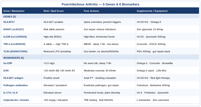
6 Key Biomarkers to Track in Post-Infectious Arthritis
Biomarkers do not tell the full story, but they tell a great deal of it. The right panel of tests can distinguish between active, resolving, and chronic inflammation; confirm which pathogen likely triggered the condition; reveal gut-immune dysfunction that keeps the response alive long after the infection is gone; and track whether any intervention is actually working. The six markers below form a progressively deeper map — starting with the standard clinical tests and building toward more specialized signals that most conventional workups skip entirely.
Biomarker 1: High-Sensitivity C-Reactive Protein (hs-CRP)
High-sensitivity CRP is a protein produced by the liver in response to inflammatory cytokines, primarily IL-6. In post-infectious arthritis, it provides a real-time snapshot of systemic inflammation — rising sharply during active flares and falling as the immune response winds down. It is one of the most reliable and responsive indicators of whether the condition is still actively progressing or beginning to resolve.
What makes hs-CRP particularly useful here is its sensitivity to intervention. If a dietary change, a supplement, or a medication is doing something meaningful, hs-CRP tends to reflect that within two to four weeks. Standard CRP assays often lack the resolution to detect low-level chronic inflammation; the high-sensitivity version picks up elevations in the 1–3 mg/L range, which represent persistent immune activation even without acute flares. This is the range where chronic post-infectious inflammation most often lives. PubMed research on CRP as an inflammatory marker in reactive arthritis.
How to Measure It
hs-CRP is measured via a standard blood draw and is available through virtually any primary care provider or direct-to-consumer lab (Quest, LabCorp in the US; comparable services across Europe). Cost: $15 to $50, often covered by insurance when ordered as part of an arthritis or cardiovascular workup. For active monitoring, testing every four to eight weeks provides useful trend data. Target range: below 1.0 mg/L is optimal; 1.0–3.0 mg/L indicates mild persistent inflammation; above 3.0 mg/L signals active immune response.
If the Score Is Bad: The Plan Without Supplements
The most impactful non-supplement intervention for elevated hs-CRP is removing the primary dietary drivers of IL-6 production: refined seed oils (sunflower, canola, soybean), ultra-processed foods, and refined sugars. Reducing these three inputs consistently moves hs-CRP in the right direction within six to eight weeks in most people. This is not a minor dietary adjustment — it requires real structural change, but the mechanism is direct.
Beyond diet, sleep duration and quality is a non-negotiable CRP driver. Sleeping under six hours per night elevates IL-6 independently of all other factors. Achieving seven to nine hours of consistently timed sleep has measurable CRP effects. Weight normalization — even a five to ten percent reduction in visceral adipose tissue — reduces the chronic TNF-α and IL-6 secretion from fat cells that sustains CRP elevation in overweight individuals. Cold exposure (brief cold immersion or contrast showers three to four times per week) has preliminary human data for reducing inflammatory cytokines and is worth adding as a low-cost adjunct.
If the Score Is Bad: The Plan With Supplements or Equipment
Omega-3 fatty acids (combined EPA+DHA): the most evidence-supported supplement for hs-CRP reduction. Dose: 2–4g EPA+DHA daily during active inflammation; reduce to 1–2g as a maintenance dose once hs-CRP normalizes below 1.0 mg/L. No cycling required — this is appropriate as a long-term daily supplement. Side effects: mild GI discomfort, fishy aftertaste (use enteric-coated forms to minimize), potential platelet effects at doses above 3g if combined with anticoagulants.
Curcumin (phospholipid-complexed form — Meriva, BCM-95, or with piperine): 500–1000mg standardized curcuminoids twice daily with meals. Human trials in arthritis have shown meaningful hs-CRP reductions over 8–12 weeks. Side effects are rare at this dose but include GI discomfort; avoid with anticoagulant therapy. Cycle 12 weeks on, 4 weeks off. PubMed research on curcumin and CRP in arthritis trials.
Boswellia serrata (standardized AKBA extract, minimum 30%): 300–500mg three times daily. Cycle 6–8 weeks on, 2 weeks off to maintain efficacy. Mild GI effects possible. The combination of omega-3, curcumin, and boswellia simultaneously addresses the arachidonic acid cascade, NF-kB signaling, and leukotriene production — three distinct inflammatory pathways that converge in reactive arthritis.
Equipment: Infrared sauna (20–30 minutes per session, 3–4 times per week at 50–60°C) has been associated with reductions in inflammatory cytokines and CRP in small human trials. It functions as a supportive rather than primary intervention, but the cumulative effect over 8–12 weeks can be meaningful.
Biomarker 2: Erythrocyte Sedimentation Rate (ESR)
ESR measures how quickly red blood cells settle in a test tube over one hour — a proxy for changes in blood protein composition driven by systemic inflammation. Unlike hs-CRP, which reflects the immediate inflammatory state and changes within hours, ESR changes more slowly and captures a broader, more sustained inflammatory burden. In post-infectious arthritis, a persistently elevated ESR (above 20 mm/h in men, above 30 mm/h in women as general reference values) often indicates the immune response has not resolved even when acute joint symptoms have partially subsided.
ESR and hs-CRP are most valuable in combination. The two markers can diverge meaningfully: a normalized CRP with a persistently high ESR typically suggests chronic smoldering inflammation — immune activity that is too low-grade to produce acute CRP but is still maintaining elevated fibrinogen and immunoglobulin levels. This pattern warrants closer attention to gut-immune drivers and possible autoimmune progression. ESR is also sensitive to confounders including anemia, infection, and hypergammaglobulinemia, so it should always be interpreted in context.
How to Measure It
ESR is a standard laboratory test available through virtually any clinical setting. Cost: $10 to $30, widely covered by insurance. Order it alongside hs-CRP for maximum interpretive value. Frequency: every four to eight weeks during active disease; every three to six months for stable monitoring.
If the Score Is Bad: The Plan Without Supplements
The non-supplement interventions that move ESR overlap significantly with those for CRP, with one important addition: structured moderate physical activity specifically reduces ESR in arthritis patients over 8–12 weeks. Walking, cycling, or swimming for 30–45 minutes four to five days per week has been studied in inflammatory arthritis with consistent ESR-reducing effects. The key is sustained moderate movement — vigorous high-intensity exercise during an active flare can temporarily worsen ESR and should be avoided until the condition stabilizes. Chronic psychological stress, acting through sustained cortisol and NF-kB activation, is also a significant ESR driver that mindfulness-based interventions specifically address (see the complementary approaches section).
If the Score Is Bad: The Plan With Supplements or Equipment
The omega-3, curcumin, and boswellia stack described for hs-CRP also consistently reduces ESR over 8–12 weeks. For persistent ESR elevation despite these measures, low-dose naltrexone (LDN) at 1.5–4.5mg taken before bed has been investigated in multiple autoimmune and inflammatory conditions with promising results, and is increasingly used in post-infectious arthritis by practitioners working at the intersection of rheumatology and functional medicine. This is an off-label protocol requiring a prescription — discuss with your physician. Side effects are generally mild (vivid dreams, initial sleep disruption) and typically resolve within two to four weeks. PubMed research on low-dose naltrexone in autoimmune and inflammatory conditions.
Biomarker 3: HLA-B27 Antigen Test
HLA-B27 is a protein encoded by the HLA-B gene that sits on the surface of nearly all nucleated cells and presents peptide fragments to CD8+ T cells. Testing positive for HLA-B27 does not mean you will develop reactive arthritis — most carriers never do. But it shifts the probability landscape substantially: approximately 60–80% of reactive arthritis patients test positive for HLA-B27, compared to roughly 6–8% of the general population in European-ancestry groups and variable rates across other ethnicities.
For someone already dealing with post-infectious arthritis, a positive HLA-B27 result carries important prognostic weight. It is associated with higher risk of axial joint involvement (affecting the spine and sacroiliac joints), a more chronic disease course, and greater risk of eventual progression toward ankylosing spondylitis. This is a one-time test — your HLA type does not change — and its value lies in risk stratification and guiding the intensity of proactive management.
How to Measure It
HLA-B27 testing is performed via flow cytometry or PCR from a standard blood draw. Cost: $100 to $300, sometimes covered when ordered by a rheumatologist with an active arthritis diagnosis. It is a single lifetime test with permanent clinical relevance. Some direct-to-consumer genomic sequencing services also report HLA-B27 status, though clinical flow cytometry is more reliable for diagnostic purposes.
If the Score Is Bad: The Plan Without Supplements
A positive HLA-B27 result should intensify rather than change the fundamentals of management. The single most important non-pharmacological intervention is a structured physical therapy program targeting axial extension, posture, and sacroiliac joint mobility. ASAS (Assessment of SpondyloArthritis International Society) guidelines specifically identify exercise as a primary disease-modifying intervention for HLA-B27-positive spondyloarthropathies — not just symptom management. Daily extension exercises, chest expansion work, and swimming or backstroke activities are the most evidence-supported movement patterns.
Smoking cessation is independently associated with more severe and progressive disease in HLA-B27-positive spondyloarthropathies. If relevant, this becomes a clinical priority, not just a general health recommendation.
If the Score Is Bad: The Plan With Supplements or Equipment
Vitamin D3 insufficiency is disproportionately common in HLA-B27-positive spondyloarthritis, and several studies associate low 25-OH vitamin D status with greater disease activity scores. Target serum 25-OH vitamin D between 40–60 ng/mL through supplementation of 2000–5000 IU D3 daily, paired with 90–200mcg vitamin K2 (MK-7 form) to maintain calcium distribution. Check serum levels every three to six months initially to dial in your dose. Side effects at these levels are rare; toxicity becomes a concern above 10,000 IU sustained for months.
Photobiomodulation (red light therapy) devices targeting affected joints at 630–850nm wavelengths have shown reductions in joint inflammation and pain in arthritis trials. Protocol: 10–20 minutes per joint area, three to five times per week, using a clinical-grade device. Home devices range from $200 to $1000. This is a low-risk, adjunctive technology that directly addresses joint-level inflammation and is particularly practical for HLA-B27 carriers managing a longer disease course. PubMed research on photobiomodulation in arthritis.
Biomarker 4: Pathogen-Specific Antibodies
Identifying which infection triggered the arthritis is not just an academic exercise — it has direct implications for treatment strategy and prognosis. Pathogen-specific serology (antibody testing) can confirm the causative organism weeks or months after the acute infection has cleared, because IgG and IgA antibodies often persist long after active symptoms resolve.
The most clinically relevant tests depend on the suspected trigger: - Anti-streptolysin O (ASO) titer and anti-DNase B: for post-streptococcal arthritis - Chlamydia trachomatis IgG/IgA: urogenital-triggered reactive arthritis - Salmonella, Shigella, Yersinia, and Campylobacter serology: enteric reactive arthritis - Anti-Borrelia antibodies with Western blot confirmation: post-Lyme arthritis - Anti-SARS-CoV-2 antibodies with anti-nucleocapsid panel: post-COVID arthritis workup
Elevated or persistently positive serology suggests ongoing immune priming from residual bacterial antigens — even without culturable organism. This is the mechanism behind molecular mimicry: antibodies generated against bacterial proteins cross-react with joint tissue components, perpetuating inflammation after the pathogen is clinically gone. PubMed research on molecular mimicry in reactive arthritis.
How to Measure It
Each pathogen requires a specific serology test, most available through standard clinical labs. Cost: $30 to $150 per pathogen-specific panel. A comprehensive reactive arthritis serology screen covering Chlamydia, the major enteric pathogens, and streptococcal markers typically costs $150–$400. Many panels are covered with appropriate arthritis or infectious disease diagnosis codes.
If the Score Is Bad: The Plan Without Supplements
If a specific pathogen is identified with persistent seropositivity, the first priority is completing pathogen eradication. In Chlamydia-triggered reactive arthritis specifically, several randomized clinical trials support a three-month course of doxycycline (or combination doxycycline plus azithromycin) to eliminate persistent intracellular infection — this approach has shown reductions in chronic disease duration in controlled trials. PubMed research on antibiotic protocols in Chlamydia-triggered reactive arthritis. Enteric forms (Salmonella, Campylobacter) do not consistently respond to antibiotic intervention once the acute phase has passed, but healing the gut barrier remains central to disrupting the ongoing immune priming.
If the Score Is Bad: The Plan With Supplements or Equipment
Bovine colostrum (500mg to 1g twice daily) provides passive immune support and has modest human trial evidence for reducing gut permeability and intestinal immune activation following bacterial GI infection. Cycle 8 weeks on, 4 weeks off. Side effects are minimal; avoid if you have a dairy sensitivity.
Berberine (500mg twice to three times daily with food) has both antimicrobial properties and gut microbiome modulating effects documented in human trials. It is particularly relevant when enteric pathogens are the trigger. Side effects: GI discomfort early on, particularly loose stools in the first week. Avoid concurrent use with medications metabolized by CYP3A4 (check interactions with your pharmacist). Cycle 8–12 weeks.
Biomarker 5: IL-17A and IL-6 Serum Levels
The IL-23/Th17 signaling axis is now established as mechanistically central to reactive arthritis and the broader spondyloarthropathy spectrum. IL-17A is produced by Th17 cells and drives joint inflammation by recruiting neutrophils, promoting osteoclast activity, and stimulating synoviocyte production of inflammatory mediators. IL-6 sits upstream, amplifying the cascade through multiple pathways including CRP production, T-cell differentiation, and bone remodeling signaling.
Measuring serum IL-17A and IL-6 is not part of a standard clinical panel, but is increasingly available through specialty reference labs and functional medicine providers. An elevated IL-17A in the context of post-infectious arthritis suggests the Th17 response has been chronically activated — which carries both prognostic implications (more likely to run a prolonged course) and therapeutic ones (these patients are better candidates for IL-17 or IL-23 pathway-targeting interventions, whether nutritional or, in severe cases, biologic). PubMed research on IL-17A in reactive arthritis and spondyloarthritis.
How to Measure It
IL-17A and IL-6 are measured via ELISA assay from a blood draw. Available through major reference labs (Quest Diagnostics, LabCorp in the US; equivalent through Cerba or Biomnis in Europe). Cost: $100 to $300 per cytokine marker. Not routinely covered by insurance without specialty referral. For most patients, testing as part of a broader autoimmune panel every three to six months during active disease is the practical approach.
If the Score Is Bad: The Plan Without Supplements
Dietary modulation of the Th17/regulatory T-cell balance is the most accessible and well-supported non-pharmacological approach to elevated IL-17A. High-fiber, plant-diverse diets fuel short-chain fatty acid (SCFA) production by the gut microbiome, which promotes regulatory T-cell differentiation at the expense of Th17 cells — directly counteracting the inflammatory pathway. A landmark Stanford randomized trial demonstrated that a diet rich in fermented foods (kefir, kimchi, sauerkraut, kombucha, yogurt) significantly reduced circulating inflammatory cytokine expression over 10 weeks in healthy adults, with effects that would be expected to be even more pronounced in a chronically inflamed population. Dietary diversity and fermentation — not supplements — are the primary levers here.
If the Score Is Bad: The Plan With Supplements or Equipment
Vitamin A (retinol), not beta-carotene, is a critical regulator of the Th17/Treg balance at the gut-immune interface: adequate retinol promotes Treg induction and suppresses Th17 differentiation through RAR signaling on dendritic cells. If dietary intake is low (limited organ meats, fatty fish, or dairy), a conservative supplement of 5000 IU retinol palmitate daily for 12 weeks is reasonable. Side effects: teratogenic at high doses — avoid in pregnancy; potential liver toxicity with chronic high-dose use. Stay under 10,000 IU unless actively monitoring liver enzymes.
Multi-strain probiotics with specific Th17-modulatory evidence — particularly Lactobacillus rhamnosus GG, Lactobacillus reuteri, and Bifidobacterium longum — have shown cytokine-modulating effects in human trials involving inflammatory conditions. A multi-strain probiotic including these organisms at 10–50 billion CFU daily, cycled 8–12 weeks, is the practical protocol. Side effects: mild bloating in the first one to two weeks.
Quercetin (500mg twice daily with food): a flavonoid with documented IL-17A and IL-23 inhibitory effects in human immune cell studies and early clinical work. Cycle 8–12 weeks on, 2–4 weeks off. Side effects minimal; avoid with quinolone antibiotics.
Biomarker 6: Fecal Calprotectin and Serum Zonulin
The gut-joint axis is not a fringe concept in post-infectious arthritis — it is mechanistically central to how the condition arises and persists. Most cases begin with enteric bacteria that breach the gut epithelium, enter systemic circulation, and trigger an immune response that misfires into joints. Two markers illuminate this pathway with particular clarity, and both are often completely absent from conventional rheumatological workups.
Fecal calprotectin is a protein released by activated neutrophils in the gut wall. It is the best available non-invasive marker of intestinal mucosal inflammation. Elevated levels (above 50 mcg/g) indicate ongoing gut mucosal immune activation — the same activation that continues to prime the joint-directed immune response even after the original pathogen is gone.
Serum zonulin (or its proxy, anti-LPS IgG antibodies) reflects gut barrier permeability. When tight junctions between gut epithelial cells are disrupted, bacterial lipopolysaccharide (LPS) enters the bloodstream, triggering systemic inflammatory signaling — including the cytokines that drive joint inflammation. Persistently elevated zonulin or anti-LPS antibodies indicate an ongoing structural problem at the gut barrier that sustains the immune response regardless of what happens at the joint level. PubMed research on gut permeability and systemic inflammation in arthritis.
How to Measure It
Fecal calprotectin is a stool test available through most standard labs and home collection kits. Cost: $40 to $80, often covered when ordered for GI investigation. Serum zonulin or anti-LPS antibodies are available through functional medicine labs (Cyrex Laboratories Array 2, Genova Diagnostics GI Effects). Cost: $100 to $200. Neither test is yet standard in rheumatological workup, but both are clinically actionable for guiding gut-directed interventions.
If the Score Is Bad: The Plan Without Supplements
The primary non-supplement intervention is eliminating gut barrier irritants: alcohol, refined seed oils, and chronic NSAID use all compromise tight junction integrity with regular use. The last item creates a genuine clinical tension — NSAIDs are standard first-line therapy for joint pain but worsen gut permeability with chronic use, potentially perpetuating the very mechanism driving the arthritis. Managing this trade-off consciously (using the lowest effective dose for the shortest duration) is more rational than ignoring it.
Time-restricted eating (14–16 hours of overnight fasting) promotes intestinal autophagy and has human trial evidence for improving gut barrier function and reducing gut-derived inflammatory signals. This is achievable without supplements or special equipment.
If the Score Is Bad: The Plan With Supplements or Equipment
L-glutamine: the primary fuel source for enterocytes (gut lining cells), essential for maintaining tight junction integrity under stress. Dose: 5–10g per day on an empty stomach, cycled 6–8 weeks. Side effects at this dose are rare; mild GI effects possible at higher doses. Effective as a standalone or as part of a gut repair stack.
Zinc carnosine (75mg twice daily): has the most consistent human trial evidence for tight junction repair specifically. Cycle 8–12 weeks. Side effects: nausea if taken on an empty stomach — take with a small meal. With long-term use beyond 8 weeks, add 1–2mg copper daily to compensate for zinc-driven copper depletion.
Collagen peptides (10–15g daily, mixed into food or drink): provide glycine and proline, the primary structural amino acids for gut lining and connective tissue repair. Very low side-effect profile; appropriate for long-term use. Additive benefit when combined with L-glutamine.
The Genetic Side: 5 Genes That Shape Your Risk and Severity
Biomarkers tell you where you are right now. Genetics helps explain why your immune system responds the way it does — and what specific interventions are most relevant to your biology. The five genes below represent the most consistently replicated genetic findings in post-infectious and reactive arthritis, with the clearest mechanistic link to the condition's pathogenesis. Genetic testing via clinical panels or direct-to-consumer services (23andMe, Nebula Genomics, SelfDecode) can reveal your status for most of them, though interpretation varies in quality — a practitioner familiar with immunogenetics can add significant clinical context.
Gene 1: HLA-B*27
What it may affect: HLA-B27 is the most important genetic risk factor for reactive arthritis and the spondyloarthropathy family it belongs to. The protein it encodes presents peptide fragments from infectious organisms to CD8+ T cells. When bacteria like Chlamydia, Salmonella, or Yersinia infect an HLA-B27 carrier, specific bacterial peptides can trigger cross-reactive T-cell responses that attack joint tissue proteins sharing structural similarity — the molecular mimicry mechanism. Positive carriers who contract a triggering infection are 20–50 times more likely to develop reactive arthritis than non-carriers. There is also evidence that HLA-B27 misfolding in the endoplasmic reticulum independently triggers inflammatory UPR (unfolded protein response) signaling, contributing to spondyloarthritis susceptibility even without external infection. PubMed research on HLA-B27 mechanisms in reactive arthritis.
Evidence level: Strong — decades of replicated human data across multiple populations and cohorts.
If the Gene Is Positive: The Plan Without Supplements
For HLA-B27-positive individuals, primary prevention of triggering infections is the highest-leverage intervention. The trigger is necessary for the condition to manifest — reducing exposure is primary prevention. Practically: thorough cooking of poultry and eggs, avoiding high-risk shellfish, safe sex practices to reduce Chlamydia exposure, and rapid treatment (not watchful waiting) for any suspected enteric or urogenital bacterial infection. The goal is minimizing the quantity of bacterial peptides that enter the HLA-B27 presentation pathway.
Daily structured extension exercises targeting the spine and sacroiliac joints (30 minutes per day), ASAS-recommended for HLA-B27-positive spondyloarthropathy patients, represent the most evidence-supported non-pharmacological disease-modifying approach for those already diagnosed.
If the Gene Is Positive: The Plan With Supplements or Equipment
Vitamin D3 + K2 (2000–5000 IU D3 with 100–200mcg MK-7 K2 daily): Vitamin D modulates HLA-B27-related self-tolerance mechanisms and suppresses the dendritic cell activation that primes cross-reactive T cells. Target serum 25-OH D at 50–60 ng/mL. Check levels every 3–6 months. Side effects minimal at these doses; retest before increasing beyond 5000 IU/day.
Omega-3 EPA+DHA (2–4g daily): long-term anti-inflammatory foundation that reduces the amplitude of the immune response when bacterial triggers do occur. Indefinite use is appropriate; periodic liver function assessment at higher doses is reasonable.
Gene 2: ERAP1 (Endoplasmic Reticulum Aminopeptidase 1)
What it may affect: ERAP1 encodes an enzyme that trims peptides inside the endoplasmic reticulum before they are loaded onto HLA molecules (including HLA-B27) for surface presentation to T cells. ERAP1 variants — particularly rs30187 (K528R) — alter which peptides get selected for presentation. The critical finding is an epistatic interaction: specific combinations of ERAP1 and HLA-B27 variants dramatically increase spondyloarthritis risk beyond what either gene contributes alone. This interaction is among the most replicated genetic findings in the field and has directly informed theories of how HLA-B27 drives disease. PubMed research on the ERAP1-HLA-B27 epistatic interaction.
Evidence level: Strong for the ERAP1-B27 interaction in ankylosing spondylitis and spondyloarthritis; early studies exist for reactive arthritis specifically.
If the Gene Is Positive: The Plan Without Supplements
ERAP1 variants alter which bacterial-derived peptides get presented to the immune system — effectively changing the signal strength of molecular mimicry. The non-supplement approach focuses on reducing the volume of bacterial peptides entering the presentation pathway: aggressive gut repair to minimize microbial translocation across the gut barrier (see calprotectin/zonulin section), rapid treatment of new bacterial infections rather than waiting for natural resolution, and long-term maintenance of gut epithelial integrity. Reducing the substrate reduces the misfire risk.
If the Gene Is Positive: The Plan With Supplements or Equipment
Zinc (15–30mg elemental zinc daily as zinc glycinate or zinc picolinate): ERAP1 is a zinc-dependent metallopeptidase — its enzymatic function requires adequate zinc cofactor. Zinc insufficiency, which is common in inflammatory conditions due to increased urinary zinc loss, directly impairs ERAP1 activity. This is one of the rare cases where a supplement directly supports the enzymatic function of the at-risk gene. Frequency: daily. Monitor serum zinc or plasma zinc at 12 weeks. Add 1–2mg copper with long-term use to prevent depletion. Side effects: nausea on an empty stomach; take with food.
Gene 3: IL23R (IL-23 Receptor, rs11209026)
What it may affect: The IL23R gene encodes the receptor through which IL-23 signals to drive Th17 cell differentiation and sustain the inflammatory response in spondyloarthropathies. The protective variant rs11209026 (R381Q, the Q allele) produces a receptor with reduced IL-23 signaling efficiency — carriers of this variant are significantly less likely to develop reactive arthritis and related conditions. The common R381 genotype, which most people carry, allows full IL-23/Th17 signaling and represents the higher-risk reference state. The clinical relevance is direct: IL-23 and IL-17 pathway inhibitors (ustekinumab, secukinumab, ixekizumab) are now approved biologics for severe spondyloarthritis, making this gene's pathway one of the most therapeutically targeted in the field. PubMed research on IL23R variants and spondyloarthritis risk.
Evidence level: Strong for spondyloarthritis broadly; the IL-17 pathway connection to reactive arthritis is well established in mechanistic and clinical studies.
If the Gene Shows High-Risk State: The Plan Without Supplements
Since IL-23 drives Th17 expansion, reducing upstream IL-23 stimuli is the primary non-pharmacological strategy. The most potent non-drug driver of IL-23 production is microbial dysbiosis and gut epithelial stress. A high-fiber, plant-diverse diet combined with regular fermented food consumption creates an SCFA-rich gut environment that suppresses IL-23 production through GPR41/GPR43 receptor signaling on intestinal dendritic cells. This is a dietary architecture that directly counteracts the genetic predisposition — not a supplement workaround, but a fundamental shift in the gut-immune environment.
If the Gene Shows High-Risk State: The Plan With Supplements or Equipment
Vitamin D3 at optimal status (same protocol as above): Vitamin D directly suppresses IL-23 production in dendritic cells through VDR (vitamin D receptor) signaling. This is mechanistically specific to the IL-23/Th17 pathway, not just a generic anti-inflammatory effect. It makes D3 optimization particularly relevant for IL23R risk carriers.
Quercetin (500–1000mg twice daily with food): has documented IL-17A and IL-23 inhibitory effects in human immune cell studies and is synergistic with curcumin. Cycle 8–12 weeks on, 2–4 weeks off. Side effects minimal; avoid concurrent use with quinolone antibiotics. This combination — vitamin D3, quercetin, curcumin — addresses the same IL-23/Th17 pathway at three distinct mechanistic points.
Gene 4: TNF-α Promoter Variant (rs1800629, −308G/A)
What it may affect: The TNF-α −308G/A variant alters the binding affinity of a transcription factor in the promoter region of the tumor necrosis factor-alpha gene, affecting how efficiently TNF-α is transcribed in response to immune stimulation. Carriers of the A allele (high-producer phenotype) generate significantly more TNF-α when their immune system encounters bacterial components — resulting in a larger-amplitude inflammatory response to triggering infections. Several studies have associated the A allele with more severe reactive arthritis, greater likelihood of chronic disease, and higher systemic inflammatory burden. TNF-α inhibitors (adalimumab, etanercept) are among the most effective treatments for severe, chronic reactive arthritis, making this gene's pathway clinically relevant from a precision medicine standpoint. PubMed research on TNF-α −308 variant in reactive arthritis.
Evidence level: Moderate for the specific variant in reactive arthritis; strong for the TNF-α pathway's role in pathogenesis broadly.
If the Gene Shows High-Producer Allele: The Plan Without Supplements
If TNF-α production is genetically amplified, minimizing the exposures that activate TNF-α transcription through NF-kB is the primary non-pharmacological approach. Chronic psychological stress is a major NF-kB activator independent of infection — making stress management a mechanistically relevant (not just generic wellness) intervention for TNF-α −308A carriers. Structured mindfulness-based stress reduction (MBSR) has demonstrated measurable NF-kB suppression in human immune cell studies. Sleep deprivation, high-intensity exercise during active flares, and excess visceral adiposity (fat cells are significant TNF-α producers) are the other modifiable drivers.
If the Gene Shows High-Producer Allele: The Plan With Supplements or Equipment
Curcumin (phospholipid-complexed, 500–1000mg twice daily) has the strongest mechanistic case for TNF-α −308A carriers specifically: its primary mechanism of action is direct NF-kB inhibition — the same transcription factor whose binding affinity is altered by this variant. This makes curcumin more specifically relevant here than for a generic inflammatory picture. Cycle 12 weeks on, 4 weeks off. Avoid with anticoagulants.
EGCG from green tea extract (400–600mg standardized EGCG, twice daily): documented NF-kB inhibitory effects with human evidence in autoimmune and inflammatory contexts. Avoid if iron-deficient (EGCG impairs dietary iron absorption). Cycle 8 weeks. Do not exceed 800mg EGCG daily due to potential hepatotoxicity at very high doses. Side effects: mild nausea possible, especially on an empty stomach.
Gene 5: TLR4 Variants (rs4986790, rs4986791 — D299G and T399I)
What it may affect: TLR4 (Toll-Like Receptor 4) is the primary innate immune sensor for gram-negative bacterial lipopolysaccharide (LPS) — the cell wall component of Salmonella, Shigella, Yersinia, Campylobacter, and Chlamydia, which together account for the majority of reactive arthritis triggers. The D299G and T399I variants reduce TLR4 sensitivity to LPS, altering the innate immune signature generated during these bacterial infections. The downstream effect on reactive arthritis risk is complex and somewhat population-dependent — some data suggest altered susceptibility or altered disease character — but TLR4 variants consistently appear in genetic studies of spondyloarthropathy and reactive arthritis cohorts. PubMed research on TLR4 variants and reactive arthritis susceptibility.
Evidence level: Early to moderate; human data exists but effect sizes and directionality vary across populations. Worth tracking if you carry these variants and have confirmed gram-negative triggers.
If the Gene Shows Risk Variants: The Plan Without Supplements
TLR4 responds to LPS entering the systemic circulation — gut barrier integrity is the primary determinant of how much LPS gets through. The non-supplement approach is exactly what's described in the calprotectin/zonulin section: rigorous gut barrier maintenance, minimizing NSAID overuse, eliminating alcohol and refined seed oils, adequate sleep, and targeted management of any known gut permeability issues. For TLR4 risk carriers, this is a mechanistic priority, not just a general health recommendation.
If the Gene Shows Risk Variants: The Plan With Supplements or Equipment
Palmitoylethanolamide (PEA) (400–600mg twice daily): an endogenous fatty acid amide with documented TLR4-attenuating and anti-inflammatory effects in human trials involving neuropathic pain and inflammatory conditions. It does not broadly suppress the immune system but attenuates downstream amplification of TLR4 signaling. Cycle 8–12 weeks; reassess. Side effects: rare; good safety profile with long-term use.
The gut repair stack (L-glutamine, zinc carnosine, collagen peptides) described in the calprotectin biomarker section is doubly relevant for TLR4 risk carriers — reducing LPS translocation across the gut wall directly addresses the mechanism that the TLR4 variant modifies.
What Tom O'Bryan's Molecular Mimicry Framework Teaches About Post-Infectious Arthritis
Tom O'Bryan, a clinician and researcher who has spent decades synthesizing human research on autoimmune mechanisms, wrote The Autoimmune Fix (Rodale, 2016) as an accessible but research-grounded framework for understanding why the immune system attacks the body after environmental and infectious triggers — and what can be done about it. The book draws on hundreds of peer-reviewed studies and offers a coherent model that applies directly to post-infectious arthritis. Here are the ten most clinically useful insights from that framework.
1. Autoimmunity Is a Spectrum, Not an On/Off Switch
O'Bryan documents that tissue-specific antibodies appear in the blood years — sometimes decades — before clinical symptoms emerge. Post-infectious arthritis does not appear suddenly from nowhere: it represents a threshold event in a process that likely began earlier. This means you can move backwards on the spectrum if you remove the inputs that sustain immune activation.
2. Three Factors Must Coexist for Autoimmunity to Manifest
Drawing on Alessio Fasano's research, O'Bryan argues that three conditions are required simultaneously: genetic susceptibility (genes like HLA-B27), an environmental trigger (the infection), and intestinal permeability (the gateway for bacterial antigens). Remove any one of the three and the reaction cannot fully manifest. This framework makes gut repair a primary intervention, not a secondary one.
3. Molecular Mimicry Is the Central Mechanism
Bacterial proteins share structural sequences with human joint proteins. Antibodies generated to fight Klebsiella, Chlamydia, or Yersinia can cross-react with cartilage, collagen, or proteoglycan components. O'Bryan explains this with sufficient mechanistic depth to make clear why the original infection is gone but the immune reaction continues — it is no longer chasing the bacteria, it is chasing proteins that look like them.
4. The Gut Barrier Is the Gatekeeper, Not the Joint
O'Bryan argues systematically that treating only the affected joint without addressing gut permeability is like mopping the floor without turning off the tap. Lipopolysaccharide, undigested bacterial peptides, and food-derived antigens crossing a compromised gut barrier continuously reprime the immune response. This directly supports the calprotectin and zonulin biomarker section above.
5. Gluten's Unique Role in Permeability
O'Bryan reviews the research showing that gliadin (the immunogenic fraction of gluten) activates zonulin release in virtually all individuals regardless of celiac status, transiently increasing gut permeability. In the context of post-infectious arthritis — where the gut barrier may already be compromised by the original infection — gluten becomes a recurrent permeability insult. This finding challenges the idea that gluten removal is only relevant to those with celiac disease.
6. Cyrex-Type Antibody Testing Reveals What Standard Panels Miss
O'Bryan advocates for testing tissue-specific antibodies (joint, connective tissue, neural) to map which systems are under cross-reactive immune attack. Standard rheumatological panels (CRP, ESR, RF, anti-CCP) often miss the full picture. Cyrex Array 5 (multiple autoimmune reactivity panel) and similar advanced panels can identify tissue targets that explain symptoms that don't fit classical diagnostic criteria.
7. The 4R Gut Repair Protocol
O'Bryan outlines a systematic gut healing protocol: Remove (eliminate reactive foods, pathogens, irritants), Replace (digestive enzymes, stomach acid support if needed), Reinoculate (probiotics and prebiotics), Repair (glutamine, zinc carnosine, collagen). This maps directly onto the biomarker-based supplement protocols described in this article and provides a structured sequence rather than a scattershot approach.
8. Every Small Trigger Adds to the Load
O'Bryan uses the metaphor of a rain barrel: each exposure (gluten, stress, sleep deprivation, environmental toxin, refined foods) adds a small amount to the barrel. Post-infectious arthritis is the moment the barrel overflows. Recovery requires reducing the total load across all inputs, not just targeting the most obvious one.
9. Lab Testing Can Reveal Autoimmune Reactivity Years Before Diagnosis
The presence of anti-CCP antibodies, anti-nuclear antibodies, or tissue-specific antibodies on advanced panels can identify subclinical autoimmune priming before structural joint damage occurs. O'Bryan argues this window of opportunity — between immune activation and tissue damage — is when intervention has the highest return on investment.
10. The Immune System Can De-Escalate
Perhaps the most important insight for someone dealing with post-infectious arthritis: the immune response is not permanently locked in. With sufficient reduction in gut permeability, removal of molecular mimicry triggers, normalization of gut microbiome, and reduction of the total inflammatory load, antibody levels can decline and tissue reactivity can diminish. This is not a cure promise — O'Bryan is careful to distinguish between remission and reversal — but it represents a meaningful departure from the "wait and see with NSAIDs" default.
Complementary Approaches With Clinical Evidence
The strategies above address root mechanisms through measurable biology. Several complementary approaches also have meaningful human evidence for reducing inflammation, improving immune regulation, and managing pain in arthritis conditions — and some are particularly well-suited to the gut-immune dynamics of post-infectious arthritis specifically.
Microbiome-Directed Therapies
Post-infectious arthritis is among the conditions most directly connected to gut microbiome disruption. The triggering infection itself — whether Salmonella, Campylobacter, or Chlamydia — alters gut microbial composition, and many patients show persistent dysbiosis long after the acute infection resolves. This dysbiosis maintains gut permeability and sustains LPS translocation, feeding the systemic immune activation that drives joint inflammation. Microbiome-directed therapies aim to restore compositional and functional gut ecology in ways that break this cycle.
A randomized controlled trial published in Cell (Wastyk et al., 2021) demonstrated that high-fermented-food diets significantly increased microbiome diversity and reduced circulating inflammatory cytokine levels — including IL-6, IL-12, and IL-17 — over 10 weeks in human participants. This finding has direct relevance to post-infectious arthritis given the cytokine overlap. Fecal microbiota transplantation (FMT) has also been investigated in reactive arthritis with preliminary positive signals in case series. PubMed research on fermented foods, microbiome diversity, and inflammation.
To apply this realistically: introduce fermented foods gradually (starting with 1–2 servings daily and increasing over 4–6 weeks to avoid initial GI discomfort from rapid microbiome shifts), pair with prebiotic fiber from diverse plant sources (targeting 30+ different plant foods per week), and consider a multi-strain probiotic during and after any antibiotic course. This is not a quick fix — meaningful microbiome restoration takes 8–16 weeks of consistent dietary intervention.
The Autoimmune Protocol (Sarah Ballantyne, PhD)
Sarah Ballantyne's Autoimmune Protocol (AIP), detailed in The Paleo Approach (2014) and subsequent peer-reviewed research, is an elimination-based dietary and lifestyle protocol specifically designed for autoimmune and inflammatory conditions. Its core logic — removing foods that contribute to intestinal permeability, hormone disruption, or immune activation, while maximizing nutrient density and gut healing — maps directly onto the mechanisms of post-infectious arthritis. Given that reactive arthritis involves molecular mimicry, cross-reactive immune responses, and gut barrier compromise, AIP addresses multiple causal pathways simultaneously.
A pilot study published in Inflammatory Bowel Diseases (Konijeti et al., 2017) showed clinically significant improvements in inflammatory bowel disease activity and quality of life scores on the AIP, with reduction in inflammatory markers — the first randomized trial of the protocol. While not specific to reactive arthritis, the inflammatory mechanisms overlap substantially. PubMed: Pilot RCT on the Autoimmune Protocol in inflammatory bowel disease (Konijeti et al., 2017).
Practically: the AIP elimination phase (4–8 weeks) removes grains, legumes, dairy, eggs, nightshades, alcohol, refined seed oils, and certain seeds and nuts — all foods with evidence for gut barrier disruption or lectin-mediated immune activation. It is not meant as a permanent diet but as a therapeutic elimination-reintroduction protocol. The reintroduction phase, starting with the least reactive foods, is as important as the elimination. People with post-infectious arthritis should work through this with a practitioner familiar with the protocol, as it requires careful nutritional planning to avoid deficiencies during elimination.
Low-Level Laser Therapy (Photobiomodulation)
Low-level laser therapy (LLLT), also called photobiomodulation, uses specific wavelengths of red and near-infrared light (typically 630–1000nm) to reduce inflammation and promote tissue repair at the cellular level by stimulating mitochondrial activity and modulating inflammatory signaling cascades — particularly reducing prostaglandin E2 and COX-2 expression locally. For arthritis, its appeal lies in the ability to target joint inflammation directly without systemic drug exposure, making it a well-tolerated adjunct to any anti-inflammatory protocol.
A Cochrane-adjacent systematic review of 22 randomized controlled trials in rheumatoid arthritis found that LLLT significantly reduced pain and morning stiffness compared to placebo, with an acceptable safety profile. The most consistent results were achieved with near-infrared wavelengths (780–860nm) and energy densities in the 1–4 J/cm² range applied directly over affected joints. PubMed research on LLLT in arthritis randomized trials. Evidence is most robust for chronic arthritis; direct trials in reactive arthritis specifically are limited.
For practical application: home devices with 660nm and 850nm dual-wavelength output at clinically appropriate irradiance are now widely available for $200–$600. Protocol: 10–20 minutes per treatment area, 3–5 times per week, held 2–5cm from skin surface. Avoid direct eye exposure. This is most useful as a pain and local inflammation management tool during flares, paired with systemic strategies.
Mindfulness-Based Stress Reduction (MBSR)
Psychological stress is not a peripheral factor in post-infectious arthritis — it is a mechanistic amplifier. Chronic stress activates the hypothalamic-pituitary-adrenal (HPA) axis and NF-kB signaling in immune cells, increasing transcription of TNF-α, IL-6, and other pro-inflammatory cytokines. For individuals carrying TNF-α −308A variants (high-producer genotype), stress is particularly potent as an inflammatory amplifier. MBSR — the structured 8-week program developed by Jon Kabat-Zinn — is the most rigorously studied mind-body intervention for chronic inflammatory conditions, with a growing body of data showing measurable effects on inflammatory biomarkers.
A landmark randomized trial by Creswell et al. (2012) demonstrated that MBSR training produced significant reductions in NF-kB-related inflammatory gene expression in peripheral blood mononuclear cells compared to controls. A subsequent meta-analysis of mindfulness interventions in arthritis populations found significant reductions in pain, fatigue, and psychological distress. PubMed research on MBSR and inflammatory gene expression.
The standard MBSR protocol involves 8 weekly two-hour group sessions plus a one-day intensive retreat, with 30–45 minutes of daily home practice. Formal training through a certified MBSR instructor is the evidence-based approach; app-based programs (Insight Timer, Waking Up) can extend practice between sessions. For post-infectious arthritis specifically, the pain management and stress regulation benefits are most relevant. Allow 8 weeks to assess clinical impact.
Tai Chi
Tai chi is a mind-body movement practice characterized by slow, flowing movements, controlled breathing, and sustained attention. For arthritis patients, its particular value lies in maintaining joint mobility and proprioception while avoiding the mechanical loading that can worsen inflammation during active disease — filling a gap that high-impact exercise cannot safely occupy during flares or in HLA-B27-positive patients managing axial involvement.
A meta-analysis of 18 randomized controlled trials of tai chi in rheumatoid and osteoarthritis populations (Lee et al., included in Arthritis Care & Research literature) found consistent improvements in pain scores, physical function, and balance, with a favorable safety profile. Tai chi also reduces cortisol and inflammatory cytokine levels over 12-week practice periods in human trials, providing a mechanism beyond mechanical joint benefit. PubMed research on tai chi in arthritis pain and inflammation.
Practically: look for classes specifically designed for arthritis or beginners, where movements are adapted for limited range of motion and flare sensitivity. Online classes (YouTube, specialized platforms) offer accessible entry points. Start with 20–30 minute sessions three times per week; build toward five times per week over 4–6 weeks. During active flares, reduce intensity but maintain gentle movement — complete rest tends to worsen outcomes in inflammatory arthritis more than carefully calibrated gentle movement.
Conclusion
Post-infectious arthritis is not a single, uniform condition. Its trajectory — whether it resolves in weeks or becomes chronic — is shaped by specific genetic variants, the nature of the triggering pathogen, the state of the gut barrier, and the amplitude of the resulting immune response. That complexity is what generic management often fails to address, and what targeted biomarker tracking and genetic awareness can help clarify.
The six biomarkers covered here — hs-CRP, ESR, HLA-B27, pathogen-specific antibodies, IL-17A, and gut permeability markers — provide a layered, real-time picture of what is actually driving your condition. The five genes covered — HLA-B*27, ERAP1, IL23R, TNF-α, and TLR4 — help explain why your immune response behaves as it does and which interventions are most relevant to your specific biology. Taken together, they move the conversation from "wait and see" to "understand and act."
The most useful next step is not attempting everything at once. Start with the biomarkers: a basic panel of hs-CRP, ESR, and HLA-B27 antigen test, ordered through your primary care physician or rheumatologist, gives you an immediate and actionable foundation. Add pathogen serology if the triggering infection was never formally identified. Discuss your results with a clinician who can contextualize them — ideally someone familiar with both conventional rheumatology and the gut-immune dimensions of post-infectious conditions. Better information does not guarantee a faster recovery, but it consistently produces better decisions — and that is a meaningful place to start.
Infectious Digestive Autoimmune
Musculoskeletal: Joint Conditions
Autoimmune: Inflammatory Conditions
Infectious: Bacterial Infections