This article contains AI generated content.
· UpdatedPost-Meniscectomy Syndrome: 7 Biomarkers and 6 Genes to Track
Introduction
If you've had a meniscectomy — partial or total — and you're still dealing with pain, stiffness, or instability months or even years later, you're facing a situation that medicine handles poorly. Doctors often frame persistent symptoms as an expected consequence: meniscal tissue is gone, the joint adapts, and pain may follow. What they rarely explain is why some people do remarkably well after the same operation while others deteriorate quickly — and what you can actually do about it beyond waiting and physical therapy.
Post-meniscectomy syndrome is not a single condition. It is a convergence of altered biomechanics, inflammatory responses, and individual biological variables that determine how your joint ages after surgery. Your personal inflammatory profile, the speed at which your cartilage degrades, and even specific genetic variants you were born with all shape your trajectory in ways that generic rehab protocols cannot address. Two people with the same surgical procedure and identical imaging findings can have wildly different outcomes — and that gap is not random.
This is where targeted tracking becomes genuinely useful. When you know which biomarkers are elevated, which biological systems are under stress, and which genetic tendencies you carry, you can make far more precise decisions — about diet, supplementation, exercise type and volume, and when to escalate care. This article is not about chasing perfect numbers. It is about understanding what is actually happening inside your joint so you can respond intelligently rather than reactively.
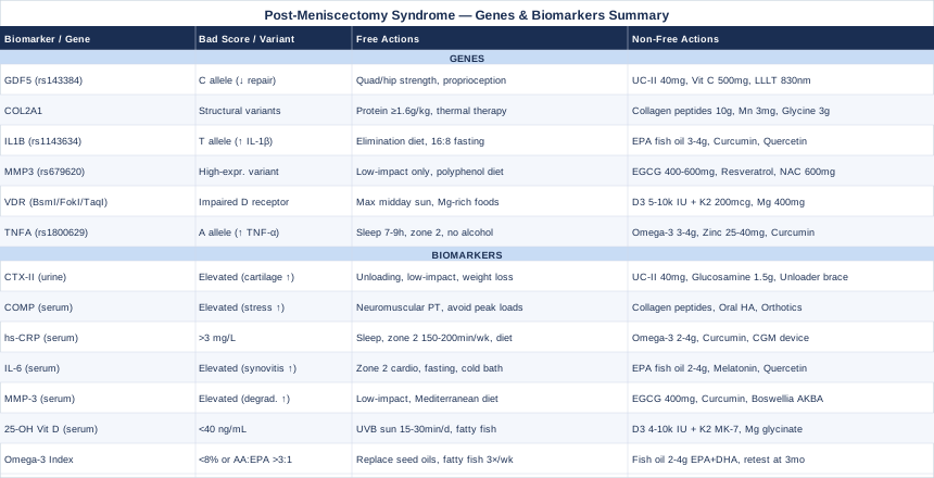
Two evidence-informed angles are covered here. First, seven key biomarkers that reveal the real state of your cartilage and inflammatory environment — with specific, actionable guidance for each one that comes back unfavorable. Second, six genes with well-established links to joint health and osteoarthritis risk, with practical protocols for compensating when a variant works against you. These two lenses complement each other: biomarkers tell you what is happening right now; genetics explain why and guide the interventions most likely to work for your specific biology. Together, they offer something that most post-meniscectomy care does not — a meaningful map.
7 Biomarkers That Reveal What's Really Happening in Your Knee
Biomarkers do not replace clinical assessment, but for post-meniscectomy syndrome they offer something imaging often cannot: a window into the biological activity happening right now. Cartilage breakdown, inflammatory cascades, and poor tissue repair can all be tracked through blood and urine tests that are increasingly accessible, affordable, and clinically meaningful. Here are the seven most informative ones.
1. CTX-II — The Gold Standard for Cartilage Degradation
What It Measures and Why It Matters
CTX-II (urinary C-terminal telopeptide of type II collagen) is a direct marker of type II collagen breakdown — the main structural protein in articular cartilage. After meniscectomy, the remaining cartilage takes on altered mechanical loads. If CTX-II is elevated in urine, it signals active cartilage degradation, often before pain worsens or X-rays show structural change. Multiple human studies have shown that high urinary CTX-II levels predict progression to radiographic osteoarthritis, making it one of the most actionable early-warning signals available for this population.
How to Measure It
CTX-II is measured via a urine sample — typically first-morning urine, which is most concentrated. It is available through functional medicine providers and direct-to-lab services; it is not yet routine in most GP offices. Cost ranges from approximately $80–$200 depending on provider and country. Reference ranges vary by lab, but values consistently in the upper quartile for age and sex are worth taking seriously, particularly when combined with clinical symptoms.
If the Score Is High: Plan Without Supplements
The most powerful lifestyle intervention for reducing CTX-II is unloading combined with low-impact conditioning. Swimming, cycling, and aquatic therapy reduce joint compressive forces while maintaining muscle mass — an important protective factor. Weight loss has a dose-response effect on CTX-II in people with elevated BMI; even 5–10% body weight reduction measurably reduces cartilage load. Proprioceptive neuromuscular training and targeted quadriceps strengthening reduce the abnormal joint mechanics that accelerate breakdown. Avoid prolonged standing on hard surfaces.
If the Score Is High: Plan With Supplements or Equipment
Undenatured type II collagen (UC-II) has been shown in human trials to reduce cartilage degradation markers, including CTX-II. A typical dose is 40 mg/day — this is not the same as hydrolyzed collagen peptides and works through a different mechanism (oral tolerance). Vitamin C (500–1000 mg/day) is essential for collagen synthesis and cofactors collagenase inhibition. Glucosamine sulfate (1500 mg/day, divided dose) has the strongest evidence among common joint supplements for slowing OA progression in some populations. For equipment, unloader knee braces shift load away from the most damaged compartment and can meaningfully reduce mechanical stress on the degrading area. Cycle these interventions in 12-week blocks with repeat CTX-II testing to determine what is working.
Research on CTX-II and cartilage degradation on PubMed
2. COMP — Cartilage Stress and Structural Damage
What It Measures and Why It Matters
COMP (Cartilage Oligomeric Matrix Protein) is released into the bloodstream when cartilage tissue is mechanically stressed or structurally degrading. After meniscectomy, altered load distribution puts greater stress on cartilage surfaces that were previously protected by intact meniscal tissue. Elevated serum COMP reflects both acute mechanical loading and ongoing structural damage. Human studies have found that COMP levels post-meniscectomy correlate with the rate of subsequent joint space narrowing and predict who develops symptomatic knee OA within five years. Unlike CTX-II, which measures fragments of degraded collagen, COMP reflects the matrix protein structure under active stress — making these two markers complementary rather than redundant.
How to Measure It
COMP is a serum (blood) test available through specialty labs and academic medical centers. Cost ranges from approximately $100–$250. Because COMP fluctuates with activity — levels rise after exercise and fall with rest — testing should be done after at least 12 hours of rest. Morning testing before any physical activity is the standard protocol for meaningful comparison between readings.
If the Score Is High: Plan Without Supplements
Structured physical therapy targeting neuromuscular control is the most evidence-backed intervention for reducing inappropriate joint loading — which is the primary driver of elevated COMP post-meniscectomy. Gait retraining, learned with a physical therapist or using biofeedback insoles, can directly alter how force is distributed across the knee during walking. Avoiding activities with high peak knee loads — running on hard surfaces, weighted squats through deep range of motion — during periods of elevated COMP is a sensible precaution. Cold water immersion after training sessions has shown modest evidence for reducing acute inflammatory markers.
If the Score Is High: Plan With Supplements or Equipment
Collagen peptides (10–15 g/day, taken 30–60 minutes before exercise with vitamin C) have shown signal in human trials for supporting cartilage matrix turnover. Oral hyaluronic acid (80–200 mg/day of high-molecular-weight HA) has a growing evidence base for improving joint lubrication and reducing cartilage stress — though effect sizes are modest. When oral options are insufficient, intra-articular hyaluronic acid injections administered by an orthopedic specialist are a more direct option, though evidence for injections is mixed and should be discussed with your clinician. For equipment, custom orthotics can reduce knee adduction moment and measurably shift compressive load — especially relevant for medial compartment involvement post-meniscectomy.
Research on COMP and knee cartilage on PubMed
3. High-Sensitivity CRP — The Systemic Inflammation Gauge
What It Measures and Why It Matters
High-sensitivity C-reactive protein (hs-CRP) is the most accessible and widely used marker of systemic low-grade inflammation. In the context of post-meniscectomy syndrome, chronic systemic inflammation is both a driver and a consequence of joint deterioration. Elevated hs-CRP is associated with faster OA progression, increased pain intensity, and worse outcomes after knee surgery in multiple observational cohorts. Critically, hs-CRP is modifiable — making it one of the highest-leverage biomarkers to track and address. It can also be elevated for reasons unrelated to the knee (metabolic dysfunction, poor sleep, visceral adiposity), meaning that treating elevated hs-CRP produces benefits for the whole body, not just the joint.
How to Measure It
hs-CRP is a standard blood test available at virtually any lab worldwide. Cost ranges from $10–$50. Most GPs will order it. Target: below 1.0 mg/L is ideal; 1.0–3.0 mg/L is intermediate; above 3.0 mg/L warrants investigation. Values above 10 mg/L usually indicate acute infection or injury and should be interpreted separately from chronic inflammation baseline.
If the Score Is High: Plan Without Supplements
Sleep quality is the single most underrated lever for hs-CRP. Seven to nine hours of quality sleep has been shown to reduce inflammatory markers significantly — and the effect is dose-dependent: one week of poor sleep can double hs-CRP. Visceral fat reduction through caloric restriction and aerobic exercise (zone 2 training, 150–200 minutes per week) produces consistent reductions in hs-CRP over 12–16 weeks. An anti-inflammatory dietary pattern — high in colorful vegetables, legumes, whole grains, and olive oil; low in refined carbohydrates and seed oils — is as effective as many supplement protocols for chronic low-grade inflammation.
If the Score Is High: Plan With Supplements or Equipment
Omega-3 fatty acids (EPA + DHA) at doses of 2–4 g/day are among the most consistently effective supplements for reducing hs-CRP, supported by multiple meta-analyses. Curcumin with piperine (500–1000 mg/day of bioavailable formulations such as theracurmin or meriva) has demonstrated hs-CRP reduction in randomized trials for inflammatory conditions. Resveratrol (150–500 mg/day) shows modest benefit in human studies for inflammatory markers. For equipment, continuous glucose monitors (CGM) — available over-the-counter in many countries — help identify blood sugar spikes that drive inflammatory cascades, making dietary adjustments far more precise. Run 12-week cycles with repeat hs-CRP testing to track progress objectively.
Research on hs-CRP and joint inflammation on PubMed
4. Interleukin-6 — The Local and Systemic Inflammatory Messenger
What It Measures and Why It Matters
IL-6 is a cytokine with a dual role: at low levels it functions as an anti-inflammatory myokine released during exercise; at chronically elevated levels it drives synovial inflammation, cartilage catabolism, and pain sensitization. Post-meniscectomy, the synovial membrane often becomes persistently inflamed, producing IL-6 locally within the joint. Systemic serum IL-6 elevation reflects the severity of this ongoing intra-articular inflammation. High IL-6 is also associated with increased pain sensitivity through central sensitization — which may explain why some post-meniscectomy patients experience pain disproportionate to what their imaging shows.
How to Measure It
Serum IL-6 is available at most reference labs, though it is less commonly ordered than hs-CRP. Cost ranges from $40–$120. Morning testing is preferred as IL-6 has a slight diurnal rhythm and is lower in the early morning. For greater precision, some functional medicine practitioners test both serum and synovial fluid IL-6, though joint fluid sampling is invasive and typically reserved for clinical decision-making rather than routine monitoring.
If the Score Is High: Plan Without Supplements
Regular aerobic exercise — particularly zone 2 cardio at a conversational pace — is one of the most potent interventions for normalizing IL-6 over time. Paradoxically, exercise transiently raises IL-6 but produces a sustained anti-inflammatory effect when done consistently over weeks to months. Time-restricted eating (16:8 window) and moderate caloric restriction also reduce circulating IL-6 in multiple human studies. Cold water immersion (10–15 minutes at 10–15°C) has shown signal for acute IL-6 reduction, though sustained effects in OA specifically are less established.
If the Score Is High: Plan With Supplements or Equipment
EPA-dominant fish oil at 2–4 g/day EPA+DHA has direct evidence for IL-6 reduction in human trials and is generally the first supplement to add. Melatonin (0.5–3 mg at bedtime) is increasingly recognized for anti-inflammatory properties and has shown IL-6 suppression in some trials — particularly relevant given its role in sleep quality. Quercetin (500–1000 mg/day with bromelain for enhanced absorption) has a growing evidence base as an IL-6 modulator through inflammasome pathway inhibition. For equipment, red light therapy panels (photobiomodulation) at 630–850 nm applied directly to the knee have shown cytokine-reducing effects in human trials for joint conditions. Typical protocol: 10–20 minutes daily, at least 5 days per week for 8 weeks.
Research on IL-6 and knee inflammation on PubMed
5. MMP-3 — The Cartilage-Dissolving Enzyme
What It Measures and Why It Matters
MMP-3 (matrix metalloproteinase-3, also called stromelysin-1) is an enzyme that degrades collagen, proteoglycans, and other extracellular matrix components in both cartilage and the synovial lining. It is upregulated in osteoarthritic joints and is a direct mediator of cartilage breakdown. After meniscectomy, abnormal joint mechanics and low-grade synovitis both independently drive MMP-3 elevation. Serum MMP-3 is a useful tracking marker because it integrates both mechanical and inflammatory contributions to cartilage breakdown in a single number. Elevated MMP-3 also predicts poor response to conservative management — making it practically useful for deciding when more aggressive intervention is warranted.
How to Measure It
Serum MMP-3 is available through specialty and reference labs. Cost ranges from $60–$150. Some functional medicine practitioners include it in comprehensive joint health panels. Repeat measurement every 3–6 months provides a meaningful trend line. Normal serum ranges vary by lab, but chronically elevated values — especially when accompanied by elevated CTX-II — indicate active matrix degradation that is outpacing repair.
If the Score Is High: Plan Without Supplements
Reducing joint mechanical stress is the primary target. Low-impact exercise modalities (cycling, swimming, water aerobics) maintain metabolic and muscular health without the peak loading forces that upregulate MMP-3 in synoviocytes. A Mediterranean-style dietary pattern — rich in polyphenols, fiber, and monounsaturated fats — has shown signal for reducing matrix metalloproteinase expression in joint tissue. Avoiding prolonged standing on hard surfaces and using appropriately cushioned footwear with good energy return reduces daily cumulative joint stress.
If the Score Is High: Plan With Supplements or Equipment
EGCG (green tea extract) at 400–800 mg/day has shown inhibitory effects on MMP-3 and MMP-13 in human chondrocytes and in some clinical studies for joint health. Curcumin (as a bioavailable form such as theracurmin or meriva, 500–1000 mg/day) directly inhibits NF-κB, the transcription factor that drives MMP-3 production. Boswellia serrata extract (AKBA fraction) at 100–200 mg/day is one of the most clinically studied anti-inflammatory supplements for OA and has shown MMP-downregulating effects in several human trials. Run a 12-week protocol before reassessing serum MMP-3. At doses above 800 mg/day, EGCG may stress the liver in susceptible individuals — take with food and monitor if using long-term.
Research on MMP-3 and joint cartilage on PubMed
6. 25-OH Vitamin D — The Underrated Regulator of Joint and Bone Health
What It Measures and Why It Matters
Vitamin D deficiency affects an estimated 40–70% of the population depending on latitude and lifestyle — and it has direct relevance to post-meniscectomy syndrome. Vitamin D receptors (VDR) are expressed in chondrocytes, synoviocytes, and immune cells throughout the joint. Deficiency impairs cartilage metabolism, promotes synovial inflammation, reduces bone density of the subchondral bone that supports the articular surface, and blunts muscle function — all of which matter acutely after meniscectomy. Multiple observational studies have linked low 25-OH vitamin D to faster OA progression and greater pain severity. Correcting deficiency is low-cost, low-risk, and directionally consistent in its benefits.
How to Measure It
25-OH vitamin D is a standard blood test available at virtually all labs worldwide. Cost ranges from $20–$60. Target for musculoskeletal health: above 40 ng/mL (100 nmol/L) at minimum; functional medicine practitioners including Peter Attia often recommend 60 ng/mL as the preferred floor for people with joint disease or inflammatory conditions. Below 20 ng/mL is frank deficiency; 20–40 ng/mL is suboptimal in this context.
If the Score Is Low: Plan Without Supplements
UVB sun exposure — approximately 15–30 minutes of midday sun on large skin areas (arms, legs, torso) — is the most natural and effective way to restore vitamin D levels. This works well in spring and summer at latitudes below 40°N but is typically insufficient at higher latitudes in winter. UVB narrowband lamps (311 nm) are a practical year-round alternative. Dietary sources (fatty fish, egg yolks, liver) contribute modestly but rarely correct frank deficiency on their own; they serve best as maintenance support on top of adequate sun or supplementation.
If the Score Is Low: Plan With Supplements or Equipment
Vitamin D3 (cholecalciferol) is the preferred form. For frank deficiency (below 20 ng/mL), a loading period of 4000–10,000 IU/day for 8–12 weeks followed by a maintenance dose of 2000–4000 IU/day is typical. Always pair D3 with Vitamin K2 (MK-7 form, 100–200 mcg/day) to ensure calcium is directed to bones rather than soft tissues — a pairing with a strong physiological rationale and wide clinical endorsement. Magnesium glycinate or malate (200–400 mg/day) is a necessary cofactor for vitamin D conversion and is itself frequently deficient. Retest 25-OH vitamin D after 12 weeks to adjust dose; toxicity is rare at these levels when K2 is included.
Research on Vitamin D and joint health on PubMed
7. Omega-3 Index and AA:EPA Ratio — The Inflammatory Fuel Gauge
What It Measures and Why It Matters
The Omega-3 Index measures the percentage of EPA and DHA in red blood cell membranes — a reliable reflection of chronic omega-3 status, unlike plasma levels that fluctuate with recent meals. The AA:EPA ratio (arachidonic acid to eicosapentaenoic acid) adds precision by showing the balance between pro-inflammatory (AA) and anti-inflammatory (EPA) signaling molecules. In post-meniscectomy syndrome, a high AA:EPA ratio means the body has more biological fuel for inflammatory prostaglandins and leukotrienes — the exact mediators that drive synovial inflammation and cartilage catabolism. An unfavorable ratio is ubiquitous in Western diets and is one of the most modifiable root causes of chronic joint inflammation.
How to Measure It
The Omega-3 Index is tested through specialized labs (OmegaQuant is a widely used reference service; home finger-prick tests are available in many countries). Cost ranges from $50–$100. Target Omega-3 Index: above 8% is optimal; 4–8% is intermediate; below 4% is associated with significant cardiovascular and inflammatory risk. Target AA:EPA ratio: below 3:1 is favorable; above 10:1 is commonly found in those eating a typical Western diet and is associated with elevated inflammatory burden across tissues.
If the Score Is Unfavorable: Plan Without Supplements
Dietary fat remodeling is the most direct intervention. Replace seed oils (sunflower, safflower, soybean, corn) — which are extremely high in omega-6 linoleic acid (LA), the metabolic precursor to arachidonic acid — with extra virgin olive oil and avocado oil for cooking and dressing. Increase fatty fish intake to at least three servings per week (sardines, mackerel, wild salmon, and anchovies are the highest in EPA+DHA per calorie and per dollar). This dietary shift alone, maintained consistently, can meaningfully move the AA:EPA ratio over 3–6 months.
If the Score Is Unfavorable: Plan With Supplements or Equipment
High-quality fish oil at 2–4 g/day of combined EPA+DHA is the primary intervention. For faster ratio correction, EPA-dominant formulas are more effective at directly reducing AA-derived inflammatory signaling. Algae-based DHA+EPA is a viable alternative for those avoiding fish products. Cycling protocol: run 3 months of supplementation, then retest the Omega-3 Index to quantify the shift. Side effects at these doses are generally limited to gastrointestinal tolerance (taking with meals resolves this in most people) and a slight increase in bleeding time that is relevant only if you are on anticoagulant medications. The investment in a repeat test is worth it — response to fish oil supplementation varies widely between individuals.
Research on Omega-3 Index and joint inflammation on PubMed
The biomarker picture above gives you a real-time view of your joint's inflammatory and structural state. Moving one level deeper — into the genetic layer — explains why some of those biomarkers tend to run high regardless of lifestyle, and which interventions are most precisely targeted for your individual biology.
6 Genes That Shape Your Susceptibility to Post-Meniscectomy Complications
Understanding your genetic profile does not mean you are locked into a predetermined outcome. Genes set tendencies, not outcomes. But knowing which variants you carry can explain why standard protocols have underperformed, guide supplementation that is genuinely appropriate for your biology, and help you prioritize interventions that compensate for your specific weak points. Consumer genetic testing platforms (23andMe, AncestryDNA) provide raw data interpretable through third-party tools such as Genetic Lifehacks, SelfDecode, or FoundMyFitness. Whole genome sequencing services are also dropping in price and provide greater detail. Here are six genes with meaningful, primarily human-level evidence for joint health and post-surgical recovery.
1. GDF5 (rs143384) — The Joint Architecture Gene
GDF5 (Growth Differentiation Factor 5) encodes a signaling protein critical for joint formation during development and for maintaining cartilage integrity in adults. The rs143384 T>C variant is one of the most replicated findings in osteoarthritis genetics — the C allele reduces GDF5 expression, impairing the joint's capacity for tissue repair after injury. This is particularly relevant post-meniscectomy, where the remaining cartilage's repair capacity becomes the primary determinant of long-term outcome. Large genome-wide association studies across multiple ethnic cohorts have confirmed that this variant increases OA risk across multiple joints, not just the knee.
If the Gene Is Unfavorable: Plan Without Supplements
Progressive resistance training with specific attention to the quadriceps and hip abductors is the most important non-pharmacological tool — muscle acts as a dynamic shock absorber, reducing peak cartilage loads that a GDF5-impaired joint cannot easily repair. Proprioceptive training (balance boards, single-leg exercises, perturbation training under physical therapist guidance) reduces the abnormal shear forces across joint surfaces. Starting this type of training before pain becomes severe — ideally within the first 6 months post-surgery — produces meaningfully better long-term outcomes than beginning after functional decline has set in.
If the Gene Is Unfavorable: Plan With Supplements or Equipment
Undenatured type II collagen (40 mg/day) may support oral tolerance mechanisms that reduce autoimmune-adjacent cartilage destruction and support the residual repair capacity that GDF5 normally facilitates. Vitamin C (500–1000 mg/day) is an essential cofactor for collagen crosslinking and synthesis, compensating in part for the reduced GDF5 signaling that normally supports this process. Silicon as choline-stabilized orthosilicic acid (5–10 mg elemental silicon/day) supports collagen matrix formation at the tissue level. For equipment, low-level laser therapy (LLLT) at 830 nm has shown signal for stimulating chondrocyte proliferation and matrix synthesis in human studies — potentially compensating in part for reduced growth factor signaling. Protocol: 3–5 sessions per week for 8 weeks, 10–20 minutes per session.
Research on GDF5 and osteoarthritis on PubMed
2. COL2A1 — The Structural Collagen Blueprint
COL2A1 encodes the alpha-1 chain of type II collagen — the primary structural protein that gives articular cartilage its tensile strength and resilience. Variants in COL2A1, ranging from rare pathogenic mutations (associated with type II collagenopathies) to more common functional polymorphisms, affect collagen fiber architecture, crosslinking efficiency, and susceptibility to enzymatic degradation. Individuals with less robust COL2A1 expression tend to have cartilage that is more vulnerable to the altered biomechanics that follow meniscectomy. This gene is particularly relevant in early-onset or familial OA patterns and in cases where cartilage degeneration progresses faster than clinical severity would predict.
If the Gene Is Unfavorable: Plan Without Supplements
Protein intake optimization — ensuring 1.6–2.0 g/kg of body weight per day from high-quality sources — provides the amino acid building blocks most needed for collagen synthesis: glycine, proline, and hydroxyproline. Reducing high-impact activities that mechanically stress cartilage with sub-optimal structural collagen is prudent. Thermal therapy (warm bath, infrared sauna at moderate temperatures) may support collagen remodeling and has shown modest anti-inflammatory benefit in musculoskeletal conditions by improving local tissue perfusion.
If the Gene Is Unfavorable: Plan With Supplements or Equipment
Hydrolyzed collagen peptides (10–15 g/day taken with vitamin C, 30–60 minutes before exercise) provide the amino acid substrate directly; several human trials have shown that specific collagen peptides accumulate in cartilage tissue at measurable concentrations. Manganese (2–5 mg/day) is a cofactor for glycosaminoglycan synthesis — a key structural component of cartilage that works in concert with type II collagen. Glycine supplementation (3–5 g/day, particularly from bone broth or as powder) provides the most abundant amino acid in collagen, which many people consistently under-consume. Avoid excessive supplemental retinol (preformed vitamin A above 5000 IU/day), as it competes with collagen synthesis mechanisms at high doses.
Research on COL2A1 and cartilage on PubMed
3. IL1B (rs1143634) — The Inflammation Amplifier
IL1B encodes interleukin-1 beta, one of the most potent drivers of cartilage destruction and synovial inflammation in the joint. The rs1143634 C>T polymorphism affects IL-1β production levels: individuals carrying the higher-producing variant mount more aggressive inflammatory responses to the same mechanical insult or pathogen load. In post-meniscectomy syndrome, this means a higher inflammatory baseline, more severe pain sensitization, and faster joint deterioration — but it also identifies a clear therapeutic target. Anything that specifically addresses the IL-1β signaling pathway becomes higher-leverage for this genetic profile than for others.
Gary Brecka, the human performance researcher who has brought genetics-based health protocols to a broad audience, has discussed how high-producing IL-1β variants contribute to chronic inflammatory conditions that are often managed symptomatically without addressing the genetic root. His approach emphasizes methylation support, targeted anti-inflammatory dietary strategies, and identifying which inflammatory cytokine is driving the clinical picture — concepts directly applicable here.
If the Gene Is Unfavorable: Plan Without Supplements
An anti-inflammatory elimination diet — removing gluten, dairy, refined sugar, and alcohol for at least 6 weeks — has the strongest dietary evidence for reducing IL-1β activity and overall inflammatory burden. Contrast hydrotherapy (alternating 3–4 minutes of warm water with 30–60 seconds of cold, repeated 4–5 cycles) has shown acute reductions in inflammatory cytokines including IL-1β. Intermittent fasting protocols (16:8 daily eating window or one 24-hour fast per week) consistently reduce circulating IL-1β in human studies with good tolerability.
If the Gene Is Unfavorable: Plan With Supplements or Equipment
EPA-dominant fish oil (3–4 g/day EPA+DHA) directly competes with arachidonic acid metabolism and reduces IL-1β production upstream. Curcumin (as theracurmin or longvida formulation, 400–1000 mg/day) is among the most studied natural IL-1β inhibitors — it suppresses NF-κB signaling, which drives IL-1β gene transcription. Quercetin (500 mg twice daily) inhibits NLRP3 inflammasome activation upstream of IL-1β secretion. Boswellia serrata AKBA fraction (100–250 mg/day) inhibits 5-LOX and has human clinical evidence for joint pain reduction through IL-1β-related pathways. Cycle curcumin in 12-week blocks with 4-week breaks to avoid adaptive tolerance. Monitor liver enzymes annually if using high-dose curcumin long-term; this precaution is especially relevant at doses above 1000 mg/day.
Research on IL1B polymorphism and joint inflammation on PubMed
4. MMP3 (rs679620) — The Matrix-Dissolving Enzyme Gene
MMP3 encodes matrix metalloproteinase-3, the same enzyme discussed in the biomarker section. At the genetic level, the rs679620 variant (linked to the 5A/6A promoter polymorphism studied in joint disease research) affects basal MMP-3 expression in joint tissue. High-expressing variants generate more MMP-3 regardless of mechanical input, meaning cartilage degradation proceeds at a faster rate even under conditions of modest inflammation or mechanical stress. This is a particularly important gene to know about if your serum MMP-3 remains persistently elevated despite comprehensive lifestyle optimization — genetics may be setting a floor that lifestyle alone cannot lower.
If the Gene Is Unfavorable: Plan Without Supplements
Systematic avoidance of high-impact loading becomes more important with this variant — the genetic floor of MMP-3 expression is already elevated, so adding mechanical insult compounds it rapidly. Pool-based exercise, elliptical training, and cycling are the preferred primary modalities. A high-polyphenol dietary pattern (berries, dark leafy greens, green tea, pomegranate) provides natural flavonoid-based MMP inhibition through multiple enzymatic pathways, and is well-tolerated indefinitely.
If the Gene Is Unfavorable: Plan With Supplements or Equipment
EGCG (green tea extract, 400–600 mg/day) is among the most potent natural MMP-3 inhibitors studied in human chondrocytes. Resveratrol (150–500 mg/day) has shown MMP-3 downregulation in synovial tissue samples. N-acetylcysteine (NAC, 600–1200 mg/day) reduces oxidative stress, which is a key upstream trigger for MMP-3 gene transcription through reactive oxygen species signaling. Avoid chronic alcohol use and chronically high-dose NSAIDs, both of which increase gut permeability and systemic inflammatory load that amplifies MMP-3 expression from synoviocytes. Cycle EGCG in 12-week blocks with a 4-week rest period; high-dose green tea extract taken on an empty stomach commonly causes nausea in sensitive individuals — always take with food.
Research on MMP3 gene variant and joint disease on PubMed
5. VDR — The Vitamin D Receptor Gateway
The VDR gene encodes the receptor through which vitamin D exerts its effects inside cells. Even with adequate serum 25-OH vitamin D, certain VDR polymorphisms — including BsmI (rs1544410), FokI (rs2228570), TaqI (rs731236), and ApaI (rs7975232) — can impair cellular responsiveness to vitamin D signaling. In joint health, this manifests as reduced chondrocyte metabolism, greater susceptibility to inflammatory gene activation, and impaired bone quality — without the blood test showing frank deficiency. This is why two people with identical serum vitamin D levels can have dramatically different joint outcomes. VDR variants are clinically relevant not just for bone health but for immune regulation, muscle function, and cartilage metabolism.
If the Gene Is Unfavorable: Plan Without Supplements
People with VDR variants that reduce signaling efficiency need to maximize sun exposure as a non-negotiable foundation — UV-B–rich midday sun exposure on large skin areas provides a form of vitamin D that bypasses some receptor efficiency concerns. Dietary patterns rich in vitamin D co-factors — magnesium (from seeds, legumes, leafy greens), vitamin K2 (from fermented foods such as natto and aged cheeses), and zinc — improve the downstream signaling efficiency even when receptor sensitivity is genetically reduced.
If the Gene Is Unfavorable: Plan With Supplements or Equipment
VDR variants often require higher than standard vitamin D3 doses to achieve adequate cellular signaling. Under appropriate medical supervision, 5000–10,000 IU/day with K2 co-supplementation (200 mcg MK-7/day) is sometimes necessary to drive serum levels to the range where receptor-impaired cells can still respond. Magnesium glycinate or malate (400 mg/day) is critical — without adequate magnesium, the enzymatic conversion of vitamin D to its active form is impaired. UVB narrowband lamps provide a supplement-independent path to generating skin-synthesized vitamin D. Retest 25-OH vitamin D every 3 months; for those with VDR variants and active joint disease, a target of 60–80 ng/mL is often pursued rather than the population-average threshold of 40 ng/mL.
Research on VDR variants and joint health on PubMed
6. TNFA (rs1800629) — The TNF-Alpha Inflammation Switch
TNFA encodes tumor necrosis factor-alpha, a master regulator of joint inflammation. The rs1800629 -308 G>A promoter variant — often called the "A allele" — significantly increases TNF-alpha production in macrophages and synoviocytes. Individuals carrying one or two copies of this allele mount stronger inflammatory responses to joint injury, synovitis, and mechanical insult. Post-meniscectomy, this can manifest as more severe and prolonged synovial inflammation, greater pain burden, and faster structural deterioration. Ali Torkamani, a leading genomics researcher at the Scripps Research Institute, has noted that TNF-alpha promoter variants are among the most clinically actionable findings in the context of chronic inflammatory conditions — particularly because they identify people for whom standard anti-inflammatory approaches are likely to underperform without additional targeting of the TNF pathway.
If the Gene Is Unfavorable: Plan Without Supplements
Sleep quality optimization is among the most effective lifestyle-level TNF-alpha modulators available — even one night of poor sleep can raise TNF-alpha production by 30% or more, and those with the high-producer variant are doubly affected. Moderate caloric restriction (a sustained 10–15% reduction from maintenance calories) reliably reduces TNF-alpha in multiple well-designed human studies. Consistent zone 2 aerobic exercise progressively reduces TNF-alpha production over months through adaptations in immune cell behavior. Eliminating alcohol is particularly important with this variant, as alcohol directly amplifies TNF-alpha production from macrophages through endotoxin translocation from the gut.
If the Gene Is Unfavorable: Plan With Supplements or Equipment
Omega-3 fatty acids (EPA+DHA, 3–4 g/day) specifically inhibit TNF-alpha at the gene expression level, competing with arachidonic acid for cyclooxygenase and lipoxygenase pathways. Zinc (25–40 mg/day with 1–2 mg/day copper to maintain mineral balance) modulates TNF-alpha secretion from macrophages through zinc finger transcription factor mechanisms. Curcumin (phytosome or nanoparticle form, 500–1000 mg/day) directly downregulates NF-κB, the primary transcription factor driving TNFA expression. Resveratrol (250 mg/day) has shown TNF-alpha reduction in human trials for inflammatory conditions. For practical monitoring, use hs-CRP or serum IL-6 as proxy readouts (serum TNF-alpha itself is more expensive and less accessible for routine tracking). Run a 12-week combined protocol before evaluating response via repeat inflammatory marker testing.
Research on TNFA variant and joint inflammation on PubMed
The genetic and biomarker frameworks above provide the what and why of your joint's biological state. What they do not always convey is the integrated, whole-system perspective that makes the difference between managing symptoms and genuinely slowing progression. That is where the following framework becomes particularly valuable.
What Peter Attia's Outlive Gets Right About Post-Surgical Joint Recovery
Peter Attia's Outlive: The Science and Art of Longevity (2023) is not a joint health book. It is a book about the biology of aging and how to act on it decades before disease becomes symptomatic. But its framework for inflammation management, metabolic health, structural fitness, and biomarker tracking is directly and practically applicable to post-meniscectomy syndrome. Here are ten of the most impactful ideas from the book applied to this specific context.
1. Zone 2 Training Is the Most Anti-Inflammatory Exercise Protocol Available
Attia draws on the work of exercise physiologists including Iñigo San Millán to argue that sustained aerobic work at a conversational pace — zone 2, approximately 60–70% of maximum heart rate — is the most effective metabolic medicine available without a prescription. For post-meniscectomy patients, choosing low-impact zone 2 modalities (cycling, swimming, elliptical) for 150–200 minutes per week produces measurable reductions in hs-CRP, IL-6, and TNF-alpha over months. The anti-inflammatory benefits accumulate slowly but durably — unlike high-intensity training, which can temporarily worsen joint inflammation in a compromised knee.
2. Muscle Is a Metabolic Organ — Build It Deliberately and Early
Attia makes the case that skeletal muscle mass is the most undervalued longevity and health variable available. For joint health specifically, muscle is the dynamic shock absorber that reduces peak cartilage loading with every step. He advocates for progressive resistance training with deliberate attention to the muscles that stabilize joints — quadriceps, glutes, hip abductors — starting in middle age or, critically, after any joint injury. Post-meniscectomy, a targeted strength program is not optional. It is the primary structural intervention available, and delaying it accelerates deterioration.
3. Protein Intake Is Almost Universally Underestimated
Attia recommends 1.6–2.0 g of protein per kilogram of body weight daily as a minimum for anyone focused on musculoskeletal health. For cartilage and collagen support specifically, the amino acid quality matters: glycine, proline, and hydroxyproline — found in collagen-rich foods and supplements — are the substrates needed for joint tissue repair. Most people eating a standard Western diet fall significantly below this threshold even when they believe they are eating enough protein, which directly limits both muscle protein synthesis and connective tissue repair capacity.
4. Sleep Is the Primary Recovery and Anti-Inflammatory Intervention
Outlive dedicates considerable space to sleep as a biological necessity rather than a lifestyle preference. Attia cites research showing that chronic sleep restriction (under 7 hours) raises IL-6, TNF-alpha, and CRP — the same markers that drive post-meniscectomy syndrome. For anyone carrying the TNFA or IL1B high-producer variants discussed above, sleep deprivation is particularly damaging. Prioritizing sleep architecture — dark, cool room; consistent timing; eliminating alcohol within 3 hours of bed — is one of the highest-leverage changes in this entire framework.
5. Blood Glucose Control Matters More Than Most Joint Patients Are Told
Attia devotes significant attention to metabolic health and glycemic variability, arguing that blood sugar fluctuations — even in people without diabetes — drive chronic inflammation, impair cellular repair, and accelerate biological aging. For joint health specifically, elevated blood glucose promotes glycation of cartilage proteins (making them stiffer and more prone to degradation) and amplifies the inflammatory cytokine signaling that damages the synovium. A CGM worn for 2–4 weeks can reveal specific dietary patterns generating inflammatory glucose spikes that a blood test alone would miss.
6. The Centenarian Decathlon Framework for Planning Joint-Friendly Activity
One of Attia's most practical frameworks is the "centenarian decathlon" — identifying the physical activities you want to perform at 100 years old and reverse-engineering the fitness required today to get there. Applied to post-meniscectomy syndrome, this reframes the goal from "manage pain" to "preserve the function I care most about." It pushes toward joint-friendly training for hiking, cycling, or playing with future grandchildren — rather than defaulting to whatever the generic rehab protocol prescribes.
7. Visceral Fat Is an Active Endocrine Organ of Inflammation
Attia emphasizes that visceral adipose tissue is not inert — it actively secretes inflammatory cytokines including IL-6 and TNF-alpha at a rate proportional to its volume. For someone with post-meniscectomy syndrome, reducing visceral fat is a direct joint treatment, not just a general health measure. A combination of zone 2 cardio, time-restricted eating, and protein-first dietary prioritization is the most evidence-consistent approach for this goal in the longevity literature.
8. Acting on Biomarkers Before Symptoms Force the Conversation
A core thesis of Outlive is that acting on biomarkers years — or decades — before disease becomes clinically apparent is what separates good long-term outcomes from poor ones. For post-meniscectomy patients, this means not waiting for severe pain or radiographic OA progression to begin tracking CTX-II, COMP, or hs-CRP. The window for meaningful intervention is open widest in the months immediately after surgery and the first few years of post-meniscectomy life. This is when biomarker trends, not symptoms, should guide decision-making.
9. Stability Comes Before Strength — and Most People Get This Backwards
Attia distinguishes carefully between strength (maximum force production) and stability (the ability to produce force without compensatory joint shear). He argues that stability deficits — extremely common after knee surgery — are the root cause of most musculoskeletal injuries and accelerated joint degeneration. Working with a qualified physical therapist or movement specialist to restore joint stability before loading it with progressive resistance training is the correct sequence. Loading an unstable joint, however well-intentioned, compounds damage rather than reversing it.
10. Emotional Health Is a Measurable Physical Variable
Attia discusses at length the evidence that psychological stress, depression, and anxiety elevate inflammatory markers and impair physical recovery through concrete biological mechanisms — HPA axis activation, elevated cortisol, suppressed immune resolution, impaired sleep architecture. For post-meniscectomy syndrome, where chronic pain commonly leads to depression and anxiety, this creates a self-reinforcing cycle. Addressing emotional health through evidence-based therapy, structured social connection, or a formal stress-reduction protocol should be built into the recovery plan as a biological intervention, not separated from it as a psychological one.
Complementary Approaches Worth Considering
The following modalities each have meaningful clinical evidence for pain, joint function, or inflammatory reduction relevant to post-meniscectomy syndrome. None replace the biomarker-informed protocols above, but each offers an additional lever with a favorable risk-to-benefit profile.
Low-Level Laser Therapy (Photobiomodulation)
Photobiomodulation (PBM) at near-infrared wavelengths (808–850 nm) penetrates joint tissue to stimulate mitochondrial cytochrome c oxidase, accelerating ATP production and reducing reactive oxygen species in chondrocytes and synoviocytes. For post-meniscectomy syndrome — where both cartilage metabolism and synovial inflammation are compromised — PBM offers a non-invasive way to support cellular energy production and modulate inflammatory cytokines locally, without the mechanical loads that can worsen joint state during a flare.
A 2015 systematic review of randomized controlled trials on LLLT for knee osteoarthritis found significant reductions in pain and morning stiffness with improvements in function lasting up to 12 weeks post-treatment (relevant RCTs and reviews on PubMed). Effect sizes are modest to moderate but consistent across trials using appropriate wavelengths and energy doses.
For practical application, home-use PBM devices — panels or targeted knee wraps — are now commercially available. Typical protocol: 830 nm wavelength, 10–20 minutes per session directly over the knee, 5–7 days per week for 8 weeks. Power density of 50–100 mW/cm² is standard for home units. Cost for a quality device ranges from $200–$600. No significant side effects at these parameters; avoid direct eye exposure.
Tai Chi
Tai chi is a movement practice combining slow, controlled motion, balance training, and breath coordination. For post-meniscectomy syndrome, it is uniquely suited because it improves neuromuscular control, proprioception, and periarticular muscle strength without generating the high peak loads of most exercise modalities. The mindful, slow-movement format also engages parasympathetic pathways that reduce cortisol and the inflammatory cytokine levels that drive synovial inflammation.
A well-designed RCT published in Annals of Internal Medicine compared tai chi to standard physical therapy for knee osteoarthritis and found comparable benefit for pain reduction with greater improvements in depression and quality of life in the tai chi group — directly relevant given the psychological burden of chronic post-surgical pain (view supporting research on PubMed).
For practical application, begin with a beginner yang-style tai chi class (in-person or online), 3 sessions per week of 30–45 minutes each. Most people with knee limitations can begin with seated modifications and progress gradually to standing forms. A 12-week program is typically needed before functional improvements become measurable. There is no equipment required and very low risk of aggravation when movements are appropriately paced.
Mindfulness-Based Stress Reduction (MBSR)
MBSR is an 8-week structured program combining mindfulness meditation, body scanning, and gentle movement. Its relevance to post-meniscectomy syndrome extends beyond pain management: it directly addresses the central sensitization that occurs when chronic joint pain rewires the nervous system toward amplified pain processing. For patients whose pain seems disproportionate to their imaging findings — a pattern that is genuinely common in post-meniscectomy syndrome — MBSR targets the neurological dimension that biomarker optimization and physical therapy alone cannot reach.
Multiple RCTs and meta-analyses have demonstrated that MBSR produces clinically meaningful reductions in pain intensity and pain catastrophizing in chronic musculoskeletal conditions, with effects that persist at 6-month follow-up (relevant research on PubMed). The program does not directly affect cartilage, but it changes how the brain processes and amplifies pain signals — which meaningfully changes the lived experience of the condition.
The MBSR curriculum is widely available online at low or no cost (the UCSD Center for Mindfulness and various app-based programs offer structured access). Commit to the full 8-week program before evaluating results. Daily practice of 20–45 minutes is the standard protocol; the main cost is consistency, not money.
Massage Therapy
The knee joint is surrounded by muscles — quadriceps, hamstrings, iliotibial band, calf — that become chronically tense and overloaded when compensatory gait patterns develop post-meniscectomy. This periarticular tension increases joint compression, limits range of motion, and contributes to pain independent of intra-articular changes. Myofascial release and trigger point techniques applied to these muscle groups address a component of post-meniscectomy pain that most clinical protocols systematically ignore.
A systematic review of massage therapy for knee OA found significant short-term improvements in pain and function across studies using both full-body and localized protocols (relevant research on PubMed). Evidence is primarily short-term in duration, but the low risk profile makes it a reasonable ongoing adjunct rather than a one-time intervention.
For practical application, targeted massage to the quadriceps, hamstrings, and IT band by a qualified therapist — 2–4 sessions per month — is more effective than general relaxation massage. Between professional sessions, foam rolling and massage guns at low intensity settings can maintain periarticular tissue quality. Avoid direct deep pressure over an actively inflamed joint; wait until flares resolve before resuming full intensity.
Biofeedback
Biofeedback uses real-time physiological monitoring to help patients consciously influence normally automatic processes, including muscle activation patterns, joint loading, and autonomic nervous system tone. Two types are particularly relevant post-meniscectomy: EMG biofeedback for retraining quadriceps activation (atrophy and neural inhibition of the VMO after knee surgery is both common and clinically significant) and HRV biofeedback for reducing autonomic dysregulation that amplifies pain sensitization and inflammatory signaling.
EMG biofeedback for VMO (vastus medialis oblique) activation has a specific evidence base in post-knee surgery rehabilitation, with studies showing faster and more complete quadriceps recovery compared to standard exercise alone (view EMG biofeedback research on PubMed). HRV biofeedback has been studied in chronic pain contexts and shows reductions in pain catastrophizing and stress-response reactivity that are relevant for central sensitization in post-meniscectomy patients.
For practical application, EMG biofeedback can be initiated with a physical therapist in a clinical setting; wearable EMG devices for home use are increasingly available. HRV biofeedback is accessible with consumer devices (Polar H10 sensor paired with HRV4Training or a dedicated device like Inner Balance) practiced for 10–20 minutes daily. Both modalities require consistency — expect 4–8 weeks of practice before meaningful neuromuscular or autonomic improvements become apparent.
Moving Forward
Post-meniscectomy syndrome is not a fixed destination. The cartilage that remains after surgery operates in a biological environment you can measurably influence — one defined by your inflammatory profile, your genetic tendencies, your metabolic state, and the mechanical loads you subject your joint to every day. What the biomarker and genetic frameworks in this article provide is precision: instead of guessing which interventions to try, you can identify which systems are under the most stress and act accordingly.
The most practical starting point is also the most accessible one. A basic panel including hs-CRP, 25-OH vitamin D, and an Omega-3 Index can be obtained affordably in most countries, often without a specialist referral. Data from just these three tests can drive meaningful, well-targeted changes in diet, supplementation, and lifestyle. Add CTX-II and COMP when you want a direct view into cartilage dynamics. Pursue genetic testing if your response to standard protocols has been poor or if your joint is deteriorating faster than your clinical severity would predict.
The next smart step is a simple one: choose one biomarker to measure this month, and one evidence-backed change to make based on what you find. Every data point you gather shifts you from generic protocol to personalized strategy — and that transition is where outcomes actually improve. If anything in this article raises questions or concerns, bring it to a physician, rheumatologist, sports medicine specialist, or functional medicine practitioner who can contextualize the results within your full medical history. Better information, pursued carefully, leads to better decisions.
Musculoskeletal: Joint Conditions Muscle Conditions Tendon & Ligament Conditions Sports Injuries
Autoimmune: Inflammatory Conditions Connective Tissue Conditions