This article contains AI generated content.
· UpdatedPost-Radiation Arthritis — 5 Genes And 7 Biomarkers To Track
Introduction
If you have gone through radiation therapy for cancer and now find yourself dealing with joint stiffness, aching, or a steadily shrinking range of motion, you are not imagining things and you are not alone. Post-radiation arthritis is a well-documented but underrecognized consequence of treatment, affecting joints within or near the treated field — the shoulder after breast cancer radiation, the hip after pelvic therapy, the jaw and cervical spine after head and neck treatment, or the knee and ankle after extremity radiation. What makes this condition particularly difficult is that symptoms often emerge or worsen months to years after treatment ends, precisely when most follow-up care is shifting its focus elsewhere.
The standard advice offered to people in this situation tends to be identical to what anyone with arthritis hears: try anti-inflammatories, do some gentle movement, apply heat or ice, be patient. That is not wrong, but it is incomplete to the point of being frustrating. Post-radiation arthritis is driven by a specific set of biological processes — radiation-induced fibrosis, oxidative damage from reactive oxygen species, persistent inflammatory cytokine signaling, and joint tissue hypoxia from microvascular injury. These are not the same mechanisms as in osteoarthritis from aging or rheumatoid arthritis from immune dysregulation, even when the surface symptoms look similar. Treatments and interventions calibrated to those other conditions will not address the specific biology at work here.
What changes the picture is precision: knowing which biological processes are most active in your body, and knowing your genetic susceptibility to those processes in the first place. Both pieces of information are now increasingly accessible. A targeted panel of seven biomarkers can show you how much inflammation, oxidative stress, tissue fibrosis, and cartilage degradation is currently present and measurable in your blood and urine. And a consumer genetic test, interpreted through the right lens, can reveal five key variants that explain why some people develop severe post-radiation joint complications while others with similar treatment exposure do not.
This article covers both of those directions in practical detail, along with insights from Peter Attia's Outlive framework and a set of complementary approaches with real clinical evidence for radiation-affected joints. The goal is not to replace your oncology follow-up or your rheumatologist, but to give you a clearer map of what is happening biologically and which levers are actually worth pulling. Better information consistently leads to better decisions — and for a condition as underserved as post-radiation arthritis, that matters more than usual.
Summary
Post-radiation arthritis is driven by radiation-induced fibrosis, oxidative stress, and inflammatory cytokine activity — not by the same mechanisms as typical aging arthritis. Tracking 7 targeted biomarkers (TGF-β1, hsCRP, IL-6, 8-OHdG, CTX-II/COMP, 25-OH Vitamin D, and ferritin) can reveal exactly which processes are most active and where to focus your efforts. 5 genetic variants in genes including TNF-α, TGFB1, SOD2, GSTP1, and IL-6 explain why radiation joint damage affects some patients far more severely than others — and each one has actionable compensation strategies, with and without supplements. Beyond the lab work, this article covers what Peter Attia's precision medicine framework in Outlive recommends for managing post-treatment biology, and which complementary modalities — low-level laser therapy, tai chi, microbiome support, mindfulness-based interventions, and Chinese herbal medicine — carry meaningful human evidence for this type of joint damage. More clarity means more targeted action.
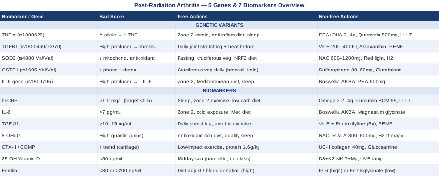
7 Biomarkers That Reveal What Is Happening in Your Joints After Radiation
Understanding your biomarker profile converts post-radiation arthritis management from guesswork into targeted action. The seven markers below reflect the core biological processes that drive joint damage after radiation: systemic and local inflammation, fibrosis signaling, oxidative DNA damage, direct cartilage breakdown, and nutritional deficiencies that compound everything else. Each one is independently measurable, independently actionable, and together they give you a biological picture that no single standard follow-up test can provide.
Biomarker 1: hsCRP (High-Sensitivity C-Reactive Protein)
Why it matters and what it reveals
High-sensitivity C-reactive protein is the most broadly used clinical marker of systemic inflammation. After radiation therapy, the inflammatory cascade triggered in treated tissues can persist long after the treatment itself ends. Elevated hsCRP reflects ongoing inflammatory signaling that accelerates joint tissue breakdown and impairs repair. In post-radiation arthritis specifically, hsCRP functions as a general warning light: if it remains elevated months or years after treatment, the inflammatory phase has not resolved and joint tissues remain under active biological stress.
Peter Attia consistently identifies hsCRP as one of the most underutilized routine blood tests, noting that values above 1.0 mg/L warrant investigation even in people who feel broadly well. For post-radiation joints, the standard is worth applying even more rigorously.
How to measure it
hsCRP is a standard blood draw, fasting preferred but not strictly required. Cost ranges from $15–$45 out of pocket; it is included in many cardiovascular risk panels and often covered by insurance. Optimal target: below 0.5 mg/L. Above 1.0 mg/L warrants investigation; above 3.0 mg/L indicates significant active inflammation requiring immediate attention.
If the score is bad, the plan without supplements
The most powerful non-supplement interventions for chronically elevated hsCRP in this context are: eliminating refined carbohydrates and seed oils from the diet, achieving consistent 7–8 hours of quality sleep, performing regular moderate aerobic exercise (150+ minutes per week at zone 2 intensity), and reducing visceral fat if applicable. For post-radiation joints specifically, gentle range-of-motion exercise in the affected joint performed consistently (5–6 days per week, 15–20 minutes per session) reduces local inflammatory signaling over time. Time-restricted eating in a 16:8 window has shown meaningful CRP reductions across multiple clinical trials.
If the score is bad, the plan with supplements or equipment
Omega-3 fatty acids (EPA+DHA): 2–4g daily of high-purity fish oil. EPA directly competes with arachidonic acid in inflammatory eicosanoid synthesis. Cycle: continuous use; reassess at 90 days. Side effects: mild GI upset; use enteric-coated form if needed; blood-thinning effect at high doses warrants caution with anticoagulants.
Curcumin (BCM-95 or Meriva formulation): 500–1000mg daily. Standard curcumin has poor bioavailability; phospholipid or enhanced-absorption formulations are required. Cycle: continuous, reassess at 12 weeks. Side effects: rare GI effects; caution with anticoagulants at high doses.
Low-level laser therapy (LLLT) device: Joint-targeted red and near-infrared light at 660–850nm, 5–10 minutes per session over the affected joint, 3–5 times per week. Reduces local inflammatory mediator production with a good safety profile.
Biomarker 2: IL-6 (Interleukin-6)
Why it matters and what it reveals
Interleukin-6 is a pro-inflammatory cytokine that sits upstream of CRP in the inflammatory cascade — it is one of the actual drivers of the inflammatory response, not just a reflection of it. In post-radiation arthritis, IL-6 is released by damaged synovial fibroblasts and injured endothelial cells within the joint capsule, and it promotes further tissue destruction while simultaneously driving fibrosis. Elevated IL-6 also produces the systemic symptoms that many radiation survivors know well: persistent fatigue, cognitive fog, and a general sense of feeling unwell that persists long after treatment ends.
Research consistently associates elevated IL-6 with worse outcomes in radiation-treated tissue, including fibrosis severity and late-onset joint stiffness. PubMed: IL-6 and radiation-induced tissue damage
How to measure it
IL-6 is measured via serum blood test. It is less routinely ordered than CRP and may require a specific request or specialty lab. Cost: $30–$80 out of pocket. Best measured fasting and in the morning to minimize circadian variation. Reference ranges vary by lab; values above 7 pg/mL are generally considered elevated in most clinical contexts.
If the score is bad, the plan without supplements
Zone 2 aerobic training — sustained cardio at a conversational pace, 30–45 minutes, 4–5 times per week — is one of the best-validated strategies for lowering chronically elevated IL-6. Brief cold water immersion after exercise (2–3 minutes at approximately 15°C) blunts the post-exercise IL-6 spike and trains the inflammatory response over time. A Mediterranean diet pattern rich in vegetables, extra-virgin olive oil, and fatty fish consistently reduces IL-6 across multiple clinical trials. Adequate sleep quality is non-negotiable: even a single night of significantly disrupted sleep can spike IL-6.
If the score is bad, the plan with supplements or equipment
Boswellia serrata (AKBA-standardized extract): 300–400mg, 3 times per day. Boswellic acids directly inhibit the 5-LOX inflammatory pathway and reduce IL-6 and related mediators. Cycle: 8–12 weeks on, 4 weeks off. Side effects: rare GI effects; generally well-tolerated.
Magnesium glycinate or malate: 300–400mg daily at night. Magnesium deficiency is common in cancer survivors and correlates with elevated IL-6. Side effects: softened stool at higher doses. Use continuously; retest magnesium RBC (not serum) at 90 days.
Full-body or joint-targeted red light panel: 660–850nm, 15–20 minutes per session, 4–5 times per week. Clinical evidence supports IL-6 reduction in musculoskeletal inflammatory conditions through photobiomodulation of inflammatory signaling pathways.
Biomarker 3: TGF-β1 (Transforming Growth Factor Beta-1)
Why it matters and what it reveals
TGF-β1 is arguably the single most important biomarker specifically linked to the mechanism of post-radiation joint damage. Radiation activates TGF-β1 in treated tissues as part of the acute injury response, and in a significant subset of patients, this activation becomes self-sustaining and chronic. TGF-β1 drives fibrosis — the progressive replacement of normal joint tissue with scar-like collagen deposits that stiffen joint capsules, constrict local vasculature, reduce cartilage nutrition, and steadily decrease range of motion. This process is the biological engine behind radiation fibrosis syndrome, and it can worsen for years after treatment ends.
What makes TGF-β1 especially valuable as a monitoring tool is that it is not just a consequence of the damage — it is part of the mechanism. Elevated plasma TGF-β1 measured at 6–12 months post-radiation predicts who will develop clinically significant fibrosis and joint stiffness long-term, giving you a forward-looking signal rather than just a retrospective one. PubMed: TGF-β1 as a predictive biomarker for radiation fibrosis
How to measure it
TGF-β1 is measured in plasma or serum. It requires specific handling protocols (activated vs. latent forms differ based on collection conditions), making it less routine than CRP. Specialty labs and functional medicine practitioners are more likely to order it as part of post-cancer monitoring panels. Cost: $50–$150 out of pocket. Elevated levels in clinical research contexts are generally considered above 10–15 ng/mL, though lab-specific reference ranges apply.
If the score is bad, the plan without supplements
Physical therapy with sustained low-load stretching of the fibrotic joint is the most evidence-supported non-pharmacological approach to active radiation fibrosis: hold each stretch for 30–60 seconds, 5–10 repetitions, daily without exception. This works by applying mechanical counter-forces against forming collagen cross-links before they fully solidify. Starting early (as soon as medically cleared, often 4–6 weeks post-treatment) is significantly more effective than starting late. Applying a heating pad to the joint for 10–15 minutes before stretching improves collagen extensibility and stretch effectiveness. Regular aerobic exercise has also been shown to lower systemic TGF-β1 over time.
If the score is bad, the plan with supplements or equipment
Vitamin E (mixed tocopherols and tocotrienols): 200–400 IU/day. The vitamin E plus pentoxifylline combination is the only intervention with positive randomized trial evidence specifically for radiation fibrosis. Vitamin E alone also shows TGF-β1 modulation. Cycle: continuous, 6-month reassessment. Side effects: at high doses, possible interaction with anticoagulant medications.
Pentoxifylline (400mg, 3 times daily — prescription required): This medication has the strongest clinical evidence for radiation fibrosis specifically when combined with vitamin E. It is not typically prescribed for this indication in standard follow-up but is used in specialized radiation oncology centers. Discuss directly with your radiation oncologist.
PEMF (pulsed electromagnetic field) device: Placed directly over the fibrotic joint, 20–30 minutes daily. PEMF has shown anti-fibrotic effects across several tissue types by reducing TGF-β1-mediated collagen upregulation in treated tissue. Home devices range from $300–$2000.
Biomarker 4: 8-OHdG (8-Hydroxy-2'-deoxyguanosine)
Why it matters and what it reveals
Radiation generates massive quantities of reactive oxygen species (ROS) that oxidatively damage DNA. 8-OHdG is one of the most widely validated markers of oxidative DNA damage in clinical research. When 8-OHdG is elevated, it indicates that the body's antioxidant systems have not matched the oxidative burden placed on them — and the same ROS that damage DNA also directly injure chondrocytes (cartilage cells), synovial cells, and periarticular bone tissue. In post-radiation joints, ongoing oxidative stress is a key driver of progressive cartilage and tissue degradation even when the radiation treatment itself ended years ago.
Measuring 8-OHdG tells you concretely whether the radiation-generated oxidative burden has been resolved or is still active. In patients who develop severe radiation-induced joint complications, 8-OHdG tends to remain elevated well beyond the acute treatment phase. PubMed: 8-OHdG and radiation-induced oxidative damage
How to measure it
8-OHdG is most commonly measured in spot or 24-hour urine samples, or in serum. Urinary 8-OHdG is more widely used in research. Available through specialty and functional medicine labs including DUTCH Test panels and Vibrant Wellness. Cost: $50–$120 out of pocket. Reduction toward lower quartiles is the clinical goal rather than a fixed threshold.
If the score is bad, the plan without supplements
Increasing dietary antioxidant density is the primary tool: dark leafy greens, berries (especially blueberries and blackberries), cruciferous vegetables, and polyphenol-rich foods including extra-virgin olive oil, green tea, and modest amounts of dark chocolate. Reducing pro-oxidant dietary inputs matters equally — refined sugars, linoleic acid-heavy seed oils, and processed meats all amplify oxidative stress. Regular moderate exercise, counterintuitively, reduces chronic oxidative stress over time even though it generates short-term ROS by upregulating endogenous antioxidant enzyme production. Quality sleep in a genuinely dark room optimizes melatonin, which is a potent endogenous antioxidant.
If the score is bad, the plan with supplements or equipment
N-Acetyl Cysteine (NAC): 600–1200mg daily as a glutathione precursor. Among the best-studied antioxidant supplements with a strong safety record. Cycle: 8 weeks on, 2–4 weeks off to avoid blunting exercise-induced antioxidant adaptation. Side effects: mild GI; take with food. Avoid continuous uninterrupted use long-term.
R-Alpha Lipoic Acid (R-ALA): 300–600mg daily. Regenerates vitamin C, vitamin E, and glutathione; crosses the blood-brain barrier. Prefer R-form over racemic ALA for better bioavailability. Cycle: continuous is generally tolerated. Side effects: can lower blood glucose — monitor if diabetic.
Molecular hydrogen (H2) therapy: Hydrogen-rich water or hydrogen inhalation device. Selectively neutralizes hydroxyl radicals and peroxynitrite — the most damaging ROS — without blocking beneficial ROS signaling. Evidence is early but promising. Protocol: 1–2 glasses of hydrogen-rich water daily, or 20–30-minute inhalation sessions 5 times per week.
Biomarker 5: CTX-II or COMP (Cartilage Degradation Markers)
Why it matters and what it reveals
CTX-II (urinary C-terminal crosslinking telopeptide of type II collagen) and COMP (Cartilage Oligomeric Matrix Protein, measured in serum) are direct markers of cartilage breakdown. While hsCRP, IL-6, and TGF-β1 reflect the processes that damage cartilage, CTX-II and COMP tell you whether active cartilage degradation is occurring right now. In post-radiation arthritis, cartilage in exposed joints is subject to direct radiation injury, nutrient deprivation from microvascular damage, and abnormal mechanical loading caused by fibrosis-induced movement compensations.
Tracking CTX-II or COMP over time also provides concrete feedback on whether your interventions are working. If the biomarker strategy and lifestyle changes are having their intended effect, these cartilage markers should trend downward over months of consistent effort — giving you objective evidence that the joint is stabilizing.
How to measure it
CTX-II is measured in second-morning urine (to account for circadian variation); COMP is a serum test. Both are available through specialty labs; COMP is more widely available in standard clinical lab settings. Cost: $60–$130 out of pocket. There is no single universal threshold — tracking the trend over time within your own results matters more than a single data point.
If the score is bad, the plan without supplements
Load management is the first priority: excess or asymmetric mechanical stress on radiation-affected cartilage accelerates breakdown. Swimming and cycling are appropriate aerobic alternatives to impact activities; gait and posture assessment by a physical therapist identifies and corrects asymmetric loading patterns caused by compensating around the stiff joint. Adequate total protein intake — 1.6–2.0g per kilogram of body weight daily — provides the substrate required for cartilage matrix repair. Weight management toward a BMI in the 22–25 range substantially reduces compressive joint forces.
If the score is bad, the plan with supplements or equipment
Type II collagen (undenatured, UC-II form): 40mg daily on an empty stomach. Works through oral tolerance — trains the immune system to reduce cartilage-attacking responses. Substantially different mechanism from hydrolyzed collagen. Cycle: continuous; assess at 90 days. Side effects: minimal.
Glucosamine sulfate (1500mg) + Chondroitin (800–1200mg): Daily, split doses. Evidence for cartilage thickness preservation in osteoarthritis; likely relevant for post-radiation cartilage support given shared mechanisms. Cycle: continuous. Side effects: mild GI in a minority; avoid glucosamine if shellfish allergy.
PEMF device applied to affected joint: 20–30 minutes daily. PEMF stimulates chondrocyte biosynthetic activity and has accumulating evidence for cartilage matrix support. PubMed: PEMF and cartilage repair
Biomarker 6: 25-OH Vitamin D
Why it matters and what it reveals
Vitamin D deficiency is remarkably common in cancer survivors, and its consequences for joint health are specific rather than vague. Vitamin D receptors are present in synovial tissue, and adequate vitamin D is required for anti-inflammatory signaling, immune regulation, calcium homeostasis, and musculoskeletal function. Deficiency is independently associated with increased joint pain, muscle weakness, and amplified inflammatory arthritis activity. In post-radiation patients, vitamin D deficiency compounds the joint-damaging effects of fibrosis and persistent cytokine activity by removing one of the key natural moderators of the inflammatory response.
Thomas Dayspring, one of the most respected lipidologists and preventive medicine specialists in the US, emphasizes that vitamin D should be measured routinely and that large portions of the population are functionally deficient even when results fall within the broadly defined standard laboratory reference range.
How to measure it
Serum 25-OH Vitamin D (calcidiol) is a standard blood test available at any laboratory. Cost: $30–$60 out of pocket; often covered by insurance with an appropriate diagnosis code. Optimal range for joint and immune function: 50–80 ng/mL (125–200 nmol/L). Below 30 ng/mL represents clear deficiency; 30–50 ng/mL is insufficient for optimal musculoskeletal and immune function.
If the score is bad, the plan without supplements
Direct midday sun exposure — bare skin, no glass between skin and sun, no sunscreen for the first 15–25 minutes — is the most effective natural vitamin D source. At latitudes above 45°, meaningful UVB exposure is unavailable from approximately October through April. Fatty fish (salmon, sardines, mackerel), egg yolks, and liver provide modest but real dietary vitamin D contribution.
If the score is bad, the plan with supplements or equipment
Vitamin D3 + K2 (MK-7 form): For deficiency below 30 ng/mL: 5000–8000 IU D3 daily paired with 100–200mcg K2 to direct calcium into bone rather than soft tissue. For insufficiency (30–50 ng/mL): 2000–4000 IU D3 daily. Retest at 90 days; adjust dose to reach 50–80 ng/mL. Magnesium (300–400mg daily) is a required cofactor for vitamin D conversion and activation — do not supplement D3 without it. Side effects: toxicity risk only with very high long-term doses; testing resolves this uncertainty.
Medical-grade UVB lamp: For individuals who cannot access adequate sun exposure, a calibrated UVB lamp used per manufacturer protocol (typically 5–15 minutes on bare skin, 3–4 times per week) generates endogenous vitamin D through the same skin synthesis pathway as natural sunlight.
Biomarker 7: Ferritin
Why it matters and what it reveals
Ferritin is typically interpreted as an iron storage marker, but its role in post-radiation arthritis monitoring is more nuanced. High ferritin in the absence of iron-loading conditions is a recognized acute-phase reactant — it rises with chronic inflammation and is independently associated with increased oxidative stress through the Fenton reaction, in which free iron catalyzes the conversion of hydrogen peroxide into extremely damaging hydroxyl radicals. Low ferritin, on the other hand, indicates iron deficiency that impairs mitochondrial energy production in joint and muscle tissue, contributes to persistent fatigue, and slows tissue repair.
In cancer survivors, ferritin interpretation can be complicated by treatment-related iron dysregulation, past transfusions, and active inflammation masking iron status. Peter Attia recommends always tracking ferritin alongside transferrin saturation and serum iron to distinguish true iron deficiency from inflammation-driven ferritin elevation.
How to measure it
Serum ferritin is a standard blood test frequently included in iron panels. Cost: $15–$40. Practical target range for post-radiation monitoring: 50–150 ng/mL. Above 200 ng/mL without clear cause (hemochromatosis, active infection, active cancer) warrants investigation. Below 30 ng/mL suggests functional iron deficiency.
If the score is bad, the plan without supplements
For high ferritin: reduce heme iron intake (particularly processed red meat), increase polyphenol-rich foods that reduce iron absorption, and address the underlying inflammation driving the elevation. Regular blood donation is a practical and accessible option for men and post-menopausal women with chronically elevated ferritin — it directly reduces total iron stores and has shown cardiovascular benefits in clinical trials. For low ferritin: increase heme iron sources (red meat, liver, shellfish) and combine plant-based iron with vitamin C to improve non-heme iron absorption.
If the score is bad, the plan with supplements or equipment
For high ferritin: IP-6 (inositol hexaphosphate, 500–1000mg daily on an empty stomach) is used clinically to reduce iron stores and has antioxidant properties. Monitor with repeat labs every 8–12 weeks; excessive iron chelation can cause deficiency. Use with medical guidance.
For low ferritin: Iron bisglycinate (25–50mg elemental iron, every other day rather than daily). Every-other-day dosing has shown superior absorption compared to daily dosing in recent trials by allowing hepcidin levels to fall between doses. Take on an empty stomach with 500mg vitamin C. Side effects: dark stools, possible mild constipation. Do not take at the same time as calcium supplements, antacids, or thyroid medications.
With a clear picture of what the biomarkers reveal, the natural next question is why your body responds the way it does — and that is where genetics enters the picture.
The 5 Genetic Variants That Shape Your Risk of Post-Radiation Joint Damage
A substantial portion of the variability in who develops severe post-radiation arthritis versus who tolerates treatment without lasting joint consequences is genetic. Consumer DNA tests like 23andMe or AncestryDNA, combined with third-party interpretation tools like Genetic Genie, Promethease, or SelfDecode, can reveal these variants with reasonable accessibility. The five genes below are the most clinically relevant for post-radiation arthritis specifically.
Gene 1: TNF-α (Tumor Necrosis Factor Alpha) — rs1800629
What it affects
The rs1800629 variant in the TNF-α gene promoter region increases baseline production of TNF-α, a master pro-inflammatory cytokine that orchestrates much of the acute and chronic inflammatory response in damaged tissues. Carriers of the A allele — and particularly AA homozygotes — mount a stronger, more sustained inflammatory response to tissue injury, including radiation injury to joints. This genotype predicts both more severe acute inflammation in the weeks after radiation and a higher risk of persistent chronic joint inflammation in the months and years that follow.
If the gene is bad, the plan without supplements
For TNF-α high expressors, anti-inflammatory dietary discipline is not optional — it is a biological compensation requirement. Specific priorities: eliminate trans fats completely, minimize dietary linoleic acid from seed oils, maximize whole-food omega-3 sources (fatty fish 3–4 times per week), and maintain a consistent 16:8 time-restricted eating window. Zone 2 aerobic exercise (4–5 sessions per week) consistently downregulates TNF-α expression at the gene level. Sleep quality is critical in this genotype — even a single night of poor sleep can meaningfully spike TNF-α in high expressors, making sleep a non-negotiable daily priority.
If the gene is bad, the plan with supplements or equipment
High-dose EPA+DHA (3–4g daily): EPA directly competes with arachidonic acid in the inflammatory eicosanoid pathway and dampens TNF-α production. Cycle: continuous; reassess at 12 weeks. Side effects: blood-thinning effect at high doses — monitor if on anticoagulants.
Quercetin (500–1000mg daily with food): One of the best-studied flavonoids for TNF-α inhibition. Pair with bromelain (200–400mg) for improved absorption. Cycle: continuous. Side effects: minimal; may slow CYP3A4 drug metabolism slightly.
LLLT over the affected joint: Shows direct TNF-α reduction in treated tissues, especially relevant when the inflamed joint is within or adjacent to the radiation field.
Gene 2: TGFB1 Gene Variants — rs1800469, rs1982073, rs1800470
What it affects
Variants in the TGFB1 gene itself alter both how much TGF-β1 is produced and how active it is in tissue. The rs1800469 variant affects promoter activity (transcription level), while rs1982073 and rs1800470 affect the signal peptide processing of the TGF-β1 protein. Individuals carrying high-production variants are significantly more prone to radiation fibrosis — this has been studied specifically in breast cancer and prostate cancer patients receiving radiotherapy, where TGFB1 genotype predicted fibrosis severity and late toxicity. PubMed: TGFB1 variants and radiation late toxicity
If the gene is bad, the plan without supplements
For TGFB1 high producers, preventing fibrosis from consolidating requires early and sustained mechanical intervention. Daily stretching of the affected joint should begin as soon as medically cleared after radiation (often 4–6 weeks post-treatment) and continue indefinitely. The protocol: sustained low-load stretching held for 30–60 seconds per repetition, 5–10 repetitions, daily. Apply a heating pad to the joint for 10–15 minutes before stretching to soften collagen. Do not skip sessions — collagen cross-linking is a continuous process, and a few days of inactivity can set progress back meaningfully in this genotype.
If the gene is bad, the plan with supplements or equipment
Vitamin E (mixed tocopherols, 200–400 IU daily): The vitamin E plus pentoxifylline combination is the only intervention with positive randomized trial evidence specifically for radiation fibrosis. Vitamin E alone shows some TGF-β1 pathway modulation as a standalone intervention. Cycle: continuous with 6-month lab reassessment.
Astaxanthin (8–12mg daily): Shows TGF-β1 pathway inhibition in early research alongside significant oxidative stress reduction. Cycle: continuous. Side effects: minor orange tint to skin at very high doses in a small minority.
PEMF device (daily, 20–30 min over affected joint): Anti-fibrotic effects documented across several tissue types; acts partially by reducing TGF-β1-mediated collagen gene upregulation in fibroblasts.
Gene 3: SOD2 — rs4880 (Ala16Val)
What it affects
SOD2 encodes mitochondrial superoxide dismutase, the primary enzyme responsible for neutralizing superoxide radicals within mitochondria. The Val/Val genotype of the rs4880 polymorphism is associated with reduced mitochondrial import of SOD2 protein and therefore impaired antioxidant protection in mitochondria — precisely the vulnerability that ionizing radiation exploits. Val/Val individuals are more susceptible to radiation-induced oxidative damage across all tissues, including synovial and cartilage cells. Gary Brecka, the human biologist and biohacker who gained significant public attention for his work on genetics-based health optimization, identifies SOD2 variants as among the most important genetic risk factors for individuals chronically exposed to oxidative stressors.
If the gene is bad, the plan without supplements
Dietary support for endogenous SOD2 function focuses on its essential cofactor: manganese. Manganese-rich foods include nuts, seeds, legumes, whole grains, and pineapple. Activation of the NRF2 transcription factor pathway — achieved through intermittent fasting, exercise, and cruciferous vegetable consumption — upregulates endogenous antioxidant enzyme expression including SOD2. Ketogenic or very low-carbohydrate diets have shown increased mitochondrial biogenesis and antioxidant enzyme upregulation in multiple studies.
If the gene is bad, the plan with supplements or equipment
NAC (600–1200mg daily): Boosts glutathione, the major downstream antioxidant system that compensates for reduced SOD2 activity. Cycle: 8 weeks on, 2–4 weeks off.
Balanced trace mineral complex with manganese (2–5mg elemental): Provides the SOD2 cofactor without the neurotoxicity risk of isolated high-dose manganese supplements.
Red light therapy panel (660–850nm): Stimulates mitochondrial cytochrome c oxidase and upregulates NRF2-driven antioxidant enzyme production. For Val/Val SOD2 individuals, this is a particularly high-leverage intervention given the mitochondrial-specific vulnerability.
Molecular hydrogen therapy: Selectively neutralizes superoxide and hydroxyl radicals — directly compensating for reduced SOD2 function at the molecular level.
Gene 4: GSTP1 — rs1695 (Ile105Val)
What it affects
GSTP1 encodes Glutathione S-Transferase Pi-1, an enzyme central to phase II detoxification — the conjugation and neutralization of reactive electrophiles, oxidized lipids, and oxidative damage byproducts. The rs1695 Val/Val genotype is associated with significantly reduced GSTP1 enzymatic activity. In a post-radiation context, this means reduced capacity to clear oxidative damage products from joint tissues, leading to their accumulation and progressive amplification of tissue injury. GSTP1 variants are extensively studied in oncology pharmacogenomics because they also affect how chemotherapy agents are metabolized.
If the gene is bad, the plan without supplements
The most effective dietary compensation for reduced GSTP1 activity is robust upregulation of its expression through sulforaphane-generating cruciferous vegetables: broccoli, cauliflower, Brussels sprouts, kale, and cabbage. Aim for 1–2 cups daily, lightly cooked (briefly steamed or stir-fried — overcooking destroys myrosinase enzyme needed for sulforaphane conversion). Avoiding chronic use of acetaminophen at high doses is specifically important for Val/Val individuals, as it depletes glutathione stores aggressively.
If the gene is bad, the plan with supplements or equipment
Sulforaphane from broccoli sprout extract (30–60mg daily of standardized sulforaphane): Robustly activates NRF2, which drives GSTP1 transcription and compensates for reduced baseline activity. Cycle: continuous. Side effects: mild GI in a minority; take with food.
Liposomal glutathione (250–500mg daily): Direct supplementation with improved bioavailability. Alternatively, 20–30g daily of high-quality whey protein provides cysteine, the rate-limiting glutathione precursor.
Infrared sauna (20–30 min, 3–4 times per week): Heat stress upregulates heat shock proteins and supports glutathione regeneration. Infrared penetrates more deeply into tissue than conventional sauna and is better tolerated for those with post-treatment fatigue.
Gene 5: IL-6 Gene Variant — rs1800795 (-174G/C)
What it affects
The -174G/C variant in the IL-6 promoter region (rs1800795) affects baseline transcription of IL-6. High-production genotypes carry a baseline-elevated inflammatory drive, meaning that even without specific injury, their systemic IL-6 levels run higher than average. After radiation injury to joint tissues, this genotype produces an amplified and more sustained IL-6 surge, increasing the risk of progressive joint inflammation and fibrosis. This variant has been studied in multiple contexts of inflammatory joint disease and post-radiation tissue reactions. PubMed: IL-6 gene variants and inflammatory disease
If the gene is bad, the plan without supplements
The strategy is identical to the IL-6 biomarker reduction plan — zone 2 aerobic exercise, Mediterranean diet, quality sleep, and stress management — but the consistency requirement is higher. In high-IL-6 producers, these lifestyle factors must be habitual and reliable, not occasional, because the inflammatory baseline is genetically elevated and will reassert quickly when lifestyle factors slip.
If the gene is bad, the plan with supplements or equipment
Boswellia serrata (AKBA, 300–400mg, 3 times daily): 5-LOX inhibitor with specific IL-6 modulating effects. Cycle: 8–12 weeks on, 4 weeks off.
PEA (Palmitoylethanolamide, 600–1200mg daily in split doses): An endocannabinoid-like endogenous compound with specific evidence for reducing IL-6-driven neuroinflammation and musculoskeletal pain without significant drug interactions. Cycle: continuous for at least 8 weeks before assessing response. Side effects: minimal, one of the best-tolerated options in this category.
The biomarker and genetic layers together create a coherent picture. What follows is a framework that ties both to broader post-treatment health management.
What Outlive by Peter Attia Teaches About Managing Biology After Cancer Treatment
Peter Attia's Outlive: The Science and Art of Longevity is one of the most systematic and evidence-grounded frameworks available for thinking about chronic disease prevention and precision medicine. While it is not written specifically about radiation-induced arthritis, its approach to biomarker-guided personalized medicine maps directly onto post-radiation joint health — and its 10 most relevant insights are worth internalizing carefully.
1. Post-treatment survivors are not returning to a prior baseline
Attia argues throughout the book that past health states are not restored by surviving disease or treatment — the body is operating from a new physiological condition that requires its own fresh assessment. Post-radiation biology differs fundamentally from pre-treatment biology in inflammation levels, vascular integrity, and tissue composition. Joint health, inflammatory tone, and metabolic function all need direct measurement rather than optimistic assumptions.
2. Zone 2 training is the foundational medicine most people skip
Attia describes zone 2 cardio — sustained low-intensity aerobic exercise at conversational pace, 30–45 minutes, 4–5 times per week — as one of the highest-leverage interventions available without a prescription. It reduces systemic inflammation, lowers IL-6, increases mitochondrial density in damaged tissues, and improves insulin sensitivity. For joint-impaired patients, swimming, cycling, and elliptical training accomplish the same aerobic goal without impact loading. Target: 150–200 minutes per week.
3. VO2 max predicts long-term outcomes more powerfully than most biomarkers
Attia cites data showing that moving from the lowest VO2 max quintile to the second quintile reduces all-cause mortality risk by approximately 50% — a larger effect than most pharmaceutical interventions. For post-radiation patients who have lost aerobic fitness during and after treatment, even modest improvements in aerobic capacity substantially change long-term biological trajectory and tissue repair capacity.
4. Inflammatory monitoring requires more resolution than a single CRP value
Attia criticizes standard medical panels for providing inadequate resolution on inflammatory status. He advocates tracking hsCRP alongside IL-6, oxidized LDL, and other markers to build a meaningful picture. This is precisely the argument behind the 7-biomarker strategy above — a single CRP value misses the fibrosis signal (TGF-β1), the oxidative damage signal (8-OHdG), and the direct cartilage signal (CTX-II/COMP).
5. Insulin resistance amplifies every chronic disease process
Attia devotes substantial attention to demonstrating that insulin resistance amplifies the downstream damage from nearly every chronic disease process, including inflammation and oxidative stress. For post-radiation patients, elevated fasting insulin or impaired glucose tolerance amplifies every biological mechanism described in this article. Measuring fasting insulin (target below 6 µIU/mL) and HOMA-IR (target below 1.5) reveals this vulnerability; addressing it through diet and exercise reduces the amplification across the board.
6. Protein intake is systematically underestimated in post-treatment populations
Attia recommends 1.6–2.2g of protein per kilogram of body weight daily for tissue maintenance and repair — substantially above conventional guidelines. Post-radiation patients often under-eat protein due to treatment-related appetite suppression, taste changes, or fatigue. Adequate protein provides the amino acid substrate for collagen synthesis, chondrocyte function, and muscle repair around affected joints. It is a zero-cost, no-prescription intervention with significant leverage.
7. Sleep is where tissue repair biology actually executes
Attia frames sleep not as a soft wellness priority but as the primary window during which growth hormone is secreted and tissue repair programs run — including cartilage and ligament regeneration. Post-radiation patients who sleep poorly are doubly disadvantaged: repair rate is impaired while inflammatory drive is simultaneously increased. Sleep quality tracking (using wearables like Oura or Whoop) and sleep hygiene optimization deserve the same priority as dietary and exercise interventions.
8. Strength training is protective for joints, not risky
Attia makes a clear case that resistance exercise protects against arthritis progression rather than accelerating it. Appropriately loaded cartilage stimulates proteoglycan synthesis and chondrocyte biosynthetic activity. For post-radiation joints, progressive loading with correct form — introduced gradually and ideally under physical therapist supervision initially — is far superior to prolonged rest, which allows further deconditioning and fibrosis.
9. N-of-1 thinking replaces population statistics
Attia's central argument for personalized medicine is that population reference ranges tell you what is average, not what is optimal for your specific biology. A "normal" ferritin or vitamin D result on a standard panel may be suboptimal for you. Tracking your own biomarkers sequentially over time — watching the direction of change in response to your interventions — is more informative than a single comparison to a reference range.
10. Cancer treatment side effects deserve their own systematic management strategy
Attia argues that cancer treatment creates long-term biological changes that require their own ongoing management program, distinct from and following cancer surveillance. Post-radiation arthritis fits precisely into this category — it is not a minor nuisance side effect but a chronic condition with identifiable, measurable, and partially modifiable biology. Treating it with the same systematic attention given to other chronic diseases is the appropriate frame.
Complementary Approaches With Specific Evidence for Radiation-Affected Joints
The following five modalities have clinically meaningful evidence in conditions that overlap directly with post-radiation arthritis. They are most useful as structured adjuncts to the biomarker-guided and lifestyle-based strategies above.
Low-Level Laser Therapy / Photobiomodulation
Photobiomodulation uses red and near-infrared light to penetrate joint tissue and stimulate mitochondrial energy production, reduce inflammatory cytokine output, and support tissue repair at the cellular level. The mechanism — absorption of photons by cytochrome c oxidase in the mitochondrial electron transport chain — makes it specifically relevant to post-radiation arthritis, where both mitochondrial dysfunction and persistent inflammatory signaling are primary drivers of ongoing joint damage. It is also one of the few modalities with evidence for directly reducing TGF-β1-mediated fibrosis in treated tissues.
A systematic review in Lasers in Medical Science confirmed significant pain reduction and functional improvement in arthritis patients treated with low-level laser therapy compared to sham treatment. For radiation-induced tissue damage specifically, photobiomodulation has shown improvements in fibrotic tissue mobility and joint range of motion in case series and small controlled trials. PubMed: LLLT and arthritis systematic review
Practical protocol: 660nm (red) and 850nm (near-infrared) wavelengths applied directly over the affected joint. 5–10 minutes per site per session, once daily, 5 days per week, for a minimum 8-week trial before assessing response. Home devices (Joovv, BioMax, RedRush, or clinical-grade panels) range from $300–$2000. For radiation-fibrosis-specific applications, initial sessions with a physiotherapist trained in photobiomodulation are preferable for protocol calibration.
Tai Chi
Tai chi is a slow, flowing movement practice that combines continuous low-impact joint loading with regulated breathing and focused attention. For post-radiation arthritis specifically, its value is in delivering consistent, gentle joint movement that stimulates synovial fluid circulation and cartilage nutrition without the abrupt loading forces that can irritate fibrotic tissue. The weight-shifting patterns also improve proprioception and neuromuscular control, which are frequently compromised when joint mechanics are altered by radiation fibrosis.
A 2016 randomized controlled trial published in Annals of Internal Medicine found that tai chi was non-inferior to physical therapy for knee osteoarthritis, producing comparable improvements in pain and function at 12 and 52 weeks. While this study addressed osteoarthritis rather than radiation-induced joint disease, the shared mechanisms — synovial fluid circulation, cartilage loading stimulation, reduction of movement-driven inflammatory mediators — make the evidence directly applicable. PubMed: Tai chi versus physical therapy for arthritis RCT
For realistic application: begin with a beginners' tai chi class (in-person or video-based), practicing 3–5 sessions per week, 30–45 minutes each. The Yang style 24-form is the most studied and accessible entry point. Movements affecting the radiation-impacted joint should be adapted to avoid end-range stress; an instructor familiar with rehabilitation or oncology populations can provide appropriate modifications. Allow 8–12 weeks before evaluating functional change.
Mindfulness Meditation / MBSR
Mindfulness-Based Stress Reduction is an 8-week structured program that integrates body scanning, sitting meditation, and gentle yoga-based movement. For post-radiation arthritis, its relevance extends beyond pain perception. Chronic psychological stress elevates cortisol, which directly promotes both systemic inflammation and bone resorption — compounding radiation-induced joint damage at the biological level. Multiple trials have documented reductions in IL-6 and CRP in chronic disease populations following MBSR, making it a physiologically relevant intervention, not simply a coping strategy.
A meta-analysis in Arthritis Care and Research found that mindfulness-based interventions produced significant reductions in pain, fatigue, and psychological distress in arthritis patients. For cancer survivors specifically, MBSR has been studied extensively and shown to improve sleep quality, reduce inflammatory marker levels, and enhance quality of life across multiple clinical trials. PubMed: MBSR in cancer survivors and inflammatory markers
In practice: the Jon Kabat-Zinn MBSR curriculum is available through the UCSD Center for Mindfulness and other certified online programs. Begin with 10–15 minutes of guided body scan meditation daily, bringing specific attention to the sensation — not pain narrative — in the radiation-affected joint. Gradually progress toward 30–45-minute sessions over 4–6 weeks. The goal is improved pain regulation and reduced stress-driven inflammatory amplification, not symptom elimination.
Microbiome-Directed Therapies
The gut-joint axis — the bidirectional relationship between intestinal microbiome composition and systemic joint inflammation — is increasingly well-characterized in arthritis research. Radiation therapy, particularly pelvic or abdominal field radiation, disrupts the gut microbiome substantially, reducing microbial diversity and increasing intestinal permeability. This allows lipopolysaccharide (LPS) from gram-negative bacteria to enter systemic circulation, where it activates TLR4 receptors and amplifies IL-6, TNF-α, and CRP — worsening joint inflammation even in joints anatomically remote from the radiation field.
Probiotic interventions in arthritis populations have shown modest but consistent reductions in CRP and inflammatory joint scores in several randomized trials. Emerging research in microbiome dysbiosis post-radiation indicates that targeted microbial restoration may partially reverse the systemic inflammatory consequences of treatment-related gut disruption. PubMed: Gut microbiome and radiation-induced inflammation
A practical approach: begin with dietary diversity — aim for 30+ different plant food varieties per week (the approach advocated by Tim Spector and the Zoe Health research team, with substantial trial data behind it). Add a multi-strain probiotic containing Lactobacillus acidophilus, Bifidobacterium longum, and L. rhamnosus (10–50 billion CFU daily) for an initial 8–12-week trial. Integrate fermented foods daily (100–200ml of plain kefir, unsweetened yogurt, or kimchi). For patients who received abdominal or pelvic radiation, a stool microbiome test (Viome, Genova Diagnostics GI Effects, or similar) provides personalized guidance on which microbial populations are most depleted and which dietary and probiotic interventions are most targeted.
Chinese Herbal Medicine
Traditional Chinese Medicine offers several formulas with pharmacologically characterized anti-inflammatory and anti-fibrotic properties relevant to post-radiation arthritis. The TCM framing of radiation-induced joint disease as a combination of Bi syndrome (joint obstruction) and Xue yu (blood stasis — the conceptual analog to radiation-induced fibrosis and microvascular damage) guides herb selection toward formulas designed to resolve stagnation and reduce inflammatory obstruction. Tripterygium wilfordii (Thunder God Vine) has the strongest clinical trial evidence, including a direct comparison to methotrexate in rheumatoid arthritis that showed comparable efficacy.
A 2014 Cochrane review of Chinese herbal medicine for rheumatoid arthritis found evidence of benefit in pain outcomes and functional measures for several specific formulas, while noting that evidence quality varied substantially across individual preparations. Tripterygium wilfordii is also associated with significant immunosuppressive and hepatotoxic potential at high doses and is not appropriate for self-administration outside of supervised clinical use. PubMed: Chinese herbal medicine and arthritis Cochrane review
The practical path for interested patients is to work with a licensed Traditional Chinese Medicine practitioner who is fully aware of your radiation history and current medications. Topical formulations are generally safer than oral preparations for self-directed use. Acupuncture, typically integrated with herbal medicine in clinical TCM practice, has separate and substantial evidence for musculoskeletal pain modulation and is worth discussing directly with an integrative oncologist or radiation oncologist familiar with acupuncture use in cancer survivors.
Conclusion
Post-radiation arthritis is not a vague or untreatable consequence of cancer care — it is a condition with identifiable biological drivers, measurable markers, and a growing set of evidence-based intervention options. The combination of fibrosis signaling, oxidative DNA damage, and persistent cytokine activity that characterizes this condition is distinct enough from other arthritis types that precision monitoring genuinely changes what is worth doing.
The seven biomarkers covered here — hsCRP, IL-6, TGF-β1, 8-OHdG, CTX-II or COMP, 25-OH Vitamin D, and ferritin — give you a structured monitoring framework that reflects the actual biology of the condition and responds to the interventions most likely to help. The five genetic variants offer a layer of explanation for your individual susceptibility and a targeted compensation roadmap. The framework from Outlive and the complementary modalities — particularly photobiomodulation, tai chi, mindfulness-based stress reduction, microbiome support, and Chinese herbal medicine — add meaningful options that most standard oncology follow-up does not routinely address.
The most productive immediate step is straightforward: bring this biomarker framework to a physician, functional medicine practitioner, or integrative oncologist who is willing to interpret your results through the lens of your specific radiation history. From there, the decisions are grounded in data rather than assumptions — and that is where real progress becomes possible.
Musculoskeletal: Joint Conditions
Autoimmune: Inflammatory Conditions Connective Tissue Conditions