This article contains AI generated content.
Post-Transplant Arthropathy - 6 Genes and 7 Biomarkers to Track
Introduction
Joint pain that shows up months or years after a transplant rarely gets the attention it deserves. Your transplant team is focused, correctly, on graft function and rejection risk. Your rheumatologist, if you have one, may not know your tacrolimus trough level or your immunosuppression history. You are left holding pieces of a puzzle that no single specialist is assembling for you, while the pain in your hands, knees, hips, or shoulders keeps interfering with the recovery you worked so hard for.
Generic joint-pain advice — lose weight, stretch more, try turmeric — was not written with your situation in mind. It does not account for the fact that your uric acid may be climbing because of the exact drug keeping your organ alive, or that your bone may be thinning faster than a typical person your age because of years of corticosteroids, or that an old iron-overload condition might be quietly re-emerging in your joints. Post-transplant arthropathy has real, identifiable drivers, and most of them are things you can actually measure.
This article takes a different approach. Instead of one-size-fits-all lifestyle tips, it walks through the specific biomarkers that explain most cases of post-transplant joint disease, what each one reveals, how to test it, and what a realistic improvement plan looks like — with and without supplements or equipment. It also covers the pharmacogenes that shape how your body handles anti-rejection medication, a science-based reframe of long-term joint and bone health, and a short list of complementary approaches with actual clinical evidence behind them.
None of this replaces your transplant team. But better information changes the questions you ask them, and better questions tend to lead to better decisions. There is real, grounded hope here — not in a miracle fix, but in the fact that most of the levers behind post-transplant arthropathy are known, trackable, and at least partly modifiable.
Summary
Post-transplant joint pain is rarely random. In most cases it traces back to a small set of measurable culprits: uric acid rising under calcineurin inhibitors, corticosteroids thinning bone and starving the femoral head of blood supply, magnesium and vitamin D falling out of range, iron quietly re-accumulating in the joints, or low-grade inflammation smoldering in the background. This article breaks down the seven biomarkers worth tracking, explains what "bad" numbers actually mean, and lays out concrete plans — with and without supplements — for each one, always with frequency, cycling, and side-effect notes attached.
It also covers six genes that shape how your body processes anti-rejection drugs, urate-lowering medication, and vitamin D, a distilled set of ten ideas from a longevity-focused book that reframes how joint and bone health should be tracked over decades, and five complementary approaches — tai chi, yoga, massage, mindfulness, and photobiomodulation — that have actual clinical trial evidence behind them for joint pain. By the end, you should have a much clearer map of what to ask your transplant and rheumatology teams to check next.
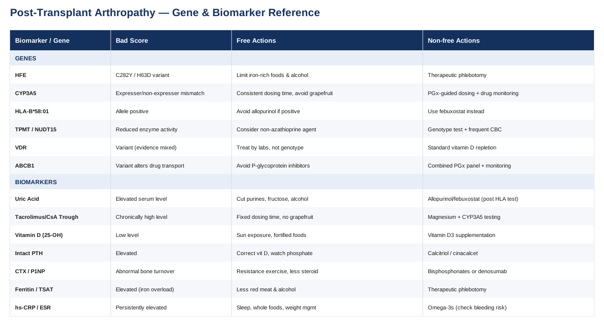
7 Biomarkers to Track for Post-Transplant Arthropathy
Post-transplant arthropathy is not one disease — it is a cluster of overlapping mechanisms: drug-induced hyperuricemia, corticosteroid-related bone damage, mineral and electrolyte disturbance, iron overload, and immune-mediated inflammation. The good news is that each of these mechanisms leaves a fingerprint in routine or semi-routine blood work. The following seven biomarkers cover the large majority of what drives joint symptoms after solid organ transplantation, and each one comes with a realistic improvement plan.
1. Serum Uric Acid
Hyperuricemia is extremely common after transplant, largely because calcineurin inhibitors — cyclosporine more than tacrolimus — reduce the kidney's ability to clear urate. This is well documented in kidney transplant recipients, where new-onset gout has even been linked to worse graft outcomes over time, as shown in a large cohort study on gout as an independent risk factor after kidney transplantation. Gout flares in transplant recipients often look atypical — more joints involved, less classic big-toe presentation — which is part of why they get misdiagnosed as generic "arthritis."
How to measure it
A simple serum uric acid test, usually bundled into a comprehensive metabolic panel your transplant clinic already draws. Standalone cost if paid out of pocket is roughly 10 to 20 dollars. Recheck every 3 to 6 months, or sooner if a joint flares.If the score is bad: the plan without supplements or equipment
Cut back on purine-dense foods (organ meats, shellfish, beer), reduce fructose-sweetened beverages, increase water intake, and address excess weight gradually. Ask your transplant physician whether a switch from cyclosporine to tacrolimus is appropriate, since tacrolimus tends to raise uric acid less. Review diuretic use, since thiazide and loop diuretics both raise uric acid independently.If the score is bad: the plan with supplements or equipment
Tart cherry extract has modest observational support for reducing flare frequency; a common protocol is 8 ounces of juice or an equivalent capsule dose daily, cycled around periods of higher risk rather than used indefinitely. If flares recur, urate-lowering therapy such as allopurinol may be prescribed — but only after HLA-B*58:01 testing in patients of Southeast Asian or African ancestry, given the risk of severe skin reactions (see the genetics section below). Colchicine for acute flares must be dose-reduced in patients on calcineurin inhibitors, since both drugs share the same metabolic pathway and combining them at full dose has caused severe muscle toxicity — this decision belongs to your physician, not self-directed dosing.2. Tacrolimus or Cyclosporine Trough Level
Your immunosuppressant blood level is not usually thought of as a "joint" biomarker, but it drives several arthropathy pathways at once: chronically high levels contribute to magnesium wasting (a known trigger for pseudogout, or CPPD), nerve and kidney toxicity, and possibly bone turnover changes, as reviewed in a comprehensive review of calcineurin inhibitor toxicity. Calcium pyrophosphate deposition disease driven by CNI-related hypomagnesemia has been specifically documented in transplant recipients, per a clinical review of CPPD diagnosis and treatment.
How to measure it
A trough blood draw taken immediately before your morning dose — this is already part of standard transplant follow-up. Out-of-pocket cost, if needed, runs roughly 50 to 150 dollars depending on the assay.If the score is bad: the plan without supplements or equipment
Take the medication at the same time every day, avoid grapefruit and pomelo entirely (both strongly inhibit the enzyme that clears these drugs and can push levels into toxic range), and never start an herbal supplement — including St. John's Wort, which does the opposite and can push levels dangerously low — without checking with your transplant pharmacist first.If the score is bad: the plan with supplements or equipment
If trough levels are chronically high and magnesium is low, oral magnesium glycinate or magnesium oxide (typically 200 to 400 mg daily, adjusted to bowel tolerance) can help correct hypomagnesemia-driven joint symptoms, rechecked every 4 to 6 weeks until stable. Pharmacogenomic testing for CYP3A5 (roughly 100 to 300 dollars where not covered) can explain persistent over- or under-exposure and guide your transplant physician's dosing rather than leaving it to trial and error.3. 25-Hydroxyvitamin D
Vitamin D deficiency is close to universal in the first year post-transplant, driven by reduced sun exposure during recovery, corticosteroid-accelerated catabolism, and, in kidney recipients, impaired conversion to the active hormone. Low vitamin D worsens secondary hyperparathyroidism and directly contributes to bone pain and myopathy that can be mistaken for joint disease.
How to measure it
A standard 25(OH)D blood test, roughly 40 to 80 dollars if not bundled into your existing bone or metabolic panel.If the score is bad: the plan without supplements or equipment
Sensible, non-burning sun exposure a few times a week (balanced against skin cancer risk, which is elevated in transplant recipients), dietary sources like fatty fish and fortified dairy, and regular weight-bearing movement to support bone health independent of vitamin D status.If the score is bad: the plan with supplements or equipment
Vitamin D3 supplementation, typically 1,000 to 4,000 IU daily depending on baseline deficiency severity, rechecked at 8 to 12 weeks rather than adjusted blindly. Avoid high-dose bolus regimens without physician oversight, since calcineurin inhibitors already affect calcium handling and excess vitamin D can tip patients into hypercalcemia.4. Intact Parathyroid Hormone (PTH)
Elevated PTH — secondary or, in longstanding cases, tertiary hyperparathyroidism — is extremely common after kidney transplant and drives bone resorption, periarticular calcification, and diffuse aches that patients often describe as joint pain rather than bone pain. Vitamin D receptor variation appears to modulate how much PTH rises for a given vitamin D level, discussed further in the genetics section.
How to measure it
Intact PTH blood test, roughly 50 to 100 dollars, usually ordered alongside calcium and phosphate. Recheck every 3 to 6 months in the first two years post-transplant, less often once stable.If the score is bad: the plan without supplements or equipment
Correcting vitamin D deficiency and monitoring phosphate intake are the first steps, since both feed directly into PTH regulation. Weight-bearing exercise supports bone quality independent of hormone levels.If the score is bad: the plan with supplements or equipment
Active vitamin D analogs (calcitriol or paricalcitol) are prescription-only and physician-dosed, as is cinacalcet for refractory tertiary hyperparathyroidism. These are not self-directed supplements — they require calcium monitoring on a schedule your nephrology or transplant team will set, typically every 4 to 8 weeks during dose adjustment.5. Bone Turnover Markers: CTX and P1NP
C-terminal telopeptide (CTX) reflects bone resorption and procollagen type 1 N-terminal propeptide (P1NP) reflects bone formation. Both respond faster than a DXA scan to changes in bone metabolism, which matters given how quickly corticosteroids can accelerate bone loss and, separately, drive femoral head osteonecrosis — a major and underrecognized cause of post-transplant hip and shoulder pain, with incidence around 4 percent in kidney recipients according to a large cohort study on avascular necrosis after kidney transplantation, and mechanistically explained in a review of glucocorticoid-induced avascular bone necrosis. Reference ranges and clinical use of these markers are summarized in a StatPearls overview of biochemical markers of osteoporosis.
How to measure it
A fasting morning blood draw is important, since both markers show daytime variation. Cost is typically 80 to 150 dollars per marker and is less consistently covered by insurance than standard labs, so ask your clinic whether it is bundled into transplant bone-health monitoring.If the score is bad: the plan without supplements or equipment
Resistance and weight-bearing exercise two to three times weekly, adequate dietary protein, smoking cessation, and — the single highest-leverage step — working with your transplant team to minimize cumulative corticosteroid dose wherever medically possible, since steroid exposure is the single biggest modifiable driver of both bone loss and osteonecrosis.If the score is bad: the plan with supplements or equipment
Calcium (dietary preferred, supplemented only to close a documented gap) and vitamin D as above. If fracture risk is high, bisphosphonates or denosumab may be prescribed; these are physician-managed with specific dosing intervals (denosumab every 6 months, oral bisphosphonates typically weekly or monthly) and require dental clearance beforehand given rare jaw osteonecrosis risk. Recheck bone turnover markers at 3 to 6 months to confirm response.6. Ferritin and Transferrin Saturation
Iron overload arthropathy is a distinct and often missed cause of post-transplant joint pain, classically affecting the second and third knuckles and sometimes overlapping with CPPD (chondrocalcinosis). It matters most in two groups: liver transplant recipients whose original diagnosis was hereditary hemochromatosis, where iron re-accumulation can recur in the new liver and elsewhere, and any transplant recipient with a history of multiple pre-transplant blood transfusions. The joint and organ manifestations of iron overload are detailed in the GeneReviews chapter on HFE-related hemochromatosis.
How to measure it
Two simple blood tests — ferritin and transferrin saturation — together costing roughly 20 to 40 dollars. If both are elevated, genetic testing for HFE variants typically follows.If the score is bad: the plan without supplements or equipment
Reduce red meat and avoid taking vitamin C alongside iron-rich meals (vitamin C boosts iron absorption), stop any multivitamin containing iron, and moderate alcohol intake, which independently increases iron absorption and liver iron deposition.If the score is bad: the plan with supplements or equipment
Therapeutic phlebotomy is the standard treatment where graft function and hemoglobin allow — typically weekly or biweekly sessions until ferritin reaches target, then quarterly maintenance draws. Iron chelation (deferasirox) is reserved for patients who cannot tolerate phlebotomy and requires close renal monitoring given the added burden on a transplanted kidney; this is a physician-directed therapy, not a self-managed one.7. High-Sensitivity CRP and ESR
These nonspecific inflammatory markers are less about diagnosing a specific cause and more about triage: in an immunosuppressed patient, new joint swelling could be gout, CPPD, osteonecrosis, or — critically — a septic joint or systemic infection that mimics arthritis. A markedly elevated CRP or ESR should always prompt an infection workup before being treated as routine "arthritis."
How to measure it
A simple blood test, roughly 15 to 40 dollars, often already included in transplant follow-up panels.If the score is bad: the plan without supplements or equipment
Prioritize sleep, address any dental or skin infection sources promptly (common entry points for infection in immunosuppressed patients), maintain a diet built around vegetables, fiber, and unprocessed foods, and manage weight, since visceral fat is itself a source of low-grade inflammatory signaling.If the score is bad: the plan with supplements or equipment
Omega-3 fatty acids (1 to 2 grams combined EPA/DHA daily) have modest anti-inflammatory evidence and are generally well tolerated, though they can add to bleeding risk when combined with other medications — flag this to your transplant pharmacist. Avoid self-prescribed NSAIDs for joint pain relief; they are a common and preventable cause of kidney injury in transplant recipients. If CRP or ESR is persistently elevated without an infectious or gout explanation, that pattern itself is useful information for your rheumatology team.Taken together, these seven values give a far more precise picture than "your joints hurt because you're on immunosuppressants." The next layer — the genes that shape how you process the drugs behind several of these biomarkers — adds useful context for the conversations you have with your transplant team.
6 Genes Worth Knowing About
Genetics in post-transplant care is less about optimizing wellness scores and more about avoiding specific, well-documented drug reactions. The following genes do not have a "reverse the mutation" fix — the plan in each case is testing and monitoring rather than supplementation, and that distinction matters.
HFE (Hemochromatosis)
Variants in HFE, most commonly C282Y and H63D, cause excess intestinal iron absorption and are the classic cause of iron-overload arthropathy, as described in the HFE-related hemochromatosis GeneReviews chapter. This matters most for liver transplant recipients whose original disease was hemochromatosis-related, where iron overload can recur.If the gene is bad: the plan without supplements
Moderate dietary iron and alcohol, avoid iron-containing supplements, and monitor ferritin and transferrin saturation on the schedule described above rather than waiting for symptoms.If the gene is bad: the plan with supplements or equipment
Therapeutic phlebotomy remains the primary "equipment" intervention, scheduled by ferritin trend rather than genotype alone, since penetrance of HFE variants is incomplete and not everyone with the variant develops overload.CYP3A5
CYP3A5 "expresser" genotypes metabolize tacrolimus faster and are prone to under-exposure at standard doses, while "non-expresser" genotypes are prone to overexposure and toxicity, as detailed in a PharmVar review of CYP3A5. This directly affects trough-level stability discussed earlier.If the gene is bad: the plan without supplements
Strict, consistent dosing timing and avoidance of grapefruit and unapproved herbal supplements that alter CYP3A4/5 activity.If the gene is bad: the plan with supplements or equipment
Pharmacogenomic-guided dosing, established through genotyping plus therapeutic drug monitoring, allows your transplant physician to set a starting dose closer to target on day one rather than titrating blind.HLA-B*58:01
This allele carries a very strong association with severe, sometimes fatal cutaneous reactions (Stevens-Johnson syndrome and toxic epidermal necrolysis) to allopurinol, the most common gout medication — an association with odds ratios above 80 in a systematic review and meta-analysis. Given how often post-transplant hyperuricemia leads to allopurinol use, this test matters.If the gene is bad: the plan without supplements
If the allele is present, allopurinol should generally be avoided; febuxostat or other alternatives are discussed with rheumatology instead.If the gene is bad: the plan with supplements or equipment
A one-time genetic test before starting allopurinol, especially relevant for patients of Southeast Asian, Han Chinese, or African ancestry where allele frequency is higher.TPMT and NUDT15
Reduced activity in either enzyme raises the risk of severe bone marrow suppression from azathioprine, still used in some transplant regimens, as summarized in NCBI's Medical Genetics Summaries entry on azathioprine, TPMT and NUDT15.If the gene is bad: the plan without supplements
Discuss switching to mycophenolate or another alternative agent if a high-risk variant is identified before starting therapy.If the gene is bad: the plan with supplements or equipment
Genotype or enzyme-activity testing before initiating azathioprine, followed by more frequent complete blood counts (often weekly at first, then monthly) if a reduced-activity variant is confirmed and the drug is continued at a lower dose.VDR (Vitamin D Receptor)
VDR polymorphisms have been linked to differences in secondary hyperparathyroidism severity and bone density after renal transplantation, as explored in research on vitamin D receptor polymorphisms and bone health after kidney transplantation, though evidence is mixed and not yet strong enough to guide individualized dosing on its own.If the gene is bad: the plan without supplements
Treat based on measured 25(OH)D and PTH levels rather than genotype, since actual blood levels remain the more actionable number.If the gene is bad: the plan with supplements or equipment
Standard vitamin D repletion as described earlier, monitored by labs rather than adjusted for genotype, since clinical trials guiding VDR-genotype-specific dosing do not yet exist.ABCB1 (MDR1 / P-glycoprotein)
This transporter gene affects absorption and cellular efflux of tacrolimus and several other drugs, and interacts with CYP3A5 status to explain some of the dose variability seen between patients on the same weight-based regimen.If the gene is bad: the plan without supplements
Avoid known P-glycoprotein inhibitors (certain antifungals, verapamil, some antibiotics) without checking with your transplant pharmacist first, since combining them with tacrolimus can unpredictably raise blood levels.If the gene is bad: the plan with supplements or equipment
Combined ABCB1/CYP3A5 pharmacogenomic panels, paired with therapeutic drug monitoring, give the clearest picture when trough levels are unexpectedly erratic despite consistent dosing.With the biology and the pharmacogenetics covered, it helps to zoom out to a broader framework for tracking joint and bone health over the decades that follow a transplant — which is where a widely read longevity book offers a useful, if imperfect, lens.
10 Ideas From Outlive That Apply Here
Peter Attia's book Outlive: The Science and Art of Longevity was not written for transplant recipients specifically, but its core argument — that most people are screened and treated too late, after damage has already accumulated — maps unusually well onto post-transplant arthropathy, where bone and joint damage from corticosteroids and immunosuppressants often develops silently for years before symptoms appear.
1. The "Centenarian Decathlon" reframes the goal
Attia argues that the real target isn't lifespan alone but the physical tasks you want to still do decades from now — carrying groceries, getting up off the floor, playing with grandchildren. For a transplant recipient, this reframes joint and bone health from "manage the pain" to "protect the specific movements that matter to you," which changes what you choose to monitor and train for.2. VO2 max is one of the strongest predictors of long-term function
Cardiorespiratory fitness correlates with all-cause mortality more strongly than almost any other single marker Attia discusses. For transplant recipients managing fatigue and reduced exercise tolerance, this is an argument for gradually building aerobic capacity rather than avoiding exertion out of caution alone.3. Muscle mass and strength directly protect joints
Attia is emphatic that strength training is not optional for healthy aging — muscle stabilizes joints, protects against falls, and offsets the sarcopenia that corticosteroids accelerate. This is one of the more directly transferable ideas: stronger muscles around a joint reduce load on damaged cartilage and bone.4. Zone 2 training builds an aerobic base without joint strain
Long, low-intensity aerobic sessions (roughly 180 minus age for heart rate, adjusted individually) build mitochondrial capacity with far less joint impact than high-intensity intervals — a practical starting point for someone rebuilding fitness after transplant surgery and steroid-related deconditioning.5. "Medicine 3.0" means testing before symptoms appear
Attia's central critique of conventional medicine is that it waits for disease to become symptomatic before acting. This is the same philosophy behind tracking the seven biomarkers above proactively, rather than waiting for a joint flare to prompt the first blood draw.6. Uric acid gets flagged as an underrated metabolic marker
Attia discusses uric acid as a marker worth watching even outside classic gout risk, given its ties to metabolic and cardiovascular health — reinforcing the same biomarker highlighted earlier in this article as central to post-transplant joint disease.7. Bone density deserves earlier, more frequent attention
Attia recommends earlier and more frequent DEXA scanning than typical guidelines suggest, given how much easier it is to prevent bone loss than reverse it — directly relevant to steroid-exposed transplant recipients, where bone loss often starts within the first year.8. Chronic low-grade inflammation ("inflammaging") drives multiple diseases at once
Attia treats systemic inflammation as a shared root cause across metabolic, cardiovascular, and joint disease, rather than a separate problem — the same logic behind tracking hs-CRP as a general-purpose warning marker rather than a disease-specific one.9. Sleep is treated as a non-negotiable pillar, not an afterthought
Poor sleep worsens inflammation, pain perception, and metabolic control — all of which compound the joint symptoms transplant recipients already contend with, making sleep quality a legitimate target alongside any lab value.10. Emotional health is called the "fifth horseman" for a reason
Attia argues that without psychological stability and social connection, none of the other interventions stick long enough to matter — a point that applies directly to the long-term discipline required to manage complex post-transplant medication and monitoring schedules.These ideas are a useful mental model, not a replacement for transplant-specific guidance — but they reinforce a theme running through this entire article: track early, track specifically, and act on trends rather than waiting for pain to force the issue. That same philosophy extends to a smaller set of complementary approaches with real trial evidence behind them.
Complementary Approaches With Real Evidence
None of the following replace immunosuppression management or biomarker tracking, and any new modality should be cleared with your transplant team, particularly regarding infection risk, bleeding risk, and interaction with medication schedules. These five were chosen because they have actual clinical trial support for joint pain and are low-risk enough to reasonably combine with post-transplant care.
Tai Chi
Tai chi is a low-impact, meditative movement practice built around slow, weight-shifting postures, which makes it unusually well suited to people managing joint pain, reduced bone density, and deconditioning at the same time — three issues that frequently overlap in post-transplant arthropathy. Because it loads joints gently and improves balance, it addresses both pain and fall risk in the same session, which matters for anyone with steroid-related bone fragility.A randomized comparative-effectiveness trial found that a structured tai chi program produced improvements in knee osteoarthritis symptoms similar to a standard course of physical therapy, and an overview of systematic reviews concluded that tai chi is now recommended by multiple major arthritis guidelines, as summarized in an overview of systematic reviews on tai chi for knee osteoarthritis.
For post-transplant patients, a realistic starting point is a beginner or "gentle" tai chi class twice weekly for 8 to 12 weeks, ideally taught by an instructor experienced with medically complex participants, with progression paced by joint response rather than a fixed schedule.
Yoga
Modified yoga programs designed around joint-friendly alignment can improve pain and function in osteoarthritic joints without the higher-impact loading of many conventional exercise programs, making it a reasonable option for transplant recipients rebuilding strength and range of motion cautiously.A randomized controlled trial of a biomechanically adapted yoga program for knee osteoarthritis found meaningful improvements in pain and physical function compared to a no-exercise control, as reported in a study on a biomechanically-based yoga exercise program for knee osteoarthritis.
Because certain deep-flexion or high-load poses can stress vulnerable joints (especially the hip, given osteonecrosis risk), post-transplant patients should look specifically for therapeutic or "gentle" yoga classes, avoid unsupported deep squats or extreme twists, and treat the first several sessions as an assessment period rather than pushing intensity.
Massage Therapy
Massage therapy targets muscle tension and circulation around affected joints and has one of the more robust evidence bases among complementary options for knee osteoarthritis specifically, with additional appeal for transplant recipients dealing with steroid-related muscle stiffness and reduced mobility.A randomized dose-finding trial found that a 60-minute weekly Swedish massage protocol produced significantly greater improvement in pain and function scores than usual care over eight weeks, as detailed in the randomized dose-finding trial of massage therapy for knee osteoarthritis.
A practical approach is a weekly session for 6 to 8 weeks, tapering to biweekly or monthly maintenance based on response; deep-tissue techniques over areas of active gout or CPPD inflammation should be avoided during a flare, and any therapist should be told about your transplant and medication history in advance.
Mindfulness Meditation / MBSR
Mindfulness-Based Stress Reduction does not change joint biology directly, but it changes how pain is processed and tolerated, which matters enormously for a population already managing a heavy medication and monitoring burden — chronic pain catastrophizing and poor sleep both amplify perceived joint symptoms independent of actual tissue damage.A randomized controlled trial found that structured mindfulness meditation produced measurable reductions in chronic pain outcomes compared to control conditions, as reported in a study on the effects of mindfulness meditation on chronic pain.
The standard protocol is an 8-week program, roughly 2 hours of group instruction weekly plus daily home practice of 20 to 30 minutes; it carries essentially no physical risk and can be layered on top of any of the other approaches here without concern for drug interaction.
Low-Level Laser Therapy / Photobiomodulation
Low-level laser therapy (LLLT), also called photobiomodulation, applies specific light wavelengths to affected joints to reduce localized inflammation and pain — an appealing option for transplant recipients who want to avoid adding oral anti-inflammatory drugs on top of an already complex medication list.An early meta-analysis found meaningful pain reduction from LLLT in osteoarthritis and rheumatoid arthritis, and a more recent umbrella review found the strongest supporting evidence specifically for osteoarthritis-related disability among the conditions studied, though it also noted that certainty of evidence remains low to moderate and protocols are not yet standardized, per the meta-analysis of low-level laser therapy for osteoarthritis and rheumatoid arthritis.
A reasonable trial run is two to three sessions weekly for 4 to 6 weeks at a clinic experienced with joint applications, tracking pain scores before and after to judge whether it is worth continuing — since response varies considerably between individuals and the evidence base, while promising, is still maturing.
Conclusion
Post-transplant arthropathy usually has a traceable cause, not a mysterious one. Uric acid, immunosuppressant trough levels, vitamin D and PTH, bone turnover markers, iron studies, and inflammatory markers explain the majority of joint symptoms that develop after transplant, and each has a concrete, monitorable improvement path. The genetics behind drug metabolism add precision to that picture, particularly around dosing safety and avoiding rare but serious drug reactions. Neither replaces the judgment of your transplant team, but both give you sharper questions to bring to them.
The single most useful next step is practical: ask your transplant clinic which of these seven biomarkers are already in your recent labs, request the ones that are missing, and bring any pattern you notice — a rising uric acid, a falling vitamin D, an unexplained CRP bump — to your next appointment as a specific talking point rather than a vague complaint of joint pain. That shift, from "my joints hurt" to "here is what my labs suggest," is where better decisions tend to start.