This article contains AI generated content.
· UpdatedPost-Traumatic Arthritis: 6 Genes And 7 Biomarkers To Track
Introduction
If you have had a significant joint injury — a fracture, a ligament tear, a dislocation — and months or years later that joint still aches, stiffens, or fails to feel right, you are not imagining it. Post-traumatic arthritis is the specific form of joint degeneration that follows trauma, accounting for an estimated 12% of all osteoarthritis cases according to landmark research on post-traumatic osteoarthritis. The original injury may have healed structurally, but inside the joint, inflammatory and catabolic processes can continue running — quietly degrading cartilage and remodeling bone for years after the event itself.
The standard approach after a joint injury covers the basics: rest, physical therapy, pain management, and imaging when things worsen. That protocol has merit, but it treats everyone identically regardless of the biological mechanisms driving their specific progression. Two people with the same tibial plateau fracture can look identical on an X-ray at two years and have completely different inflammatory environments inside the joint. One is stabilizing; the other is actively deteriorating. Standard clinical care rarely distinguishes between them until the gap is already large.
The reason for this divergence is measurable. Specific blood and urine markers can quantify whether cartilage is actively breaking down, whether inflammatory cytokines are running above the threshold that drives chronic joint damage, and whether key regulatory nutrients are at adequate functional levels. Genetic variants in the same inflammatory and tissue-remodeling pathways can explain individual vulnerability — and suggest targeted compensations. Neither lens alone tells the full story, but together they offer something that imaging and symptom tracking cannot: a window into the biological process before it reaches an irreversible state.
This article works through both approaches. The biomarker section covers seven markers that anyone can track with the right lab panels, each with a concrete plan for when numbers come back out of range. The genetic section covers six variants most consistently associated with post-traumatic joint outcomes, with specific lifestyle and supplement protocols for each. Better information, acted on early, does not guarantee a different outcome — but it dramatically changes the range of options available.
7 Biomarkers That Reveal What Is Actually Happening in Your Joint
Biomarkers give you something clinical exams and X-rays rarely provide: a real-time signal of the processes either protecting or eroding your joint. The seven markers below were selected because they are either the most clinically validated for post-traumatic joint environments, the most mechanistically relevant to cartilage degeneration and inflammation, or both. Costs listed are approximate US figures for reference; specialty panels vary by lab and insurance coverage.
Biomarker 1: High-Sensitivity C-Reactive Protein (hsCRP)
hsCRP is the most accessible systemic inflammation marker and the one most physicians already run on standard panels. The high-sensitivity version matters here because it detects low-grade chronic inflammation — the kind that runs below the threshold of obvious illness but above the level at which cartilage remains protected. In post-traumatic arthritis, even mild persistent elevation of CRP correlates with faster cartilage volume loss and greater synovial thickening over time. The target for joint health is below 0.5 mg/L; values consistently above 1.0 mg/L indicate enough inflammatory load to merit intervention.
How to Measure It
Standard blood draw through any primary care physician or direct-to-consumer lab (LabCorp, Quest, Ulta Lab Tests). Cost: approximately $10–$30 without insurance. Request high-sensitivity CRP specifically, not standard CRP, which misses low-grade elevation. Retest every 3–6 months when actively intervening.
If the Score Is Bad, the Plan Without Supplements
The single most effective lifestyle intervention for chronically elevated hsCRP is improving sleep quality and duration to 7–9 hours per night — sleep deprivation is a direct CRP driver. Beyond sleep, an anti-inflammatory dietary pattern (Mediterranean-style, emphasizing oily fish, olive oil, vegetables, and legumes while removing ultra-processed foods and seed oils) consistently lowers CRP by 0.3–0.8 mg/L over 12 weeks. Moderate aerobic exercise at Zone 2 intensity (a pace at which you can hold a conversation) 3–4 times per week has a documented anti-inflammatory effect, while high-intensity exercise without adequate recovery can paradoxically raise CRP short-term. Reducing psychological stress through structured relaxation or breathing work also matters, since cortisol dysregulation perpetuates low-grade systemic inflammation.
If the Score Is Bad, the Plan With Supplements or Equipment
Omega-3 fatty acids (EPA+DHA): 2–4 g/day with a meal, no cycling required for maintenance use. Blood-thinning effect becomes relevant above 4 g/day in those on anticoagulants. Curcumin (BCM-95 or Longvida formulation specifically — standard turmeric powder has negligible bioavailability): 500–1,000 mg/day with food; some practitioners cycle 8 weeks on / 2 weeks off. May mildly potentiate blood-thinning medications. GI discomfort is possible at higher doses. Magnesium glycinate: 200–400 mg at bedtime; no cycling needed; loose stools possible above 500 mg. Infrared sauna, 3–4 sessions per week, has emerging evidence for CRP reduction — 20–30 minutes per session at 140–160°F.
Biomarker 2: COMP (Cartilage Oligomeric Matrix Protein)
COMP is a structural protein found almost exclusively in cartilage and tendon. When cartilage is under stress or actively degrading, COMP fragments are released into the bloodstream. It is one of the most joint-specific markers available, making it considerably more informative than general inflammation markers for tracking actual cartilage status. A pivotal study on COMP release after joint injury is available in the NIH literature. Elevated serum COMP after joint trauma is associated with progression to clinical post-traumatic arthritis, and repeated measurements over time can indicate whether an intervention is stabilizing cartilage turnover or not.
How to Measure It
Serum COMP is available through specialty labs (Hospital for Special Surgery, certain academic medical centers, or via practitioners ordering through ARUP or Mayo Clinic Laboratories). Cost: approximately $100–$200. Not on standard panels — requires a specific request. Reference ranges vary by lab; a value persistently above the lab's 95th percentile for age and sex warrants attention. Retest every 6 months when monitoring.
If the Score Is Bad, the Plan Without Supplements
Sustained mechanical overloading of the joint is the primary driver of elevated COMP. For someone with a history of knee or ankle trauma, this means auditing loading patterns: eliminating high-impact activity on the affected joint (running on hard surfaces, jumping) and replacing it with swimming, cycling, or water aerobics that provide cardiovascular benefit without compressive loading. Weight management is critical — each pound of bodyweight adds approximately 3–4 pounds of force across the knee; losing 10 lbs reduces COMP-releasing compressive stress meaningfully. Physical therapy focused on periarticular muscle strengthening (particularly quadriceps for knee PTA) offloads the articular surface during daily activity.
If the Score Is Bad, the Plan With Supplements or Equipment
Hydrolyzed collagen peptides (type I/III, 10 g/day in liquid): taken simultaneously with vitamin C (500–1,000 mg) to maximize collagen synthesis — the vitamin C is essential for this protocol to work. No cycling required; minimal side effects. Glucosamine sulfate (not glucosamine hydrochloride): 1,500 mg/day; 12-week minimum before reassessing; mild GI effects possible; shellfish allergy caution. Pulsed electromagnetic field (PEMF) therapy devices — used 20–30 minutes/day over the joint — have a small but positive evidence base for cartilage metabolism and are available as home devices (Bemer, OrthoFix, or similar).
Biomarker 3: Urinary CTX-II (C-Telopeptide of Type II Collagen)
uCTX-II is a breakdown product of type II collagen, the structural collagen that makes up articular cartilage. When cartilage is degraded, CTX-II fragments are excreted in urine, making this marker a direct readout of collagen catabolism rate rather than a general inflammation signal. Research published in the NIH database has established uCTX-II as a marker of cartilage degradation and a predictor of radiographic joint space narrowing. For post-traumatic arthritis specifically, elevated uCTX-II in the first 12 months after injury is among the strongest available predictors of long-term joint deterioration.
How to Measure It
Second-morning void urine sample, corrected for creatinine concentration. Available through specialty labs and some rheumatology-focused practitioners. Cost: approximately $100–$250. Results are expressed in ng/mmol creatinine; reference ranges are sex- and age-adjusted. This is not a standard lab test, but it can be ordered through naturopathic physicians, functional medicine practitioners, or rheumatologists familiar with research biomarkers.
If the Score Is Bad, the Plan Without Supplements
Elevated uCTX-II responds most directly to joint unloading and weight normalization. Even a 5% reduction in bodyweight has been shown to reduce uCTX-II in overweight individuals with knee involvement. Low-impact movement is protective: regular walking, cycling, and swimming maintain synovial fluid circulation (which delivers nutrients to cartilage) without the compressive peaks that drive collagen fragment release. Avoiding prolonged static loading — sitting with the joint in one position for hours — also matters; brief movement breaks every 30–45 minutes help maintain the fluid exchange that cartilage depends on.
If the Score Is Bad, the Plan With Supplements or Equipment
Boswellia serrata (5-Loxin or AprèsFlex, standardized to 30% AKBA): 100–200 mg/day; benefits typically emerge at 8–12 weeks; well-tolerated; rare GI effects. Cycle 8 weeks on / 2 weeks off for long-term use. Avocado-Soybean Unsaponifiables (ASU): 300 mg/day; long-term protocol; minimal side effects; this combination has Level 1A evidence in EULAR guidelines for structural effects in knee OA. Glucosamine sulfate: 1,500 mg/day alongside ASU for complementary mechanism (sulfate delivery vs. cell signaling modulation).
Biomarker 4: Serum IL-6 (Interleukin-6)
Interleukin-6 is the key cytokine connecting acute injury response to chronic synovitis in post-traumatic arthritis. It spikes immediately after joint trauma and, in predisposed individuals, fails to fully resolve — establishing a sustained inflammatory environment that drives both cartilage catabolism and pain sensitization. Serum IL-6 above 3 pg/mL in a resting, non-acutely-ill individual suggests active inflammatory signaling. The gene encoding IL-6 (discussed in the genetics section) creates a biological predisposition in some people to produce more of it — but serum level reflects the actual output, not just the risk.
How to Measure It
Blood draw through specialty labs or academic medical centers. Not always on standard inflammatory panels — request specifically. Cost: approximately $50–$150. Some functional medicine practitioners include it in broader cytokine panels. Retest every 3–4 months when actively intervening to monitor response.
If the Score Is Bad, the Plan Without Supplements
IL-6 production is heavily influenced by sleep quality (deprivation of even one night raises IL-6 meaningfully), psychological stress (through HPA axis activation), and adipose tissue (fat cells are a major IL-6 source). Structured relaxation, 7–9 hours of sleep with consistent timing, and reductions in visceral adiposity through diet and Zone 2 exercise all address the root production drivers. Cold exposure (cold showers or brief cold water immersion, 2–3 minutes at under 60°F, 3–4 times per week) has a documented short-term IL-6 modulating effect through autonomic pathways.
If the Score Is Bad, the Plan With Supplements or Equipment
Omega-3 fatty acids: 2–4 g EPA+DHA/day — directly modulates the eicosanoid balance that regulates IL-6 transcription. Ginger extract (standardized to 5% gingerols): 500–1,000 mg/day with food; cycle 8 weeks on / 2 off; mild GI effects; mild anticoagulant effect at higher doses. Palmitoylethanolamide (PEA): 300–600 mg twice daily; no cycling required; excellent safety profile across multiple clinical trials; no known significant drug interactions. PEA works through a distinct anti-neuroinflammatory mechanism and complements omega-3s well.
Biomarker 5: 25-OH Vitamin D
Vitamin D is not a vitamin in the conventional sense — it is a steroid hormone that regulates hundreds of genes, including those governing joint inflammation, cartilage metabolism, and immune resolution. Low vitamin D status is strikingly common in post-traumatic arthritis populations, and research has linked vitamin D status to osteoarthritis outcomes. Below 40 ng/mL (100 nmol/L), the anti-inflammatory and anabolic cartilage effects of vitamin D signaling are substantially impaired. Peter Attia's approach to vitamin D in his longevity medicine practice targets 50–70 ng/mL as the functional range, notably higher than the 20 ng/mL threshold most standard labs flag as sufficient.
How to Measure It
Standard blood draw measuring 25-hydroxyvitamin D (not 1,25-dihydroxyvitamin D, which reflects current activation but not storage status). Available through any primary care physician or direct-to-consumer lab. Cost: $30–$80. Retest every 3–6 months when supplementing to dial in the appropriate dose.
If the Score Is Bad, the Plan Without Supplements
Solar UVB exposure remains the most physiologically natural way to raise vitamin D levels. Fifteen to thirty minutes of midday sun (when UVB penetrates the atmosphere, roughly 10am–3pm) with arms and legs exposed — without sunscreen on those areas — is sufficient to produce 10,000–20,000 IU equivalent on light skin in peak summer. Darker skin tones require significantly more time. Geographic latitude, season, and cloud cover all reduce UVB availability substantially — northern latitudes above 40°N produce essentially no vitamin D from sun between October and March. Foods high in magnesium (pumpkin seeds, leafy greens, dark chocolate, almonds) support the enzymatic conversion of vitamin D to its active form.
If the Score Is Bad, the Plan With Supplements or Equipment
Vitamin D3 (cholecalciferol): 2,000–5,000 IU/day as a starting dose for most adults below 40 ng/mL; some individuals with VDR variants (see genetics section) require 5,000–8,000 IU. Always combine with vitamin K2. Vitamin K2 (MK-7 form): 100–200 mcg/day with a fat-containing meal — essential alongside D3 to direct calcium to bone rather than soft tissue. Magnesium glycinate: 200–400 mg/day — required cofactor for vitamin D hydroxylation; many people supplementing D3 see no serum rise because of underlying magnesium depletion. Monitor serum 25-OH D every 3 months; upper safety limit for most people is around 100 ng/mL.
Biomarker 6: Serum MMP-3 (Matrix Metalloproteinase-3)
MMP-3 (also called stromelysin-1) is an enzyme that degrades proteoglycans, fibronectin, and type II collagen — the structural components of articular cartilage. Elevated circulating MMP-3 signals that the joint environment has shifted toward active enzymatic destruction of the cartilage matrix. Research has linked MMP-3 levels to destructive joint disease progression, and its measurement adds specificity to standard inflammation markers because it reflects degradative enzymatic activity rather than general immune activation. A serum level consistently above 10 ng/mL in someone with known joint injury warrants systematic intervention.
How to Measure It
Available through specialty labs (ARUP Laboratories, Mayo Medical Laboratories) or academic rheumatology centers. Some functional medicine practitioners include it in advanced joint panels. Cost: approximately $80–$200. Not a standard primary care test — requires specific ordering. Used primarily for monitoring disease activity rather than single-point diagnosis.
If the Score Is Bad, the Plan Without Supplements
MMP-3 activity is strongly upregulated by pro-inflammatory cytokines (particularly IL-1β and TNF-α — both covered in the genetics section). Any intervention that durably lowers systemic IL-6, CRP, and TNF-α will secondarily reduce MMP-3 expression. Practically: an anti-inflammatory dietary pattern, management of visceral adiposity, and elimination of smoking (nicotine directly upregulates MMP expression) are the highest-yield lifestyle levers. Weight loss of 5–10% in overweight individuals consistently reduces MMP-3 levels in synovial fluid and serum.
If the Score Is Bad, the Plan With Supplements or Equipment
EGCG (green tea extract, standardized to 45–50% EGCG): 400–600 mg/day; always taken with food to prevent GI irritation; cycle 8–12 weeks on / 2 weeks off; may reduce non-heme iron absorption — space away from iron-rich meals; avoid in iron-deficiency states. Curcumin BCM-95: 500–1,000 mg/day, 8-week cycles. Sub-antimicrobial dose doxycycline (20 mg twice daily): a prescription option with a growing evidence base specifically for MMP inhibition — doctor-supervised, 3 months on / 1 month off cycling, mild GI effects possible, photosensitivity caution in sun-exposed activities.
Biomarker 7: Omega-6/Omega-3 Ratio
The omega-6 to omega-3 ratio in cell membranes determines the balance between pro-inflammatory and anti-inflammatory eicosanoid signaling at the cellular level. The modern Western diet pushes this ratio to 15:1 or higher; a ratio at or below 4:1 is associated with significantly reduced inflammatory marker output, including within joint tissues. For post-traumatic arthritis, where inflammatory signaling is already sensitized by prior injury, a chronically high omega-6/omega-3 ratio functions as a persistent amplifier that keeps the joint environment pro-inflammatory regardless of other interventions. This is one of the most modifiable root drivers of joint inflammation and one of the least discussed in standard orthopedic care.
How to Measure It
Specialty whole-blood fatty acid tests (OmegaQuant, Cleveland HeartLab's OmegaCheck, or Genova's NutrEval) measure the actual cell membrane ratio rather than estimating intake. Cost: approximately $50–$150 depending on the panel. Consumer options like OmegaQuant's home kit (fingerstick) are available without a physician order. Target is a ratio at or below 4:1; optimal for anti-inflammatory purposes is 1:1 to 3:1. Retest after 3–4 months of dietary or supplemental intervention.
If the Score Is Bad, the Plan Without Supplements
The dietary approach is two-directional: reduce omega-6 intake (by eliminating seed oils — sunflower, corn, soybean, safflower, cottonseed — which are the dominant source in processed foods) and increase omega-3 intake through fatty fish. Replacing seed oil-based cooking fats with olive oil, avocado oil, or butter significantly drops omega-6 load. Eating oily fish (sardines, mackerel, wild salmon, herring) 3–4 times per week meaningfully improves the ratio over 8–12 weeks without supplements.
If the Score Is Bad, the Plan With Supplements or Equipment
Triglyceride-form fish oil: 2–4 g EPA+DHA/day with meals, no cycling for long-term maintenance, refrigerate after opening, mild fishy taste (flavored options available). Choose brands with IFOS (International Fish Oil Standards) certification: Carlson, Nordic Naturals, OmegaBrite, and Viva Naturals are consistently third-party tested. Blood-thinning effect is relevant above 4 g/day in those on anticoagulants. For those unable to tolerate fish oil: algae-derived omega-3 provides DHA primarily and is suitable for plant-based approaches, though EPA is limited in most formulations.
Tracking these seven markers over time — rather than waiting for symptoms to dictate action — puts you in a fundamentally different position with respect to your joint's trajectory. Each marker tells a distinct part of the story, and together they reveal whether your post-traumatic biology is trending toward stability or toward progressive deterioration. The next layer of analysis asks not just what your numbers are, but why they are what they are.
What Your Genetic Profile Reveals About Post-Traumatic Arthritis Risk
Genetics does not determine destiny in post-traumatic arthritis — but it does establish the terrain. Two people with the same injury and the same initial inflammatory response can diverge significantly at 5 and 10 years depending on whether their genetic variants favor aggressive inflammatory signaling, rapid cartilage matrix degradation, or impaired repair. The six variants below are among the most consistently studied in joint inflammation and osteoarthritis genetics research. Consumer genomic tests (23andMe, AncestryDNA) and clinical DNA panels provide access to most of these; others require targeted clinical genotyping. The plans here are additive to the biomarker interventions described above.
Gene 1: IL-6 (rs1800795) — The Inflammation Amplifier
The IL-6 gene encodes interleukin-6, the primary driver of systemic acute-phase inflammation. The rs1800795 C allele is associated with increased IL-6 transcription, meaning carriers produce more IL-6 in response to any inflammatory stimulus — including joint trauma. Research on IL-6 polymorphisms confirms their role in inflammatory diseases. For post-traumatic arthritis, this variant creates a primed baseline: the joint's inflammatory response is more intense and slower to resolve, accelerating synovial degradation of cartilage. The C/C genotype represents the highest-production profile; C/G is intermediate; G/G is lower production.
If the Gene Is Bad, the Plan Without Supplements
Priority one is eliminating the most potent IL-6 production drivers: poor sleep, psychological stress, excessive visceral fat, and hyperglycemia. A sleep protocol targeting 7–9 hours with consistent timing reduces baseline IL-6 production substantially. A Mediterranean dietary pattern, specifically one that reduces refined carbohydrates, provides measurable IL-6 reduction through multiple pathways. Cold water immersion 3–4 times per week (2–3 minutes at below 60°F) engages autonomic anti-inflammatory regulation through vagal tone improvement. Endurance training at moderate intensity (Zone 2) consistently reduces circulating IL-6 over 8–12 weeks in adherent individuals.
If the Gene Is Bad, the Plan With Supplements or Equipment
Omega-3 (EPA+DHA, 3 g/day), curcumin BCM-95 (500 mg twice daily with food), and PEA (300 mg twice daily) form an evidence-informed stack targeting IL-6 at different levels of production and downstream signaling. No cycling required for omega-3 or PEA; curcumin can run 8-week cycles. Blood-thinning caution with combined omega-3 and curcumin in those on anticoagulants. PEA is notably free of drug interactions. Infrared sauna sessions (20 minutes, 3×/week) are a useful adjunct for those with high CRP and IL-6 who want non-pharmaceutical options.
Gene 2: TNF-α (rs1800629) — The Cartilage Destruction Amplifier
Tumor necrosis factor-alpha (TNF-α) is a master inflammatory cytokine that, when chronically elevated in joint tissue, directly induces chondrocyte apoptosis and cartilage matrix breakdown. The rs1800629 A allele is associated with higher TNF-α transcription, and studies examining TNF-α polymorphisms confirm associations with arthritis susceptibility and severity. In post-traumatic arthritis, individuals with this variant can experience a more aggressive destructive phase after injury, with faster cartilage space narrowing than those with the G/G genotype. The A allele is relatively common — approximately 25–30% of European populations carry at least one copy.
If the Gene Is Bad, the Plan Without Supplements
TNF-α is the primary target of some of the most expensive biologic drugs in rheumatology. Lifestyle approaches work through the same upstream pathways: low-carbohydrate or ketogenic diets have the strongest dietary evidence for TNF-α reduction, particularly in those with elevated fasting insulin. Intermittent fasting (a 16:8 time-restricted eating window) reduces TNF-α expression in multiple clinical studies. Prioritizing deep sleep (stages 3 and 4) matters specifically here — TNF-α rises with sleep deprivation at a magnitude that measurably increases joint catabolic activity within days.
If the Gene Is Bad, the Plan With Supplements or Equipment
Omega-3 (3–4 g EPA+DHA/day, long-term), boswellia serrata AKBA-standardized at 100–200 mg/day (cycle 8 weeks on / 2 weeks off; well-tolerated; mild GI effects at higher doses), and tart cherry concentrate (480 mg anthocyanins/day; continuous use; excellent safety; documented effect on both TNF-α and uric acid) form a complementary approach. The three work through different mechanisms and can be combined safely. For those with significantly elevated serum TNF-α alongside joint symptoms, a rheumatology consultation is appropriate to assess whether biologic therapy meets the threshold for clinical benefit.
Gene 3: IL-1β (rs16944) — The Cartilage Catabolic Driver
Interleukin-1 beta is arguably the single most important driver of cartilage catabolism in osteoarthritis — it directly stimulates chondrocytes to produce MMPs, inhibits proteoglycan synthesis, and drives apoptosis of cartilage cells. The rs16944 T allele is associated with higher IL-1β production, which in a post-traumatic joint environment means a more aggressively catabolic milieu from the time of injury onward. Much of the cartilage degradation that characterizes early post-traumatic arthritis is IL-1β-mediated — which explains why diacerhein (an IL-1β modulator) has shown structural benefits in clinical trials.
If the Gene Is Bad, the Plan Without Supplements
Blood glucose control is directly relevant here: acute hyperglycemia triggers NLRP3 inflammasome activation, the cellular machinery that processes and releases mature IL-1β. A low-glycemic diet with consistent meal composition, adequate protein (to blunt postprandial glucose spikes), and elimination of sugar-sweetened beverages substantially reduces this activation pathway. Cold water immersion and resistance training both suppress NLRP3 signaling through different mechanisms. Intermittent fasting also reduces inflammasome activity — a 16:8 window is sufficient for effect without requiring extended caloric restriction.
If the Gene Is Bad, the Plan With Supplements or Equipment
Avocado-soybean unsaponifiables (ASU, 300 mg/day): the most specifically documented IL-1β-modulating supplement, long-term use, minimal side effects, Level 1A EULAR evidence. PEA (600 mg/day): continuous, no cycling required, addresses downstream neuroinflammatory effects of IL-1β. Diacerhein (50 mg twice daily with food, prescription required in some countries): an IL-1β converting enzyme inhibitor with published RCT data for structural effects in OA; causes yellow-orange discoloration of urine (harmless, from metabolite excretion) and mild GI effects in the first 2–3 weeks that typically self-resolve; not appropriate in inflammatory bowel disease; requires physician oversight.
Gene 4: MMP-3 (rs679620) — The Matrix Degradation Variant
The MMP-3 gene encodes stromelysin-1, the enzyme that degrades multiple cartilage matrix components directly. The rs679620 variant affects promoter activity and is associated with higher baseline MMP-3 transcription in joint-relevant tissues. For post-traumatic arthritis, this variant creates a joint environment where cartilage matrix degradation proceeds faster following any inflammatory signal — the destructive machinery is running at higher baseline activity. Evidence supports MMP-3 as both a driver and a measurable marker of joint destruction. Elevated serum MMP-3 and this genetic variant together identify a particularly high-risk subgroup for rapid post-traumatic progression.
If the Gene Is Bad, the Plan Without Supplements
Joint protection strategies become more important for rs679620 carriers than average: avoiding repetitive mechanical loading on the injured joint, using appropriate footwear and orthotics that distribute load away from the affected compartment, and building periarticular muscle strength to reduce joint contact stress. Physical therapy focused on neuromuscular training (balance, proprioception, controlled loading) effectively reduces the stress events that trigger MMP-3 release. Eliminating smoking is critical — nicotine directly upregulates MMP-3 expression through NF-κB pathways.
If the Gene Is Bad, the Plan With Supplements or Equipment
EGCG (400–600 mg/day, with food, 8–12 week cycles): consistently shown to inhibit MMP-3 expression in joint tissue studies; space away from iron-containing meals; avoid in iron-deficiency states. Curcumin BCM-95 (500 mg twice daily, 8-week cycles) addresses upstream NF-κB signaling that drives MMP-3 transcription. Sub-antimicrobial doxycycline (20 mg twice daily, physician-supervised, 3 months on / 1 month off): inhibits MMP activity through mechanisms independent of antibiotic action; photosensitivity caution; mild GI effects possible; gut microbiome monitoring recommended during cycles.
Gene 5: GDF5 (rs143384) — The Cartilage Repair Deficit
Growth differentiation factor 5 (GDF5) plays a critical role in joint formation during development and in the maintenance and repair capacity of adult cartilage. The rs143384 T allele reduces GDF5 expression in joint tissues, impairing the baseline repair signaling that ordinarily counterbalances cartilage catabolism. Genetic analysis of GDF5 confirms its functional role in joint tissue. This means that for rs143384 T allele carriers, the post-traumatic repair response is biologically blunted — the joint degrades at the same rate or faster, but rebuilds more slowly. The T/T genotype represents the highest-risk profile; T/A is intermediate.
If the Gene Is Bad, the Plan Without Supplements
Mechanical loading is one of the most potent GDF5-upregulating signals — the gene responds to appropriate joint stress. Resistance training 3–4 times per week with progressive overload (particularly exercises that eccentrically load the joint: eccentric leg press, step-downs for knee involvement) stimulates GDF5 expression through mechanotransduction pathways. Adequate dietary protein (1.6–2.0 g/kg bodyweight per day) provides the amino acid substrate for the cartilage matrix repair that GDF5 signaling initiates. Recovery from exercise must be complete — rs143384 carriers appear to have reduced resilience to training overload.
If the Gene Is Bad, the Plan With Supplements or Equipment
Hydrolyzed type II collagen (10 g/day) with vitamin C (1,000 mg at the same time): the vitamin C co-administration is mechanistically necessary for collagen hydroxylation — this is not optional. Take 30–60 minutes before exercise to maximize delivery to the joint under loading. No cycling required; well-tolerated. Glucosamine sulfate (1,500 mg/day, 12-week minimum assessment window) supports proteoglycan synthesis in the cartilage matrix that GDF5 repair signaling targets. Platelet-rich plasma (PRP) injection is a clinical procedure option for GDF5 T/T carriers with significant post-traumatic joint involvement: it provides concentrated growth factors locally, partially compensating for the reduced endogenous GDF5-mediated repair signaling. Evidence is still accumulating for specific post-traumatic arthritis indications, but the growth factor delivery rationale is particularly well-matched to this genetic deficiency.
Gene 6: VDR (FokI / BsmI Polymorphisms) — The Vitamin D Resistance Variant
The vitamin D receptor (VDR) gene determines how efficiently cells respond to active vitamin D. Several common polymorphisms — particularly FokI (rs2228570) and BsmI (rs1544410) — alter the receptor's binding affinity and transcriptional activity. Individuals with low-efficiency VDR variants effectively need higher circulating 25-OH vitamin D levels to achieve the same biological effect in joint tissue, cartilage metabolism, and immune regulation. This is Gary Brecka's key insight applied to joint health: a standard vitamin D level of 30 ng/mL that looks acceptable on paper may be functionally insufficient for someone with a low-efficiency VDR genotype, leaving anti-inflammatory and cartilage-supportive vitamin D pathways underactivated.
If the Gene Is Bad, the Plan Without Supplements
Maximize the body's natural vitamin D production: consistent midday solar UVB exposure (15–30+ minutes depending on skin tone and latitude), with arms and legs exposed. Beyond sun, prioritize magnesium-rich foods daily — magnesium is the rate-limiting cofactor for vitamin D hydroxylation, and VDR variants that reduce efficiency make this cofactor even more critical. Oily fish (sardines, mackerel, wild salmon) provide not only vitamin D but also omega-3s that synergize with vitamin D receptor-mediated anti-inflammatory signaling.
If the Gene Is Bad, the Plan With Supplements or Equipment
Vitamin D3 at higher therapeutic doses (3,000–8,000 IU/day) — the appropriate dose for VDR variant carriers should be guided by serum response rather than a fixed number; target 60–70 ng/mL for functional adequacy in low-efficiency genotypes. Vitamin K2 MK-7 (200 mcg/day): non-negotiable when taking D3 above 2,000 IU; taken with a fat-containing meal; directs calcium appropriately. Magnesium glycinate or threonate (300–400 mg/day): required cofactor for D3 conversion; threonate form has better CNS penetration for those also managing pain-related sleep issues. Monitor serum 25-OH vitamin D every 3 months; reduce dose if levels exceed 100 ng/mL. Vitamin D toxicity is rare with K2 co-administration but remains a consideration at the higher end of this range.
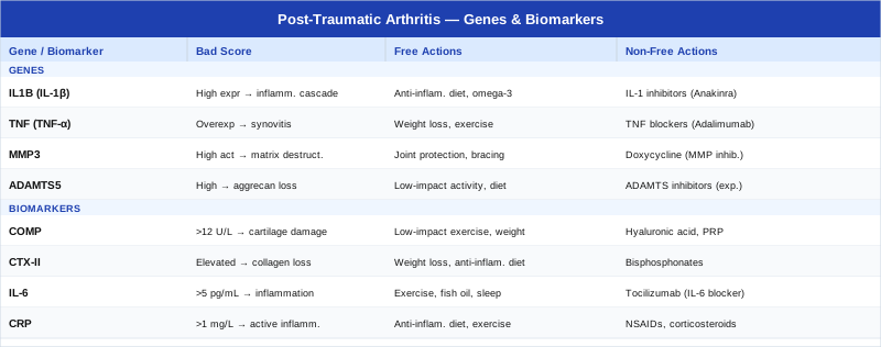
With both biomarkers and genetic variants mapped, the following table brings together the key parameters, their risk thresholds, and the primary action pathways in one consolidated reference.
The Framework That Gets Joint Inflammation Right — Peter Attia's Outlive
Outlive: The Science and Art of Longevity (2023) by Peter Attia, MD, is not a book specifically about joint health — but its framework for understanding metabolic inflammation, exercise physiology, and preventive medicine is arguably the most useful single resource for someone trying to manage post-traumatic arthritis through biology rather than symptom suppression. Attia references hundreds of peer-reviewed studies and consistently challenges the "manage symptoms until surgery is needed" mentality that dominates standard orthopedic follow-up. The ten most relevant insights for post-traumatic arthritis are outlined below.
1. Zone 2 Training Is a Systemic Anti-Inflammatory Drug
Zone 2 cardio — defined as the highest intensity at which you can sustain a full conversation, roughly 60–70% of maximum heart rate — is the intensity that most effectively improves mitochondrial function and reduces systemic inflammatory markers. Attia argues it should be the foundation of any longevity-oriented exercise protocol: 45 minutes, 3–4 times per week. For post-traumatic arthritis, the key insight is that Zone 2 in a joint-appropriate modality (cycling, swimming, elliptical — not running on concrete) delivers systemic anti-inflammatory benefit without the compressive loading that worsens cartilage damage.
2. Muscle Mass Is the Single Most Protective Factor for Joints
Attia makes the case that muscle mass and strength are the most underappreciated biomarkers in medicine — and for joint health the mechanistic argument is direct. Adequate periarticular muscle acts as a shock absorber, reducing the force transmitted to articular cartilage during every step, squat, and stair. Quadriceps weakness is one of the strongest predictors of knee OA progression. His recommendation: treat strength training as medicine, with the same prescription rigor. For post-traumatic arthritis this means progressive resistance training 2–3 times per week, even during flares (working around the joint rather than at it if necessary).
3. VO2 Max Predicts Inflammatory Status, Not Just Fitness
Higher VO2 max is not merely a fitness metric — it correlates strongly with reduced circulating inflammatory markers and with lower all-cause mortality at a magnitude that dwarfs most pharmacological interventions. Attia cites data showing that moving from the bottom to the second fitness quintile reduces mortality risk more than moving from the second to the top. For post-traumatic arthritis, this matters because systemic aerobic capacity directly determines the body's ability to resolve acute inflammatory events and prevent them from becoming chronic. Low-impact aerobic training that improves VO2 max is therefore doubly beneficial.
4. Insulin Resistance Feeds the Joint Inflammatory Loop
One of Attia's central arguments is that metabolic dysfunction — specifically insulin resistance and hyperinsulinemia — is the upstream driver of most chronic inflammatory conditions. In joint biology, insulin resistance correlates with elevated IL-6, TNF-α, and MMP activity, all of which accelerate cartilage degradation. He recommends tracking fasting insulin (not just fasting glucose) as an early metabolic health indicator. For post-traumatic arthritis patients, improving insulin sensitivity through dietary carbohydrate quality, resistance training, and time-restricted eating is one of the highest-leverage systemic interventions.
5. Sleep Is the Most Neglected Recovery Tool
Attia is emphatic that sleep deprivation is not a minor inconvenience — it is a potent activator of inflammatory pathways. One night of poor sleep (below 6 hours) measurably raises IL-6, TNF-α, and CRP the following day. For post-traumatic arthritis, this means that a person who is diligent with supplements and diet but chronically sleeping 5–6 hours is working against themselves biochemically. His specific recommendations include consistent sleep/wake timing, a cool bedroom environment (65–68°F), and addressing sleep-disordered breathing if present — sleep apnea is a potent chronic inflammatory driver.
6. Omega-3s at Therapeutic Doses, Not Sprinkle Doses
Attia distinguishes between the trace amounts of omega-3 that most people think they are getting from "a healthy diet" and the therapeutic doses (2–4 g EPA+DHA/day) that actually shift cell membrane phospholipid composition and downstream inflammatory signaling. He measures omega-3 status through whole-blood fatty acid testing (OmegaCheck or equivalent) and targets an omega-3 index of 8–12%. The implication for post-traumatic arthritis is that dietary omega-3 alone is rarely sufficient — supplemental fish oil at a dose calibrated to serum response is what moves the inflammatory dial.
7. Time-Restricted Eating Reduces Inflammatory Activation
A 16:8 eating window (16 hours fasting, 8 hours eating) reliably reduces multiple inflammatory markers without requiring caloric restriction per se. The mechanism involves autophagy induction and NLRP3 inflammasome suppression — directly relevant to IL-1β, the primary cartilage-catabolic cytokine discussed in the genetics section. Attia recommends TRE as a tool for metabolic health rather than weight loss per se. For post-traumatic arthritis patients managing cartilage degradation, the inflammasome suppression pathway makes TRE a particularly well-targeted daily practice.
8. Sedentary Time Is a Distinct Risk Beyond Activity Level
One of Attia's provocative points is that overall physical activity and daily sedentary time are independent risk variables — a person who exercises for one hour but sits for 12 hours has a materially different inflammatory and metabolic profile than one who exercises similarly but breaks up sitting frequently. For post-traumatic arthritis, prolonged static joint loading increases synovial fluid stagnation and reduces cartilage nutrient diffusion. Breaking up sitting with 2–5 minutes of movement every 30–45 minutes — joint rotations, brief walks, bodyweight squats — is both anti-inflammatory and directly cartilage-protective.
9. Protein Timing Around Exercise Amplifies Cartilage Repair
Attia emphasizes protein timing as well as quantity. For cartilage repair specifically, consuming hydrolyzed collagen with vitamin C 30–60 minutes before the exercise session that loads the joint delivers collagen precursors to the joint during the post-exercise nutrient delivery window, when blood flow to joint tissue is highest. This is the protocol used in the specific collagen research context and significantly outperforms taking collagen at random times during the day. Total daily protein at 1.6–2.2 g/kg provides the broader amino acid substrate for all tissue repair.
10. Medicine 3.0: Track Before It Hurts
Attia's overarching framework — Medicine 3.0 — is built on the premise that waiting for symptoms or X-ray changes before intervening is a systematic failure of preventive medicine. By the time radiographic joint space narrowing is visible, a large fraction of the cartilage is already gone. His philosophy applied to post-traumatic arthritis means establishing baseline biomarkers (COMP, uCTX-II, hsCRP, IL-6, vitamin D, omega-3 index) within the first weeks after a significant joint injury and tracking them every 3–6 months thereafter. The earlier the signal, the wider the window for intervention.
The complementary approaches below represent additional tools that have accumulated specific evidence for post-traumatic or osteoarthritis-related joint conditions and integrate well with the biomarker and genetic framework above.
Evidence-Backed Complementary Approaches
Tai Chi
Tai chi is a low-impact, mind-body movement practice combining slow, deliberate joint motion with breath coordination and proprioceptive training. Its relevance to post-traumatic arthritis is multifaceted: it provides controlled joint loading that builds periarticular muscle, improves proprioception (commonly impaired after joint trauma), and reduces the psychological stress that amplifies inflammatory biomarkers. Unlike high-impact exercise, it can be practiced through almost all stages of post-traumatic arthritis without risking cartilage overload.
In a rigorous clinical trial, Dr. Chenchen Wang and colleagues at Tufts Medical Center compared tai chi to standard physical therapy for knee osteoarthritis, finding equivalent or superior outcomes for pain, function, and quality of life at 12 and 52 weeks. The study used a 12-week program of two 60-minute classes per week. This level of evidence is notable — tai chi matching the gold-standard physical therapy protocol is a finding that challenges the assumption that movement therapies need to be intense to be effective for joint conditions.
Practical application: begin with a beginner-level program, ideally in-person with a qualified instructor for the first 6–8 weeks to ensure correct form that protects the injured joint. Online programs through Paul Lam's Tai Chi for Arthritis platform are widely used and well-structured. Two to three sessions per week is the evidence-supported dose. Progress to 60-minute sessions as tolerated. Avoid pushing through acute flares; modify or rest any movement that produces sharp joint pain.
Low-Level Laser Therapy / Photobiomodulation
Low-level laser therapy (LLLT), increasingly called photobiomodulation, uses red (630–700 nm) and near-infrared (800–1000 nm) wavelengths delivered at non-thermal intensities to stimulate cellular energy production (via cytochrome c oxidase in mitochondria), reduce inflammatory mediator release, and promote tissue repair. For post-traumatic arthritis, the mechanistic rationale is strong: mitochondria in chondrocytes respond to near-infrared wavelengths, and the combination of energy augmentation and anti-inflammatory signaling addresses two of the core deficits in the post-traumatic joint environment.
Systematic reviews of LLLT for musculoskeletal conditions have found meaningful short-term reductions in joint pain and improvements in function, particularly for knee osteoarthritis. The Ottawa Panel's evidence-based clinical guidelines include positive recommendations for LLLT in OA. Key protocol parameters: 830 nm near-infrared wavelength, dose of 4 joules per point over the affected joint, 3 sessions per week, for 4–8 weeks. Clinical LLLT is available through physical therapy practices; home photobiomodulation devices (Joovv, Mito Red Light, EMR-TEK) deliver sufficient power at 810–850 nm for home use.
Practical application: for home devices, use a panel delivering 20–40 mW/cm² at the surface, position 2–6 inches from the joint, 10–20 minutes per session, 5–6 days per week. This differs slightly from clinical LLLT protocols but achieves comparable cumulative dose. Do not use over active infections or malignancies. The evidence base is moderate rather than definitive — meaningful benefit for many but not universal; a 6-week trial at consistent protocol is a reasonable assessment window.
Mindfulness-Based Stress Reduction (MBSR)
Mindfulness-Based Stress Reduction is an 8-week structured program, originally developed by Jon Kabat-Zinn at the University of Massachusetts Medical School, combining seated meditation, body scan, and mindful movement practices. Its relevance to post-traumatic arthritis goes beyond stress management: pain catastrophizing — the tendency to amplify anticipated pain and feel helpless about it — is one of the most consistent predictors of functional disability and inflammatory marker elevation in chronic joint conditions. MBSR specifically targets catastrophizing through non-judgmental awareness training.
Meta-analyses of MBSR in chronic pain conditions show consistent reductions in pain intensity (typically 0.3–0.5 standard deviation effect size), improved function, and reduced psychological distress. For post-traumatic arthritis specifically, the central sensitization that often develops over months of joint pain makes pain reprocessing interventions like MBSR structurally important — not as a replacement for biomarker management, but as a complementary layer that influences how the nervous system processes and amplifies joint signals. Reducing catastrophizing has also been shown to reduce circulating inflammatory cytokines through psychoneuroimmunological pathways.
Practical application: attend a structured 8-week MBSR course in person or through a trained online instructor (search the UMass Center for Mindfulness directory for certified teachers). A viable free alternative is the Palouse Mindfulness online program, which follows the original MBSR curriculum. Daily practice of 30–45 minutes is the evidence-supported dose; even 15–20 minutes daily for adherent practitioners shows meaningful benefit. This is not a short-term intervention — sustained practice over 6–12 months produces the most durable neuroplastic changes in pain processing.
Conclusion
Post-traumatic arthritis is not an inevitable consequence of joint injury, but it is a biologically complex process that standard care rarely addresses with enough precision. The combination of seven trackable biomarkers and six genetic variants described in this article provides a more complete picture of what is driving a specific individual's joint trajectory than any single imaging study or symptom report could offer. Acting on out-of-range biomarkers — through targeted dietary, lifestyle, and supplemental interventions before symptoms become severe — represents the kind of early, informed action that moves the odds in your favor.
The practical next step is to request a baseline blood and urine panel that includes at minimum hsCRP, 25-OH vitamin D, and an omega-3 index through your physician or a direct-access lab. From there, COMP and uCTX-II add joint-specific information worth tracking at 6-month intervals. If consumer genetic testing is accessible, running your rs1800795, rs1800629, and rs143384 variants through a physician-reviewed report can guide whether your supplemental protocol should emphasize anti-inflammatory signaling, growth factor support, or vitamin D receptor optimization. Bring the results to a sports medicine physician, rheumatologist, or functional medicine practitioner who is comfortable interpreting them in the context of your specific injury history. Better information does not guarantee a different outcome, but it consistently produces better decisions.
Musculoskeletal: Bone Conditions Joint Conditions Sports Injuries
Autoimmune: Inflammatory Conditions