This article contains AI generated content.
Posterior Tibial Tendon Dysfunction — 4 Genes and 6 Biomarkers to Track
Introduction
If you are reading this, there is a good chance your arch has started to flatten, the inner side of your ankle aches after a walk that never used to bother you, or standing on one foot to rise onto your toes has quietly become impossible. Maybe a clinician mentioned posterior tibial tendon dysfunction and handed you an orthotic and a photocopied sheet of stretches. That advice is not wrong, but it can feel thin — as if the real question of why this tendon, why now, and why it is not settling down never got answered.
Generic advice treats the tibialis posterior tendon as a purely mechanical part that wore out. Sometimes that is the whole story. Often it is not. The same tendon in two people with the same shoes and the same body weight can behave completely differently, because the tissue is embedded in a metabolic and genetic terrain — blood sugar, cholesterol particles, inflammation, vitamin D, uric acid, and the collagen genes you were born with. Ignore that terrain and you are polishing the surface while the foundation shifts.
This article takes the deeper route. Instead of stopping at "wear the brace and rest," it looks at the measurable signals that quietly shape whether a tendon repairs or keeps degrading — the numbers you can actually track and change. The goal is not to replace your podiatrist or orthopedic surgeon, but to give you a smarter map to bring to them.
Here is the plan. First, the six blood markers most worth knowing about, why each one matters for tendon health, how to measure it, and what to do if it is off. Then a shorter tour of the collagen and matrix genes that stack the deck, and how to compensate for them. After that, a book that reframes how connective tissue heals, and a handful of complementary approaches with real human evidence. Better information genuinely can lead to better decisions — and for a tendon, better decisions made early are worth far more than heroic ones made late.
Summary
Posterior tibial tendon dysfunction (PTTD), also called adult acquired flatfoot or progressive collapsing foot deformity, is not only a mechanical failure of one tendon on the inside of your ankle. A growing body of research shows it travels with metabolic company: high blood sugar, unfavorable cholesterol, chronic low-grade inflammation, low vitamin D, and elevated uric acid all change how tendon cells behave and heal. That means the same numbers you might track for heart health double as a dashboard for your tendon.
In the pages below you will find the six biomarkers most worth watching — including the one metabolic marker Peter Attia keeps coming back to, and the cholesterol particle number that Allan Sniderman and Thomas Dayspring argue matters more than standard cholesterol. For each, you will learn how to measure it, roughly what it costs, and two clear plans if the score is bad: one without supplements and one with supplements or equipment, always with frequency, cycling, and side effects spelled out.
Then comes the genetic layer — four collagen and matrix genes (including COL5A1, the "stiff versus stretchy tendon" gene) that help explain why some people's tendons fray under loads others shrug off, plus what you can actually do about a bad variant. You will also get a summary of a connective-tissue book that challenges the "just rest it" doctrine, and five complementary approaches with genuine human evidence. The most useful takeaway may surprise you: the tendon on the inside of your ankle often responds less to what you rub on it and more to what your bloodwork is doing.
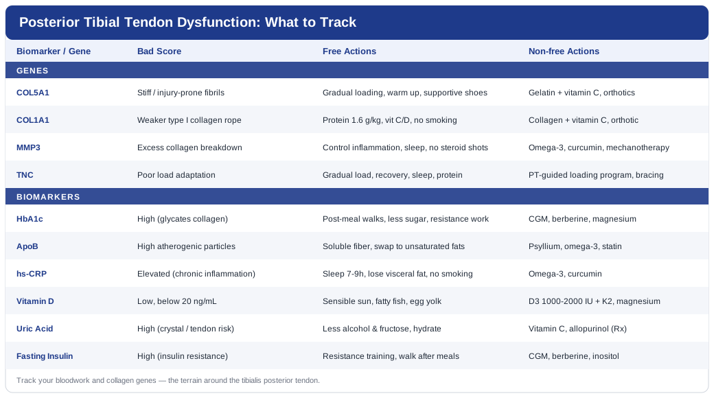
The Bloodwork That Quietly Decides How Your Tendon Heals
Before we get to individual numbers, it helps to know why blood tests belong in a conversation about a foot. Tendon is living tissue fed by small vessels and maintained by cells (tenocytes) that constantly build and break down collagen. When the internal environment turns hostile — sugary, inflamed, lipid-laden — those cells lose the plot, and repair falls behind damage. Clinical reviews of PTTD repeatedly list diabetes, obesity, and hypertension as risk factors alongside the mechanical ones, and note the condition is more common in people carrying metabolic comorbidities (Posterior Tibial Tendon Dysfunction: An Overview, PMC). So the six markers below are not a detour from your foot — they are part of it.
1. HbA1c and Fasting Glucose — the sugar that stiffens collagen
Why it matters: Chronically high blood sugar drives the formation of advanced glycation end-products (AGEs), which cross-link collagen abnormally, thicken the tendon, and reduce its ability to glide and absorb load. A review of diabetes and tendon pathology describes exactly this loss of collagen organization and blunted healing (The impact of diabetes mellitus on tendon pathology, PMC), and a companion paper shows the damage extends beyond crosslinking to the cells themselves (Effect of Diabetes on Tendon Structure and Function, PMC). HbA1c reflects your average glucose over roughly three months.
How to measure it
A fingerstick or venous HbA1c plus fasting glucose is one of the cheapest and most available tests, typically bundled into a routine panel for a few dollars to around 30 USD out of pocket. Aim to know both your HbA1c (ideally in the low-normal range, not just "under the diabetes cutoff") and your fasting glucose.If the score is bad — the plan without supplements
Prioritize a walk after your largest meals (even 10–15 minutes blunts the post-meal glucose spike), reduce refined carbohydrate and sugary drinks, and add resistance training two to three times weekly to improve insulin sensitivity. Protein and fiber before starch at a meal lowers the spike. These changes can move HbA1c over 8–12 weeks (the lifespan of red blood cells), so retest no sooner than three months. Side effects are minimal; if you are on glucose-lowering medication, coordinate changes with your clinician to avoid hypoglycemia.If the score is bad — the plan with supplements or equipment
A continuous glucose monitor (roughly 40–100 USD per two-week sensor) turns abstract advice into concrete feedback about which meals spike you. On the supplement side, evidence is modest: berberine (typically 500 mg two to three times daily with meals) can lower fasting glucose but may cause GI upset and interacts with several medications; cycle it (for example 8–12 weeks on, then reassess) rather than taking it indefinitely without monitoring. Magnesium (200–400 mg elemental at night) supports insulin signaling and is generally well tolerated aside from loose stools at higher doses. None of these replace the dietary and exercise foundation.2. ApoB (or LDL-C and the full lipid panel) — the particles that infiltrate tendon
Why it matters: Cholesterol does not just clog arteries; it accumulates in tendons. A systematic review found an association between unfavorable lipid levels — higher total cholesterol, LDL, and triglycerides, lower HDL — and altered tendon structure or tendon pain (Is higher serum cholesterol associated with altered tendon structure or tendon pain? PMC), and cross-sectional work shows cholesterol accumulation changes Achilles tendon biomechanics (The effects of cholesterol accumulation on Achilles tendon biomechanics, PMC). Lipidologists like Allan Sniderman and Thomas Dayspring argue that ApoB — a direct count of atherogenic particles — is a more reliable target than LDL-C alone, and Peter Attia has popularized measuring it routinely.
How to measure it
A standard lipid panel (total cholesterol, LDL-C, HDL-C, triglycerides) is inexpensive, often under 30 USD. ApoB is a separate, still-affordable add-on (roughly 20–40 USD) that many labs now offer directly. A non-fasting sample is acceptable for ApoB.If the score is bad — the plan without supplements
Increase soluble fiber (oats, legumes, psyllium), replace saturated fat with mono- and polyunsaturated sources (olive oil, nuts, fatty fish), lose excess visceral fat, and keep moving. These lower ApoB and triglycerides meaningfully over 6–12 weeks. Retest a lipid panel at that interval. Side effects are essentially none, though a sudden large fiber increase can cause bloating — ramp up gradually.If the score is bad — the plan with supplements or equipment
Psyllium husk (5–10 g daily with water) is a well-studied, low-risk LDL-lowering fiber; take other medications an hour apart from it. Plant sterols (about 2 g daily) modestly reduce LDL. Omega-3 (EPA/DHA, 1–2 g daily) mainly lowers triglycerides; high doses can thin blood slightly, so pause before surgery. If your ApoB is high and lifestyle is not enough — especially with a family history or tendon xanthomas — statin therapy is the evidence-based tool, and reviews note that lipid-lowering treatment has improved tendon symptoms in some patients. Discuss statins with your physician; the tendon literature around them is nuanced, not a reason to avoid a genuinely indicated drug.3. hs-CRP — the smoke detector for chronic inflammation
Why it matters: High-sensitivity C-reactive protein is a general marker of low-grade systemic inflammation, the kind that keeps tenocytes in a degradative state and slows healing. It will not diagnose PTTD, but a persistently elevated hs-CRP is a red flag that the internal environment is working against your tendon and your cardiovascular system alike.
How to measure it
hs-CRP is a cheap blood test (often 10–30 USD). Because it rises transiently with any infection or injury, retest when you are well and repeat to confirm a true baseline rather than a spike.If the score is bad — the plan without supplements
The biggest levers are losing visceral fat, sleeping 7–9 hours, quitting smoking, moving daily, and eating a whole-food, high-fiber, lower-refined-carbohydrate diet. Chronic sleep debt and abdominal fat are two of the strongest drivers. Improvements show over weeks to a few months; recheck at 8–12 weeks. No side effects — this is the healthiest possible way to lower a number.If the score is bad — the plan with supplements or equipment
Omega-3 (1–2 g EPA/DHA daily) and curcumin (500–1000 mg daily with a fat source or a bioavailability enhancer) can modestly lower inflammatory markers. Curcumin may cause GI upset and can mildly thin the blood, so cycle it around procedures and avoid combining high doses with anticoagulants without medical advice. These are supportive, not curative — a normal hs-CRP driven by supplements over an inflamed lifestyle is a false comfort.4. 25-Hydroxyvitamin D — the healing cofactor most people are low in
Why it matters: Vitamin D influences collagen quality and tendon repair. A scoping review of vitamin D in tendon healing found that deficiency was consistently associated with poorer outcomes — delayed healing, higher retear rates, weaker functional recovery — and that supplementation may help (The Role of Vitamin D in Postoperative Tendon Healing: A Scoping Review, PMC), with cohort data linking preoperative deficiency to worse rotator cuff repair results (Association of Preoperative Vitamin D Deficiency With Retear Rate, PMC). If you are heading toward surgery or serious rehab for PTTD, this is a number you want in range.
How to measure it
Ask for a 25-hydroxyvitamin D blood test (roughly 20–50 USD). Most guidelines consider deficiency below 20 ng/mL (50 nmol/L); many clinicians target a comfortable mid-range.If the score is bad — the plan without supplements
Sensible sun exposure (short, non-burning midday exposure of arms and legs several times weekly, adjusted for skin tone and latitude) and vitamin-D-rich foods (fatty fish, egg yolk, fortified products) help, but for someone truly deficient in a low-sun climate, diet and sun alone are often insufficient — which is the honest limitation of the no-supplement route here.If the score is bad — the plan with supplements or equipment
Vitamin D3 is the practical fix: a common maintenance dose is 1000–2000 IU daily, with higher short-term repletion doses sometimes used under medical guidance for significant deficiency. Take it with a fatty meal and pair it with vitamin K2 and adequate magnesium. Do not megadose blindly — vitamin D is fat-soluble and toxicity (hypercalcemia) is real at very high sustained intakes. Retest after 8–12 weeks and adjust. This is one of the cheapest, best-evidenced tendon-supportive supplements available.5. Serum Uric Acid — the crystal risk hiding in a tendon
Why it matters: Elevated uric acid is not only about gout attacks in the big toe. Research shows asymptomatic hyperuricemia is associated with tendon damage and even Achilles tendon rupture, by disrupting tendon stem/progenitor cells (Asymptomatic Hyperuricemia Is Associated with Achilles Tendon Rupture, PMC). For a tendon already under strain, a high uric acid load is one more headwind.
How to measure it
Serum uric acid is a standard, inexpensive test (often 10–25 USD), frequently included in metabolic panels.If the score is bad — the plan without supplements
Cut back on alcohol (especially beer), sugary drinks and high-fructose foods, and very high purine intake (organ meats, excessive shellfish). Stay well hydrated and lose excess weight gradually — crash dieting can transiently raise uric acid. Changes show within weeks; retest at 6–8 weeks. Side effects are none beyond the discipline required.If the score is bad — the plan with supplements or equipment
Vitamin C (500 mg daily) has a modest uric-acid-lowering effect and is low risk. Tart cherry intake is popular and may help symptoms, though evidence is limited. If uric acid is high with a history of gout or tophi, a prescription urate-lowering drug (such as allopurinol) is the definitive treatment and is far more effective than any supplement — this is a physician conversation, not a supplement experiment.6. Fasting Insulin and HOMA-IR — the early-warning metabolic marker
Why it matters: Fasting insulin rises long before glucose does, making it an early signal of insulin resistance — the metabolic soil in which tendinopathy and metabolic syndrome grow. Peter Attia frequently emphasizes measuring fasting insulin (and calculating HOMA-IR) precisely because it flags trouble years before HbA1c budges. Because metabolic syndrome blunts tendon healing, this marker connects directly to your foot.
How to measure it
Fasting insulin is a blood test (roughly 15–40 USD) drawn after an overnight fast; combined with fasting glucose it yields HOMA-IR, a simple insulin-resistance estimate. Lower fasting insulin generally indicates better metabolic flexibility.If the score is bad — the plan without supplements
The same levers that fix glucose fix insulin: build muscle with resistance training, walk after meals, prioritize protein and fiber, reduce refined carbohydrate, improve sleep, and shed visceral fat. This is the highest-leverage set of habits in the entire article because it moves several of these six markers at once. Retest after about 12 weeks.If the score is bad — the plan with supplements or equipment
A continuous glucose monitor again shines here as feedback equipment. Berberine and magnesium (dosed as under HbA1c above) can support insulin sensitivity, with the same caveats — cycle berberine and watch for GI effects and drug interactions. Inositol (2–4 g daily) is another well-tolerated option with reasonable evidence for insulin sensitivity. Treat all of these as amplifiers of, not substitutes for, training and diet.Bloodwork tells you about the environment your tendon lives in. The next layer is the blueprint the tendon was built from — your genes.
The Collagen Blueprint: Genes That Stack the Deck
Biomarkers are things you can change month to month. Genes are fixed, but knowing them explains why some people's tendons fray under loads others tolerate — and, importantly, several genetic risks can be compensated for through the very habits and markers above. Genomics figures like Ali Torkamani have argued that this kind of individualized risk information is most useful when it changes what you actually do. Here are four genes with real human evidence in tendon pathology. Note that most of this research is on Achilles and other tendons rather than the tibialis posterior specifically, so treat it as informative background, not a diagnosis.
COL5A1 — the "stiff vs. stretchy tendon" gene
COL5A1 encodes part of type V collagen, a master regulator of how collagen fibrils assemble. A well-studied variant (rs12722) in its 3'-untranslated region is associated with chronic Achilles tendinopathy and with range-of-motion and tendon stiffness differences (Polymorphisms within the COL5A1 3'-UTR and Achilles tendinopathy, PubMed), and a meta-analysis of 21 studies links it to musculoskeletal soft-tissue injuries (COL5A1 rs12722 and musculoskeletal soft tissue injuries: meta-analysis, PMC).
What it may affect: fibril structure, tendon stiffness, and injury susceptibility.
If the gene is bad — the plan without supplements
Favor gradual, progressive loading over sudden spikes in activity; a stiffer or more injury-prone tendon punishes "weekend warrior" jumps in volume. Warm up thoroughly, respect longer recovery windows, and support the arch with appropriate footwear or orthotics to reduce chronic overload on the tibialis posterior. Consistency beats intensity.If the score is bad — the plan with supplements or equipment
The gelatin-plus-vitamin-C loading strategy (see the book summary below) aims to boost collagen synthesis around targeted exercise: roughly 15 g of gelatin or hydrolyzed collagen with about 50 mg vitamin C taken 30–60 minutes before loading, a few times weekly. It is low risk and inexpensive; the main caveat is that human tendon-outcome evidence is still emerging. Well-fitted orthotics or a supportive brace are the "equipment" that most directly offsets a structurally vulnerable tendon.MMP3 — the demolition-crew gene
MMP3 encodes a matrix metalloproteinase that breaks down and remodels collagen. A variant (rs679620) is associated with Achilles tendinopathy and appears to interact with COL5A1 to modify risk (Variants within the MMP3 gene are associated with Achilles tendinopathy, PubMed).
What it may affect: the balance between collagen breakdown and rebuilding during remodeling and repair.
If the gene is bad — the plan without supplements
Because MMP activity is stoked by inflammation, the highest-value move is to keep the inflammatory markers above (hs-CRP, glucose, uric acid) in good ranges through sleep, weight management, and an anti-inflammatory diet. Avoid repeated corticosteroid injections into the tendon, which can further weaken tissue.If the score is bad — the plan with supplements or equipment
Omega-3 and curcumin (dosed as under hs-CRP) may help tilt the remodeling balance by lowering inflammation; cycle curcumin around any procedures. Structured mechanotherapy — progressive tendon loading under a physiotherapist — is the "equipment/protocol" that healthily stimulates remodeling in the right direction.COL1A1 — the main rope fiber
COL1A1 encodes type I collagen, the dominant structural protein in tendon. An Sp1-binding-site polymorphism has been studied in Achilles tendon injuries, with some variants appearing protective (Sp1-binding site polymorphism in COL1A1 and Achilles tendon injuries, PubMed).
What it may affect: the quantity and quality of the primary collagen that gives tendon its tensile strength.
If the gene is bad — the plan without supplements
Maximize the raw materials and stimulus for good collagen: adequate protein (roughly 1.6 g/kg/day spread across meals), sufficient vitamin C and vitamin D from food and sun, and progressive loading. Do not smoke — tobacco impairs collagen synthesis and tendon blood supply.If the score is bad — the plan with supplements or equipment
Collagen or gelatin plus vitamin C before loading (as above), plus ensuring vitamin D repletion, targets collagen production directly. These are low-risk. A supportive orthotic reduces the tensile demand on a potentially weaker rope while you rebuild it.TNC (Tenascin-C) — the load-response gene
TNC encodes tenascin-C, a matrix protein upregulated in response to mechanical load and involved in tendon repair; variants have been associated with Achilles tendon injury in early genetic studies. The evidence base here is smaller and less replicated than for COL5A1, so treat it as suggestive.
What it may affect: how the tendon matrix responds and adapts to mechanical loading.
If the gene is bad — the plan without supplements
Give load-adaptation genes the best chance by loading intelligently: gradual progression, enough recovery between sessions, and avoiding sudden training spikes. Sleep and protein again underpin adaptation.If the score is bad — the plan with supplements or equipment
There is no specific supplement for TNC. The practical "equipment" answer is a well-structured, physiotherapist-guided loading program and appropriate footwear or bracing to keep loads within a tolerable, adaptive range rather than a destructive one.Genes set the starting line, but connective tissue is famously responsive to how you treat it — which is exactly the argument of the book below.
The Connective-Tissue Playbook That Rewrites "Just Rest It"
If you want one resource that reframes tendon and joint problems around biology rather than resignation, Built from Broken by Scott Hogan is a strong, study-referenced choice. Its central, somewhat contrarian message is that connective tissue is not doomed to degrade with age and that targeted loading, nutrition, and habits can meaningfully rebuild it — a direct challenge to the "rest, brace, and accept decline" script many people are given. Here are ten of its most useful ideas, adapted to a foot tendon.
1. Rest is a starting point, not a treatment
Prolonged rest can calm pain but often weakens tendon, delaying real recovery. Controlled loading, not avoidance, is what rebuilds capacity.2. Tendons adapt to progressive load
Tendon responds to gradually increasing mechanical stress by remodeling and strengthening. The art is dosing load high enough to stimulate and low enough to avoid flaring.3. Collagen synthesis has a timing window
The book highlights research (associated with Keith Baar) that taking gelatin or collagen with vitamin C shortly before loading can raise collagen synthesis around the exercise window — a practical, low-cost tool.4. Inflammation is a signal, not just an enemy
Acute inflammation is part of repair; chronic, systemic inflammation is the problem. Blunting all inflammation with constant anti-inflammatories can impair healing.5. Metabolic health is tendon health
The same blood sugar, lipid, and weight factors that protect your heart protect your tendons — echoing the biomarker section above.6. Sleep is where connective tissue rebuilds
Deep sleep drives the hormonal environment for repair. Chronic sleep debt undermines everything else you do.7. Protein and specific nutrients are raw materials
Adequate protein, vitamin C, vitamin D, and minerals are non-negotiable inputs for building new collagen.8. Isometrics can reduce tendon pain
Holding a muscle under tension without movement can reduce tendon pain and let you keep loading — useful when dynamic exercise is too painful.9. Pain does not always equal damage
Understanding modern pain science reduces fear-driven avoidance, which itself worsens outcomes.10. Consistency compounds
Connective tissue changes slowly; small, consistent inputs over months beat sporadic heroic efforts. This is arguably the single most important mindset for a tendon problem.The book's loading philosophy dovetails neatly with the best-evidenced rehab research: controlled, progressive tendon loading. That principle also underlies most of the complementary options worth considering.
Complementary Approaches Worth Considering
The modalities below have at least some genuine human evidence relevant to tendon health, load tolerance, or the pain and balance problems that accompany PTTD. None is a stand-alone cure, and for a collapsing arch the mechanical foundation — loading, footwear, orthotics, and sometimes surgery — remains primary. Where evidence is thin, it is flagged honestly.
Progressive Loading and Eccentric/Heavy-Slow Resistance Exercise
This is the closest thing to a cornerstone non-surgical treatment for tendinopathy, and while most trials study the Achilles, the loading principle transfers to the tibialis posterior. Progressive resistance retrains the tendon to tolerate force rather than avoiding it.
A meta-analysis found eccentric exercise more effective than other exercise approaches for mid-portion Achilles tendinopathy (Eccentric exercise for mid-portion Achilles tendinopathy: systematic review and meta-analysis, PMC), and a randomized controlled trial showed heavy slow resistance training worked as well as eccentric training with high patient satisfaction (Heavy Slow Resistance vs. Eccentric Training for Achilles Tendinopathy: RCT, PubMed).
Practically, for PTTD this usually means physiotherapist-guided calf raises and specific tibialis posterior strengthening (such as resisted foot inversion and heel raises, often progressing to single-leg work), performed slowly and progressed over weeks. Expect some tolerable discomfort, not sharp pain, and give it two to three months. This is the intervention to prioritize if you do only one.
Low-Level Laser Therapy / Photobiomodulation
Low-level laser therapy (LLLT) uses specific wavelengths of light aiming to reduce pain and support tissue repair, and it is commonly offered in physiotherapy clinics for tendon and foot complaints.
A systematic review and meta-analysis of randomized trials found LLLT effective for reducing pain and disability in lower-extremity tendinopathy and plantar fasciitis (Efficacy of low-level laser therapy in lower extremity tendinopathy or plantar fasciitis, PMC). That said, umbrella reviews caution that overall certainty of evidence remains low to moderate.
Realistically, treat LLLT as a possible adjunct to loading, not a replacement for it — worth trying if pain is limiting your ability to exercise, ideally at clinics that follow recognized dosage guidelines. It is non-invasive and low-risk, so the main downsides are cost and time rather than harm.
Tai Chi for Balance and Foot Loading
PTTD degrades balance and alters how you load your foot, and falls become a real concern as the arch collapses. Tai chi is a gentle, weight-shifting practice that directly trains balance, proprioception, and lower-limb control.
Meta-analyses show tai chi improves balance and reduces falls in older adults (Tai Chi for fall prevention and balance improvement: systematic review and meta-analysis, PMC), and a pilot study in older adults with type 2 diabetes reported improved ankle proprioception and reduced forefoot plantar pressure after eight weeks (Tai Chi effects on somatosensation and balance in older adults with type 2 diabetes, PMC) — directly relevant to how the foot bears load.
Apply it as a low-intensity complement two to three times weekly, ideally in supportive footwear or an orthotic if standing barefoot on a flattening arch is painful. It will not correct a deformity, but it can improve stability and confidence while you address the tendon itself.
Mindfulness Meditation / MBSR for Persistent Pain
Chronic foot pain wears on mood and sleep, and stress amplifies pain perception. Mindfulness-based stress reduction (MBSR) trains attention and a less reactive relationship to pain.
Mindfulness has moderate evidence for reducing chronic musculoskeletal pain intensity and improving coping and quality of life in systematic reviews, though effects are generally modest and not tendon-specific.
Used realistically, a short daily practice (10–20 minutes) or a structured MBSR course can help you tolerate rehab, sleep better (which supports tissue repair), and avoid the fear-avoidance spiral that keeps people from loading their tendon. It is essentially risk-free and pairs well with everything else here.
Massage Therapy as a Symptom Adjunct
Soft-tissue massage around the calf and inner ankle can temporarily reduce muscle tension and discomfort, making it easier to perform rehab exercises.
The evidence for massage in tendinopathy specifically is limited and mostly short-term, so it should be framed as symptom relief rather than a structural fix.
Practically, occasional massage or self-massage can be a reasonable comfort measure between loading sessions, but spend your primary time and money on progressive strengthening and, where indicated, proper orthotic support. Avoid aggressive deep pressure directly over an acutely painful, swollen tendon.
Taken together, these approaches share a theme: they help you keep loading and stay stable while the real work — metabolic health, targeted strengthening, and mechanical support — does the heavy lifting.
Conclusion
Posterior tibial tendon dysfunction is often presented as a simple mechanical breakdown, but the fuller picture is that your tendon heals or degrades inside a body-wide environment you can actually measure and influence. Six markers — HbA1c, ApoB, hs-CRP, vitamin D, uric acid, and fasting insulin — give you a practical dashboard, and most of them respond to the same handful of habits: build muscle, walk after meals, sleep well, eat whole foods, and keep visceral fat down. Your collagen genes may stack the deck, but they mostly raise the stakes on those very same habits rather than overriding them.
The most encouraging takeaway is how much overlap there is: the choices that lower your cardiovascular risk are, in large part, the choices that give a struggling tendon its best shot at repair. That is leverage worth using.
So take the next smart step rather than the dramatic one. Get the bloodwork, note which numbers are off, start a progressive loading program with a physiotherapist, make sure your footwear and any orthotics actually support the arch — and bring this information to a qualified clinician who can weigh it against your specific foot, imaging, and stage of the condition. Better information, acted on early and consistently, is how good outcomes are made.
Musculoskeletal Endocrine & Metabolic
Musculoskeletal: Tendon & Ligament Conditions