This article contains AI generated content.
· UpdatedPosterolateral Corner Injury: 5 Genes And 6 Biomarkers To Track
Introduction
A posterolateral corner (PLC) injury is one of the most complex and frequently missed injuries in the knee. It involves a cluster of structures — the lateral collateral ligament, popliteus tendon, popliteofibular ligament, and surrounding capsule — that together provide rotational and varus stability. When this corner is compromised, the consequences extend far beyond the initial trauma: chronic instability, progressive cartilage wear, and incomplete recovery despite surgery are all too common.
What most standard rehabilitation guides miss is that two people with the same MRI report and the same surgical outcome can have radically different recovery trajectories. One rebuilds the joint fully in twelve months. The other struggles with persistent laxity, inflammation, and reinjury for years. The difference often lies beneath the surface — in how their biology generates and degrades connective tissue, manages inflammation, and responds to mechanical load.
Generic protocols rarely answer the question that actually matters: why is this specific person healing poorly, or what biological factors are quietly stacking the deck against them? That is where biomarkers and genetics become more than academic topics. They become practical navigation tools for clinicians, athletes, and patients who want to do more than wait and hope.
This article takes that deeper approach. The primary section breaks down six biomarkers that are directly relevant to PLC injury, explaining what each reveals, how to measure it affordably, and what to do when the score is unfavorable — both with and without supplements. A shorter genetics section follows, covering five genes with meaningful evidence for connective tissue vulnerability. Together, these two lenses offer a genuinely actionable framework for anyone navigating PLC injury recovery or trying to prevent recurrence.
6 Biomarkers to Track After a Posterolateral Corner Injury
Biomarkers do not replace imaging or clinical assessment, but they add a layer of biological specificity that neither an MRI nor a physical exam can provide. For PLC injuries specifically, the right panel can reveal whether systemic inflammation is actively undermining healing, whether connective tissue turnover is running in the wrong direction, or whether a key nutrient deficiency is quietly stalling collagen synthesis. The six below represent the most clinically useful and measurable options — selected for relevance to ligament biology, accessibility, and actionability.
1. High-Sensitivity C-Reactive Protein (hs-CRP)
Why it matters. CRP is the liver's primary acute-phase protein, released in response to inflammatory cytokines like IL-6 and TNF-alpha. After a PLC injury, a short-term elevation is expected and appropriate — it is part of the healing cascade. The problem arises when hs-CRP remains elevated for weeks or months, signaling that low-grade systemic inflammation is ongoing. In that state, fibroblast activity is suppressed, collagen maturation is impaired, and the mechanical quality of healing tissue is compromised. Research consistently shows that chronically elevated CRP is associated with slower soft tissue repair and higher rates of re-injury in ligamentous structures.
How to measure it. A standard hs-CRP blood draw is available at any primary care clinic or direct-to-consumer lab. Cost typically ranges from $15 to $40. Optimal is below 1 mg/L; borderline is 1–3 mg/L; elevated is above 3 mg/L. At Peter Attia's standard, values above 2 mg/L warrant investigation.
If the score is bad — the plan without supplements. The most powerful non-supplement lever for hs-CRP is sleep quality. Deep, uninterrupted sleep of 7.5 to 9 hours per night is strongly anti-inflammatory. A whole-food, low-glycemic diet — specifically one that eliminates refined seed oils, processed carbohydrates, and added sugar — reduces baseline CRP significantly within four to six weeks. Regular low-intensity movement (walking, gentle cycling) rather than prolonged rest has also been shown to reduce CRP more effectively than bed rest, even in injured individuals. Cold exposure through ice baths or contrast therapy can temporarily modulate inflammatory signaling in the injured limb.
If the score is bad — the plan with supplements or equipment. Omega-3 fatty acids (EPA + DHA) at 2–4g per day have robust human evidence for reducing hs-CRP in both healthy and injured populations. This is a daily, ongoing supplement with no cycling required; common at-scale doses can be well tolerated long-term but monitor for GI effects. Curcumin with piperine (500–1000mg of a high-bioavailability form such as Theracurmin or Meriva, daily) has shown consistent CRP-lowering effects in randomized trials. For optimal tolerability, cycle curcumin at 8 weeks on and 2 weeks off. Boswellia serrata (AKBA fraction, 100–200mg daily) adds a complementary anti-inflammatory pathway via 5-LOX inhibition with good tolerability for extended use. From an equipment standpoint, infrared sauna sessions (20–30 minutes, 3–4 times per week) have shown meaningful hs-CRP reductions in moderate-quality trials.
2. COMP — Cartilage Oligomeric Matrix Protein
Why it matters. COMP is a non-collagenous glycoprotein found in the extracellular matrix of cartilage, tendons, and ligaments. When these tissues are under mechanical stress or actively degrading, COMP is released into the bloodstream. For PLC injuries, where both the ligamentous structures and adjacent tibial cartilage are under abnormal load, serum COMP acts as a real-time stress gauge. Elevated COMP after injury can indicate excessive loading, ongoing structural breakdown, or a transition toward secondary osteoarthritis. Several studies have validated COMP as an early predictor of cartilage deterioration in knee injury populations — making it particularly valuable in PLC cases where the standard of care does not always include post-surgical biomarker monitoring.
How to measure it. COMP requires a serum blood draw and is processed at specialty or rheumatology-oriented labs. It is not a routine panel item. Cost: $60–$120. Some rheumatology-oriented joint health panels include it alongside other inflammatory markers. Reference ranges vary by lab, but values above 12 U/L (ELISA-based methods) are typically considered elevated in adult athletes.
If the score is bad — the plan without supplements. The most direct lever for COMP is load management. Reducing compressive and rotational joint loading — through activity modification, temporary use of an unloading knee brace, and gait retraining — is the first intervention. Aquatic therapy is especially useful here because it allows active rehabilitation at dramatically lower joint-reaction forces. Proprioceptive and neuromuscular training that restores PLC-specific stability reduces abnormal load distribution on the cartilage adjacent to the injured structures.
If the score is bad — the plan with supplements or equipment. Hydrolyzed collagen peptides (10–15g daily, taken with vitamin C 30–60 minutes before mechanical loading) have shown measurable improvements in cartilage and ligament tissue quality in randomized studies. This is a daily supplement with no cycling needed; long-term use is considered safe. Glucosamine sulfate (1500mg daily) has the most evidence for reducing COMP elevation in early osteoarthritic knees and in post-traumatic joint stress scenarios. A combination with chondroitin sulfate (1200mg daily) may be additive. Blood flow restriction (BFR) training is an equipment-based approach that stimulates cartilage matrix anabolism at very low mechanical loads — a critical advantage when the joint cannot tolerate conventional resistance training.
3. Interleukin-6 (IL-6)
Why it matters. IL-6 is the primary upstream signal that drives CRP production in the liver, but it carries its own clinical meaning beyond what CRP captures. In the knee joint specifically, IL-6 is produced locally by synoviocytes and macrophages after ligamentous injury, and it acts directly on fibroblasts to modulate collagen synthesis. At low concentrations following acute injury, IL-6 is anabolic and beneficial. At chronically elevated levels, it shifts toward a catabolic and anti-regenerative phenotype — reducing fibroblast proliferation, impairing tendon-to-bone healing, and promoting synovial inflammation. Elevated serum IL-6 weeks after PLC injury is a signal that the local inflammatory environment is not resolving as expected.
How to measure it. IL-6 is measurable via a standard serum blood draw. It is available at most hospital labs and through direct-to-consumer platforms. Cost: $60–$120. Optimal values are below 2 pg/mL in a resting adult; values above 5–7 pg/mL in a non-acute setting warrant investigation.
If the score is bad — the plan without supplements. Sleep is again the most impactful free lever — IL-6 follows a circadian rhythm and is strongly suppressed by adequate deep sleep. Chronic psychological stress (via cortisol) upregulates IL-6, so stress management practices such as structured breathing protocols (box breathing, 4-7-8 method practiced for 10–15 minutes daily) directly affect this marker. Reducing visceral adipose tissue through a caloric deficit and sustained low-intensity cardio is one of the most durable ways to lower baseline IL-6, since adipose tissue is a major source of IL-6 secretion.
If the score is bad — the plan with supplements or equipment. Omega-3 fatty acids work through the same pathway here as for CRP reduction — 2–4g EPA/DHA daily, ongoing. Quercetin (500–1000mg daily) has shown direct IL-6 suppression in human studies; cycle at 8 weeks on, 2 weeks off to preserve sensitivity. Magnesium glycinate (300–400mg nightly) has modest but consistent evidence for reducing inflammatory cytokines including IL-6, with the added benefit of improving sleep quality — a two-for-one intervention. Photobiomodulation (red light therapy, 630–850nm, 10–15 minutes over the injured joint, 4–5 days per week) has shown reductions in local IL-6 in soft tissue injury models.
4. Vitamin D (25-OH)
Why it matters. Vitamin D is not a vitamin in the traditional sense — it is a steroid hormone that regulates over 900 genes, including several directly involved in musculoskeletal healing. For ligament and tendon repair, vitamin D receptors are expressed in fibroblasts, tenocytes, and bone cells, making it a key regulator of the collagen synthesis pathway. Deficiency below 30 ng/mL is associated with impaired ligament-to-bone integration, reduced muscle force output (which matters enormously for PLC-supporting musculature like the biceps femoris and popliteus), and higher rates of re-injury. Multiple studies in athlete populations have found that those with vitamin D levels below 32 ng/mL have significantly higher soft tissue injury rates and slower recovery timelines.
How to measure it. A 25-OH vitamin D blood test is widely available and inexpensive. Cost: $40–$80. Most standard panels include it, or it can be ordered individually. Optimal for healing is generally considered 40–70 ng/mL; below 30 ng/mL is clinically deficient; below 20 ng/mL is severely deficient.
If the score is bad — the plan without supplements. Direct midday sun exposure of 15–25 minutes per day on large skin surface areas (arms, legs, back) can meaningfully raise vitamin D levels over 6–8 weeks in fair-skinned individuals. Dietary sources — fatty fish (salmon, mackerel), egg yolks, liver — contribute modestly but are insufficient alone to correct deficiency. Dark-skinned individuals and those at northern latitudes above 40° will find sun exposure largely ineffective from October to April.
If the score is bad — the plan with supplements or equipment. Vitamin D3 supplementation at 3,000–5,000 IU daily is the most evidence-based starting point; retest after 90 days. Critically, always pair with vitamin K2 (100–200mcg MK-7 form daily) to direct calcium to bone rather than soft tissue. This combination is safe for long-term, year-round use. At very low levels (below 20 ng/mL), a loading protocol of 10,000 IU daily for 4–6 weeks followed by a maintenance dose is sometimes used under medical supervision. Magnesium is a cofactor for vitamin D activation — deficiency in magnesium blunts the response to supplementation, making magnesium glycinate a worthwhile co-supplement.
5. Matrix Metalloproteinase-3 (MMP-3)
Why it matters. MMP-3 (stromelysin-1) is an enzyme that degrades multiple extracellular matrix components including collagen types II, III, IV, and IX, fibronectin, and proteoglycans. In normal tissue remodeling, MMP-3 plays an essential role in clearing damaged matrix to allow regeneration. In the context of a PLC injury, persistently elevated MMP-3 indicates that the remodeling phase is dysregulated — the tissue is being broken down faster than it can be rebuilt. This imbalance is a significant driver of failed ligament healing and post-traumatic joint degeneration. Serum MMP-3 has been studied as a biomarker of synovitis severity and cartilage degradation in rheumatoid arthritis and post-traumatic knee conditions, with elevated levels correlating with worse structural outcomes.
How to measure it. MMP-3 is measured via serum blood draw and is available at specialty labs and some rheumatology clinics. Cost: $80–$150. Standard reference ranges are typically below 28 ng/mL in adult females and below 59 ng/mL in adult males (ranges vary by lab and assay). Elevated levels in the context of known knee injury have prognostic significance even within the "normal" reference window.
If the score is bad — the plan without supplements. Reducing mechanical overload is the primary lever — MMP-3 activity in joint tissue is upregulated by excessive compressive and shear forces. Progressive, structured rehabilitation that gradually reintroduces load without exceeding the tissue's current capacity is the most effective free intervention. A whole-food, anti-inflammatory diet low in processed carbohydrates and high in polyphenols naturally inhibits MMP activity through multiple pathways. Intermittent fasting (16:8 protocol daily) has shown measurable reductions in MMP expression via reduced NF-κB signaling in several human metabolic studies.
If the score is bad — the plan with supplements or equipment. EGCG from green tea extract (400–600mg standardized EGCG daily, taken with food) is among the best-studied natural MMP inhibitors with relevant human trial data; cycle at 12 weeks on, 4 weeks off and monitor liver enzymes at high doses. Resveratrol (250–500mg daily with a meal) inhibits MMP-3 expression through SIRT1 activation; long-term safety is well established at these doses. Doxycycline at sub-antimicrobial doses is used in clinical practice as an MMP inhibitor (notably in periodontal disease), and while evidence for musculoskeletal application is emerging, this is a prescription-only option requiring a physician's involvement. BFR training again offers a structural benefit by stimulating matrix synthesis without the high joint forces that upregulate MMP-3.
6. CTX-II (C-Terminal Telopeptide of Type II Collagen)
Why it matters. CTX-II is a fragment released into urine when type II collagen — the primary structural collagen of articular cartilage — is cleaved by enzymes during cartilage breakdown. It is one of the most specific biomarkers of articular cartilage degradation available without invasive joint sampling. For PLC injuries, where abnormal joint mechanics place excessive stress on the lateral tibial plateau and associated cartilage, CTX-II provides a window into whether that stress is crossing the threshold into structural tissue loss. Elevated CTX-II in the months following a PLC injury is a meaningful warning sign that secondary osteoarthritic changes may be beginning — and that load management and matrix support interventions need to be prioritized immediately.
How to measure it. CTX-II is measured in a second-morning urine sample (to control for diurnal variation) and processed at specialty labs. Cost: $100–$200. It is normalized to creatinine concentration. Reference ranges vary, but persistently elevated values above the 90th percentile for age-matched populations in the context of known knee injury warrant clinical attention.
If the score is bad — the plan without supplements. Body weight management is the most direct free intervention — each kilogram of body weight adds roughly 3–4 kilograms of force across the knee during walking. A 5% reduction in body weight in overweight individuals reduces CTX-II meaningfully. Swimming and aquatic exercise allow joint movement and cardiovascular work without cartilage compression. Sleeping in a position that reduces prolonged knee joint compression can also contribute modestly.
If the score is bad — the plan with supplements or equipment. Type II collagen (undenatured, UC-II form, 40mg daily) is distinct from hydrolyzed collagen peptides and has a different mechanism — it works via oral tolerance to reduce autoimmune-like cartilage degradation. It has shown reductions in CTX-II in clinical trials for osteoarthritis; use daily, ongoing, with no cycling required. Glucosamine sulfate (1500mg daily) with chondroitin sulfate (1200mg daily) remains the combination with the broadest evidence base for reducing CTX-II excretion over 3–6 month periods. Vitamin C at 500–1000mg daily provides the critical cofactor for hydroxylation of proline and lysine in collagen synthesis — without adequate vitamin C, collagen quality is directly impaired.
What Genetics Can Reveal About Posterolateral Corner Vulnerability
Genetics does not determine destiny, but it does shape the biological landscape in which injury and recovery occur. Several gene variants have been linked to connective tissue quality, ligament laxity, and inflammatory response — all directly relevant to PLC injury risk and recovery trajectory. Understanding your genetic profile does not change the past, but it can sharply focus the interventions that will matter most going forward.
COL5A1 — The Collagen V Gene
What it affects. COL5A1 encodes the alpha-1 chain of type V collagen, which regulates the diameter of type I collagen fibrils in ligaments and tendons. Variants in this gene — particularly the BstUI and DpnII RFLP polymorphisms — have been consistently associated with ACL and other soft tissue injuries in multiple athletic populations. The mechanism is a reduction in fibril uniformity, leading to ligaments with less tensile stiffness and more susceptibility to mechanical failure under unexpected load. A 2009 study by Collins et al. in the British Journal of Sports Medicine found that the TT genotype at the 3' UTR was significantly overrepresented in athletes with chronic Achilles tendinopathy and soft tissue injury history.
If the gene is bad — the plan without supplements. Proprioceptive and neuromuscular training is the most effective compensatory strategy. Because the structural stiffness of the ligament is reduced, the neuromuscular system must take on a larger role in joint stabilization. Daily single-leg balance training (progressing to perturbation-based drills on unstable surfaces), lateral bounding, and reactive agility work are key. Avoiding sudden high-speed valgus or varus loading movements, especially in fatigue states, is critical injury prevention. Extended warm-up periods before high-demand activity improve ligament viscoelastic properties even with suboptimal baseline stiffness.
If the gene is bad — the plan with supplements or equipment. Hydrolyzed collagen peptides (15g daily with vitamin C, 30 minutes before loading) directly support collagen fibril synthesis in tendons and ligaments. Custom functional bracing during high-risk activities (cutting sports, heavy lifting) serves as an external mechanical substitute for reduced ligament stiffness. Vitamin C (500–1000mg daily) is non-negotiable as a cofactor for collagen cross-linking.
COL3A1 — The Collagen III Gene
What it affects. COL3A1 encodes type III collagen, which is the predominant collagen in the early phases of ligament healing and in the walls of blood vessels supplying healing tissue. Variants in this gene are associated with connective tissue disorders including joint hypermobility and vascular fragility. In the context of PLC injury, COL3A1 polymorphisms may impair the initial healing scaffold that bridges the gap in torn ligamentous tissue, resulting in slower and weaker early-phase repair.
If the gene is bad — the plan without supplements. The priority is protecting the early healing phase. This means strict compliance with non-weight-bearing or protected weight-bearing protocols in the first 4–6 weeks post-injury, avoiding the temptation to accelerate return to load. Progressive tension through graded exercise (isometric → isotonic → dynamic) follows a longer timeline than standard protocols.
If the gene is bad — the plan with supplements or equipment. Vitamin C (1000mg daily, ongoing) is essential for hydroxyproline formation in type III collagen. Proline (1–2g daily) as a free amino acid supplement directly supplies the substrate for collagen synthesis and is safe long-term. Silica supplementation from bamboo extract (300mg daily of organic silica) has some early human evidence for supporting collagen cross-linking — lower-evidence but low-risk.
TNXB — Tenascin-X Gene
What it affects. Tenascin-X is an extracellular matrix glycoprotein encoded by TNXB. It facilitates the proper assembly and spacing of collagen fibrils in connective tissue. Haploinsufficiency or functional variants in TNXB are a well-documented cause of hypermobility Ehlers-Danlos syndrome (hEDS) and related joint hyperlaxity phenotypes. Even partial loss-of-function variants produce a milder hypermobility phenotype that significantly increases the risk of multi-ligament injuries — including PLC injuries, which can occur more readily when the joint lacks baseline restraining stiffness.
If the gene is bad — the plan without supplements. Stability-focused resistance training is central to compensation. Strengthening the popliteus, biceps femoris (long and short heads), and lateral gastrocnemius — the key dynamic stabilizers of the posterolateral corner — provides neuromuscular substitution for structural laxity. Exercise selection should minimize end-range loading in vulnerable positions. Tape and bracing during high-demand activity provides meaningful mechanical restraint when intrinsic stiffness is reduced.
If the gene is bad — the plan with supplements or equipment. Magnesium (300–400mg glycinate form nightly) and boron (3–6mg daily from whole food or supplement) have some evidence for supporting extracellular matrix synthesis. Hyaluronic acid supplementation (150–200mg daily of oral high-molecular-weight form) supports the joint matrix environment in which tenascin-X normally operates.
MMP3 Gene Variants
What it affects. Beyond the enzyme itself (tracked as a biomarker), the MMP3 gene has a well-characterized promoter polymorphism at position -1171 (5A/6A) that affects transcriptional activity. The 5A allele is associated with higher MMP-3 expression, meaning individuals carrying two 5A alleles degrade their connective tissue matrix more aggressively than those with the 6A/6A genotype. This has been studied in ACL and tendon injury populations, where 5A/5A individuals show both higher injury rates and slower structural recovery.
If the gene is bad — the plan without supplements. Anti-inflammatory lifestyle factors — high sleep quality, low-glycemic diet, controlled exercise volume — are more impactful in MMP3 5A carriers because the basal level of matrix degradation activity is elevated. Avoiding overtraining states, which flood the joint with catabolic cytokines, is particularly important.
If the gene is bad — the plan with supplements or equipment. EGCG from green tea extract (400–500mg daily, 12 weeks on / 4 weeks off) directly inhibits MMP-3 transcriptional activity and is well matched to this specific variant. Curcumin reduces NF-κB-driven MMP-3 upregulation — relevant for both the gene variant and the chronically elevated enzyme. Use 500–1000mg of a high-bioavailability form daily, cycling as noted above.
IGF1 — Insulin-Like Growth Factor 1 Gene
What it affects. IGF1 is the primary anabolic growth factor for tendon, ligament, and cartilage repair. Variants in the IGF1 gene and its promoter — particularly the (CA)n microsatellite repeat — affect circulating IGF-1 levels and tissue responsiveness. Lower IGF-1 signaling capacity is associated with impaired collagen synthesis, slower satellite cell recruitment in muscle, and reduced fibroblast proliferation in ligament tissue. In the context of PLC recovery, this translates to slower structural healing and reduced final tissue quality.
If the gene is bad — the plan without supplements. Sleep is the primary driver of endogenous IGF-1 production — the large pulse of growth hormone during slow-wave sleep is the main stimulus. Optimizing sleep duration and quality (dark room, no screens after sunset, consistent sleep and wake times) is the most powerful free intervention. Resistance training — even at low loads — significantly upregulates IGF-1 production locally in exercised tissues; this is available to anyone regardless of genotype.
If the gene is bad — the plan with supplements or equipment. Zinc (15–25mg daily as zinc glycinate or picolinate) is a direct cofactor for IGF-1 production and receptor signaling, and deficiency is common in active individuals. Creatine monohydrate (3–5g daily, no loading required, no cycling needed) has modest but consistent evidence for amplifying IGF-1-driven anabolic signaling in musculoskeletal tissue. Leucine-rich protein intake (whey or essential amino acid blend with at least 3g leucine per dose, 2–3 times daily) maximally stimulates mTORC1 and downstream IGF-1 signaling in healing tissue.
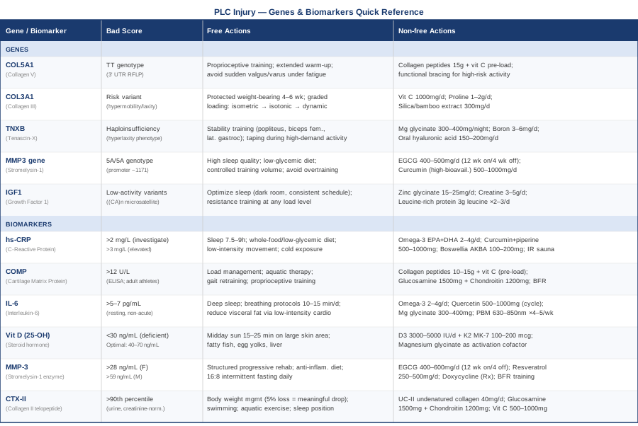
Quick Reference: Genes and Biomarkers at a Glance
What Andrew Huberman's Work on Connective Tissue Recovery Can Change for You
The Huberman Lab podcast episode titled "Improving Your Gut Microbiome Health" is not the relevant episode here — but his two-part series on "Tools to Optimize Your Connective Tissue, Tendons and Ligaments" synthesizes a surprisingly actionable body of science that most orthopedic rehabilitation programs never mention. What makes this material particularly valuable is that it draws on human mechanobiology studies rather than cell culture models alone, and the recommendations are immediately applicable without a clinic.
1. Collagen synthesis has a narrow timing window
Huberman highlights work from Keith Baar's lab showing that collagen synthesis in tendons and ligaments peaks approximately 1 hour after a loading stimulus, then falls back toward baseline within 6 hours. This means that consuming 15g of hydrolyzed collagen with vitamin C 30–60 minutes before rehabilitation exercise — not after — dramatically increases the amino acid availability during the synthesis window. This single timing shift has measurable effects on tissue remodeling that most patients and even physios are unaware of.
2. Low-load, high-frequency stimulation outperforms conventional rehab
Rather than the standard 3-times-per-week rehabilitation session model, the mechanobiology evidence favors shorter, lighter sessions 2–3 times per day. Ligament and tendon fibroblasts respond to brief mechanical stimulation, then require a recovery window of 4–6 hours before they are responsive again. Splitting a 60-minute session into three 20-minute sessions spaced throughout the day is not impractical — and it can meaningfully accelerate matrix remodeling over a 12-week protocol.
3. Isometrics first, always
The evidence Huberman summarizes is consistent: isometric contractions at 70–80% of maximum voluntary contraction, held for 30–45 seconds, 5 repetitions, suppress pain in tendinopathic tissue (via cortical inhibition of the painful response) and provide a mechanical stimulus without the shear forces of dynamic loading. For PLC recovery where the posterolateral structures are in early-phase healing, isometric popliteus and LCL-loading exercises can begin earlier and more safely than isotonic programs.
4. Sleep is not optional — it is structural
Huberman is unusually direct on this: deep sleep is the period when IGF-1 and growth hormone peak, collagen gene expression is upregulated, and inflammatory clearance in joint tissue is most active. Sleeping fewer than 7 hours or with disrupted sleep architecture — measurable via wearable devices like the Oura ring — is the most underdiagnosed cause of stalled ligament healing in active individuals.
5. Blood flow restriction training is the bridge to full loading
BFR training — applying a cuff at 40–80% limb occlusion pressure while performing very low-load resistance exercise — produces metabolic stress and muscle activation patterns that normally require 70–80% of one-rep-maximum loads. For PLC recovery where the joint cannot yet tolerate heavy loading, BFR allows the athlete to maintain or rebuild muscle mass and stimulate cartilage anabolic responses without exceeding the tissue's structural tolerance.
6. Inflammation is a tool — not an enemy
One of the most counter-intuitive points in Huberman's synthesis of injury biology is that suppressing inflammation too aggressively in the acute phase actually delays tissue remodeling. NSAIDs taken immediately after injury and for the first 1–2 weeks may reduce pain but appear to blunt the fibroblast recruitment and angiogenesis that are essential for healing. This does not mean avoiding all anti-inflammatory strategies — it means timing them appropriately, prioritizing resolution-phase interventions (omega-3s, curcumin) rather than blanket inflammatory suppression during the first week.
7. Eccentric loading is where strength is rebuilt — eventually
Once the joint tolerates isotonic movement (typically 6–10 weeks post-injury for conservatively managed PLC, later post-surgery), eccentric loading of the biceps femoris, popliteus, and lateral gastrocnemius produces the highest tensile strain on the posterolateral structures and drives the most robust collagen fiber alignment. Starting with very low-amplitude eccentrics on a decline board or leg curl machine and progressing weekly is the evidence-based path.
8. Thermal cycling accelerates connective tissue remodeling
Heat exposure (sauna, 15–20 minutes at 80–90°C) following BFR or light loading sessions increases local blood flow, upregulates heat shock proteins, and may support extracellular matrix turnover. Huberman cites Finnish epidemiological data and mechanistic work suggesting that regular sauna use is associated with reduced musculoskeletal injury rates over time — a low-cost, low-risk addition.
9. The role of the microbiome in systemic inflammation is real
Emerging research that Huberman has discussed with guests including Sonnenburg and Snyder shows that gut microbiome diversity correlates inversely with systemic inflammatory markers — including those directly relevant to ligament healing. Fermented foods (kefir, kimchi, yogurt), high-fiber diets, and minimizing antibiotic exposure support the microbiome environment that suppresses chronic low-grade inflammation. This is not an alternative to the targeted interventions above — it is a complementary layer.
10. Monitoring matters more than guessing
Across all Huberman's conversations with sports medicine and rehabilitation researchers, the consistent message is that individual variability in recovery is enormous — and that the same protocol works dramatically better for some people than others. Tracking biomarkers (hs-CRP, Vitamin D, COMP), using wearables to monitor sleep quality and HRV, and adjusting protocols based on objective data rather than subjective pain perception is the standard that separates effective from ineffective rehabilitation.
Complementary Approaches With Real Evidence
Low-Level Laser Therapy (Photobiomodulation)
Photobiomodulation (PBM) uses red and near-infrared light (typically 630–1000nm) to penetrate tissue and stimulate mitochondrial function, reduce oxidative stress, and modulate inflammatory cytokines at the cellular level. For PLC injuries, which involve deep posterolateral structures including the popliteus tendon and popliteofibular ligament, near-infrared wavelengths (820–850nm) can penetrate to meaningful tissue depth (2–5cm) and influence the local healing environment. The mechanistic rationale is strong: PBM reduces IL-6 and TNF-alpha in injured soft tissue while increasing fibroblast proliferation and collagen synthesis.
A 2017 randomized controlled trial published in Photomedicine and Laser Surgery demonstrated that PBM applied to knee injury sites significantly reduced pain and inflammation markers compared to sham treatment over a 4-week protocol. A meta-analysis on PBM for tendon and ligament healing (Oliveira et al., 2017) concluded that near-infrared light applied 3–5 times per week shows consistent benefit for soft tissue repair in both acute and subacute phases. Evidence for PLC specifically is extrapolated from ligament and tendon studies rather than PLC-dedicated trials.
For practical application: use a device providing 100–200mW at 830–850nm wavelength. Apply over the lateral knee for 60–120 seconds per point, 3–5 times per week. A dedicated clinic session or a high-quality home device (Joovv, LightPath, or similar medical-grade options) can deliver this. Avoid use directly over areas of active hematoma in the first 48 hours post-injury. No significant side effects have been reported at standard therapeutic doses.
Massage Therapy
Soft tissue massage in the context of PLC injury is not primarily about the injured ligamentous structures themselves — it addresses the secondary muscle guarding, fascial restriction, and neuromuscular inhibition that predictably develop around a significant knee injury. The biceps femoris, iliotibial band complex, and lateral gastrocnemius frequently develop tightness and trigger points that alter joint kinematics and increase stress on the healing PLC structures. Manual soft tissue work targeting these areas supports more normal movement patterns during the critical remodeling phase.
A 2015 review in the Journal of Athletic Training found that soft tissue mobilization combined with structured rehabilitation improved knee range of motion and functional outcomes more rapidly than rehabilitation alone for ligament injury populations. Trigger point therapy to the biceps femoris specifically has been shown to reduce lateral knee pain and improve popliteal fossa mobility in individuals with posterolateral knee dysfunction.
Apply massage therapy at 1–2 sessions per week during the subacute and remodeling phases (from approximately weeks 3–4 onward post-injury). Focus on myofascial release of the biceps femoris, ITB, and lateral gastroc rather than direct pressure over the injured PLC structures. Deep transverse friction over healing ligament tissue may be appropriate after week 8–10 once early repair tissue has stabilized, but only under the guidance of a qualified physiotherapist or sports medicine clinician. Self-massage with a foam roller and targeted lacrosse ball work can supplement clinic sessions.
Biofeedback
Biofeedback in the context of PLC injury addresses one of the most clinically significant but underappreciated aspects of complex knee injury: neuromuscular inhibition. After significant ligament trauma, the joint's neural feedback mechanisms are disrupted — proprioceptive afferents are damaged or silenced, and the central nervous system often responds with muscle inhibition as a protective mechanism. The popliteus and peroneal nerve-innervated structures of the lateral knee are particularly vulnerable. Surface EMG biofeedback allows real-time monitoring of muscle activation patterns, helping patients overcome inhibition and retrain correct co-activation sequences.
Research on biofeedback for knee rehabilitation is predominantly in the ACL and patellofemoral literature, where multiple randomized trials have demonstrated faster recovery of neuromuscular control and better functional outcomes compared to conventional exercise alone. The neuromuscular inhibition mechanisms are directly analogous in PLC injury, making the evidence reasonably transferable. A 2012 study in Physical Therapy in Sport found that EMG biofeedback-assisted training improved vastus medialis oblique and lateral knee stabilizer activation more effectively than standard therapy.
Practically, surface EMG biofeedback requires either clinic sessions with a physiotherapist trained in this approach or a dedicated home device (Neurpace, Delsys portable EMG, or similar). Begin with simple single-joint activation exercises for the biceps femoris and popliteus, using visual or auditory feedback to confirm activation. Progress over 4–6 weeks to multi-joint movement patterns (single-leg squat, step-down) with real-time feedback. Two to three sessions per week of 20–30 minutes is a practical protocol that has been used in the rehabilitation literature.
Conclusion
Posterolateral corner injury sits at the intersection of complex anatomy, individual biology, and rehabilitative science — and standard protocols only address part of that picture. The six biomarkers covered here — hs-CRP, COMP, IL-6, vitamin D, MMP-3, and CTX-II — give you a measurable window into the inflammatory and structural processes that determine whether healing goes well or goes sideways. The five genetic factors provide a framework for understanding why some individuals face a harder recovery baseline, and how to compensate intelligently.
None of this replaces orthopedic assessment, imaging, or working with a qualified physiotherapist. But it does give you the tools to ask better questions, catch problems earlier, and make targeted decisions rather than generic ones. The next smart step is a simple one: request an hs-CRP and 25-OH vitamin D test at your next medical visit, assess where you stand, and build from there. Better information rarely solves everything — but it reliably points toward better decisions.
Musculoskeletal: Joint Conditions Tendon & Ligament Conditions Sports Injuries
Autoimmune: Inflammatory Conditions Connective Tissue Conditions